##
Workup for corticotropin (ACTH)-independent hypercortisolism is required in any patient with an adrenal mass, regardless of symptoms of hormone excess. Adrenalectomy is the treatment of choice for a unilateral cortisol-secreting adrenal mass. Secondary adrenal insufficiency develops in 50%–100% of patients after adrenalectomy and needs to be properly treated. Glucocorticoid withdrawal syndrome occurs in the majority of patients; its severity depends on the degree and duration of hypercortisolism
CASE REPORT
A 28-year-old woman was referred for the evaluation of elevated blood pressure, episodic diaphoresis, and tachycardia. One year before the current visit, she was initiated on lisinopril, and several months ago, metoprolol was added. As she continued to have episodic hypertension and diaphoresis, workup for secondary causes of hypertension was performed, including workup for catecholamine excess and primary aldosteronism (both negative). She was referred to endocrinology to investigate the reason for her unexplained symptoms.
Her medical history was positive for fibromyalgia and hypertension. In addition, she reported an incidental finding of adrenal adenoma 3 years ago. She did not recall any particular evaluation at that time. Medications at the time of referral included lisinopril 5 mg daily, metoprolol 50 mg daily, and duloxetine 60 mg daily. She also reported a 40-pound weight gain over the last several years, noted easier bruising, hair loss, and irregular menses. She denied hirsutism or acne.
On physical examination, her blood pressure was 126/87 mmHg and body mass index (BMI) was 28.68 kg/m 2 . She had no striae or proximal myopathy but did have a mild dorsocervical pad, supraclavicular pads, and mild rounding of the face.
INVESTIGATIONS
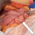
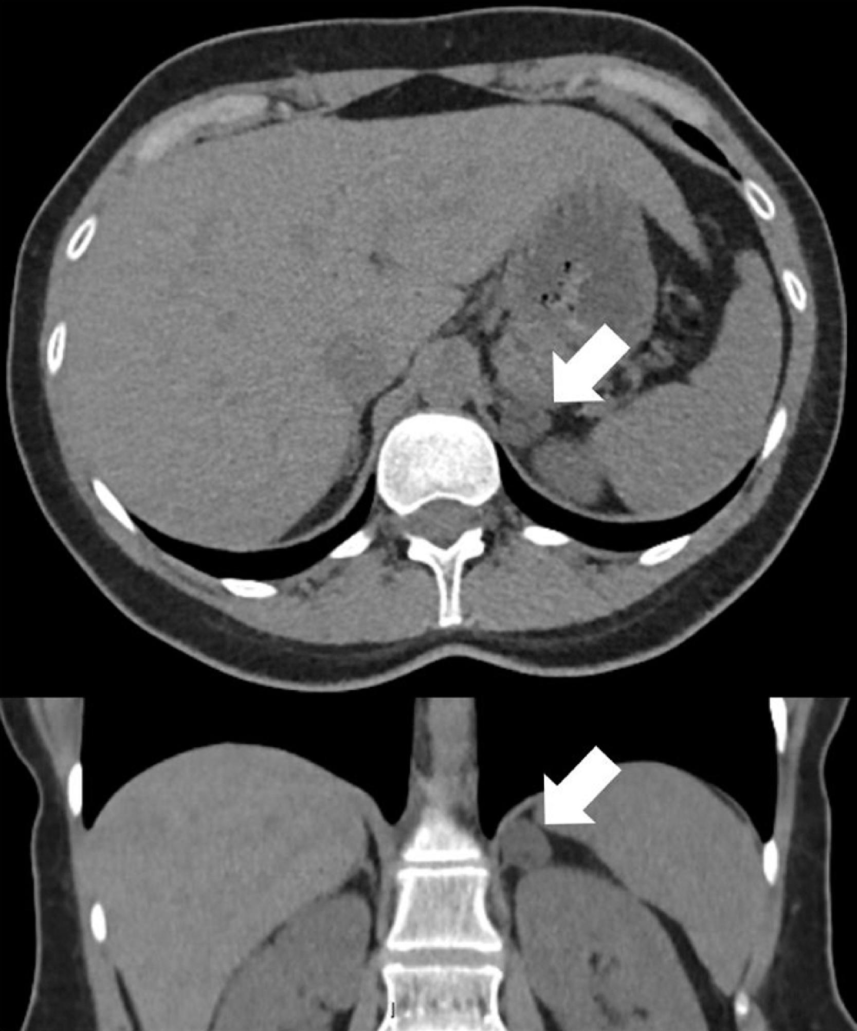
Unenhanced computed tomography (CT) of the abdomen was performed and demonstrated a 1.6-cm left adrenal mass consistent with a diagnosis of adrenal cortical adenoma (unenhanced CT attenuation was 6 Hounsfield units [HU]) ( Fig. 16.1 ). The right adrenal gland was slightly atrophic. Hormonal workup for hypercortisolism was performed, including morning measurement of ACTH and dehydroepiandrosterone sulfate (DHEA-S), 1-mg overnight dexamethasone suppression test, and 24-hour urinary free cortisol (UFC) excretion. The laboratory data were consistent with ACTH-independent hypercortisolism ( Table 16.1 ). Laparoscopic adrenalectomy was recommended.
| Biochemical Test | Result | Reference Range |
| 1-mg overnight DST, mcg/dL | 11 | <1.8 |
| Morning cortisol, mcg/dL | 13 | 7–25 |
| ACTH, pg/mL | <5 | 7.2–63 |
| DHEA-S, mcg/dL | <15 | 18–284 |
| Urine free cortisol, mcg/24 h | 108 | 3.5–45 |
Stay updated, free articles. Join our Telegram channel

Full access? Get Clinical Tree


