1 Introduction to peripheral blood smear examination
Wedge smear preparation
Making the peripheral blood smear
Although some automated analyzers prepare and stain blood smears according to established criteria, manual blood smear preparation is still used in many places. The wedge smear is a convenient and commonly used technique for making peripheral blood smears. This technique requires at least two 3 × 1-inch (75 × 25-mm) clean glass slides. High-quality, beveled-edge microscope slides are recommended. One slide serves as the blood smear slide and the other as the spreader slide. These can then be reversed to prepare a second smear. A drop of ethylenediaminetetraacetic acid (EDTA) anticoagulated blood about 3 mm in diameter is placed at one end of the slide. Alternatively, a similar size drop of blood directly from a finger or heel puncture is acceptable. The size of the drop of blood is important. Too large a drop creates a long or thick smear, and too small a drop often makes a short or thin smear. In preparing the smear, the technician holds the pusher slide securely in front of the drop of blood at a 30- to 45-degree angle to the smear slide (Figure 1-1, A). The pusher slide is pulled back into the drop of blood and held in that position until the blood spreads across the width of the slide (Figure 1-1, B). It is then quickly and smoothly pushed forward to the end of the smear slide, creating a wedge smear (Figure 1-1, C). It is important that the whole drop of blood is picked up and spread. Moving the pusher slide forward too slowly accentuates poor leukocyte distribution by pushing larger cells, such as monocytes and granulocytes, to the very end and sides of the smear. Maintaining a consistent angle between the slides and an even, gentle pressure is essential. It is frequently necessary to adjust the angle between the slides to produce a satisfactory smear. For higher than normal hematocrit, the angle between the slides must be lowered so that the smear is not too short and thick. For extremely low hematocrit, the angle must be raised. A well-made peripheral blood smear (Figure 1-2) has the following characteristics:
1. About two-thirds to three-fourths of the length of the slide is covered by the smear.
2. It is slightly rounded at feather edge (thin portion), not bullet shaped.
3. Lateral edges of the smear should be visible. The use of slides with chamfered (beveled) corners may facilitate this appearance.
4. It is smooth without irregularities, holes, or streaks.
5. When the slide is held up to light, the feather edge of the smear should have a “rainbow” appearance.

Figure 1–1 Wedge technique of making a peripheral blood smear. A, Correct angle to hold spreader slide. B, Blood spread across width of slide. C, Completed wedge smear.
(From Rodak BF, Fritsma GA, Keohane EM: Hematology: clinical principles and applications, ed 4, St. Louis, 2012, Saunders.)
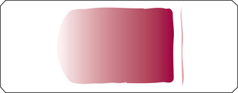
Figure 1–2 Well-made peripheral blood smear.
(From Rodak BF, Fritsma GA, Keohane EM: Hematology: clinical principles and applications, ed 4, St. Louis, 2012, Saunders.)
Figure 1-3 shows examples of unacceptable smears.
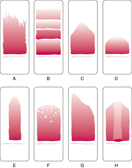
Figure 1–3 Unacceptable peripheral blood films. Slide appearances associated with the most common errors are shown, but note that a combination of causes may be responsible for unacceptable films. A, Chipped or rough edge on spreader slide. B, Hesitation in forward motion of spreader slide. C, Spreader slide pushed too quickly. D, Drop of blood too small. E, Drop of blood not allowed to spread across the width of the slide. F, Dirt or grease on the slide; may also be caused by elevated lipids in the blood specimen. G, Uneven pressure on the spreader slide. H, Time delay; drop of blood began to dry.
(From Rodak BF, Fritsma GA, Keohane EM: Hematology: clinical principles and applications, ed 4, St. Louis, 2012, Saunders.)