Vulvar Cancer


Vulvar Cancer


Before the mid-1980s, it was commonly believed that radiation therapy (RT) had no role in the curative management of vulvar cancer. Then in 1986, Homesley et al.1 reported the results of a randomized trial that demonstrated improved survival when adjuvant RT was given to patients with node-positive vulvar cancer. Since then, clinicians have sought to refine and, where appropriate, expand the indications for RT. However, there continue to be many sources of controversy concerning the relative roles and optimal treatment guidelines for surgical and radiotherapeutic management. Several features contribute to these management challenges.


  • Vulvar cancer is uncommon. In the United States, fewer than 6,000 new cases are diagnosed each year. Although multidisciplinary practices vary, generally no more than one-third of patients with vulvar cancer have an indication for RT. This means that vulvar cancer comprises an even smaller proportion of RT practice than vaginal cancer.


  • There is very little level I evidence to guide treatment. Since the Homesley trial,1 most studies relevant to the radiotherapeutic management of vulvar cancer have been retrospective. Many of the patients included in these studies were treated before modern diagnostic and therapeutic standards were available, limiting the relevance of their data to current practice.


  • The external beam techniques used to treat vulvar cancer are challenging. As discussed in this chapter and in Chapter 5, poor visualization of many vulvar tumors on standard diagnostic imaging studies makes accurate target volume definition difficult. The shape and orientation of the targets and close proximity of critical structures also pose special treatment planning challenges.


  • Management of the short-term side effects of surgery and radiation is particularly challenging for patients with vulvar cancer. Postoperative complications, infection, and radiation effects can all lead to treatment delays that may reduce the effectiveness of RT. Skilled management of treatment-related side effects plays an important role in achieving optimal results.


  • The long-term effects of treatments can be severe. Many factors, including postoperative complications, radiation effects, infection, and tissue damage caused by the primary vulva cancer, can have lasting effects on the well-being of patients with locoregionally advanced vulvar cancer. These effects can be minimized through careful integration of multidisciplinary treatments. However, clinicians are still searching for the best ways to balance these treatments, particularly for patients with locally advanced disease.


PRETREATMENT EVALUATION

Careful pretreatment evaluation helps to define the extent of disease and to determine whether definitive surgical resection can be accomplished without sacrificing organ function. The primary goals of pretreatment evaluation are as follows:


  • To determine the extent and resectability of local and regional disease.


  • To rule out metastatic disease that would change the treatment goal from curative to palliative.


  • To rule out other primary genital tract cancers. About half of vulvar cancers are human papilloma virus (HPV) related, and these patients are at increased risk for cervical, vaginal, and anal cancers.


  • To determine whether the patient is medically fit for treatment. All patients require a careful history and complete physical examination before initiation of treatment.

Pretreatment studies should be selected to answer these questions in the most cost-effective way possible. Studies that can help to guide treatment include the following:


  • Physical examination: Pelvic examination should include careful inspection and palpation of the vulva and perianal region including rectovaginal examination to determine the extent of local disease and proximity of tumor to the urethra, clitoris, anus, and vagina. The cervix and vagina should be carefully examined, ideally with colposcopy, to rule out invasive or preinvasive lesions in those sites. If patient discomfort prevents an adequate assessment in clinic, examination under anesthesia (EUA) should be performed; if there is periurethral disease, cystourethroscopy should be performed. In cases of locally advanced disease, both the gynecologic oncologist and radiation oncologist should be present at EUA if possible. In selected cases (particularly those with vaginal involvement), interstitial placement of fiducial markers can assist in subsequent radiation treatment planning (CS 12.3). All clinical findings should be carefully documented, with detailed descriptions of the location and morphology of abnormal lesions and estimates of tumor diameters and the distances between tumor and perineal structures. Examination should also include an assessment of the groins, although even extensive lymphadenopathy can be missed on physical examination (CS 12.8).


  • Ultrasound: In experienced hands, ultrasound combined with ultrasound-directed fine needle aspiration (FNA) cytology has a better than 90% sensitivity for detecting nodal metastasis (Chapter 4).2 In one study of 40 patients, Moskovic et al.3 correctly identified metastatic nodal disease in 11 of 13 groins; the two false negatives were in nodes with metastatic deposits <3 mm. However, the sensitivity of ultrasound undoubtedly varies with the experience of the ultrasonographer and, even in the most experienced hands, a negative result is insufficient to rule out the need for groin treatment in patients with invasive cancers. Ultrasoundguided FNA may, in selected cases, help to guide the dose of radiation given to equivocal nodes; however, if there is any doubt, suspicious nodes that have not been removed should be treated as if they were positive, because the price for error is high (CS 12.1). Ultrasound is also a noninvasive, inexpensive method for following the groins of patients after negative results for sentinel node biopsy if a complete dissection has not been performed (CS 12.10).


  • Contrast-enhanced computed tomography (CT): Although CT is generally superior to physical examination for the detection of lymphadenopathy, it cannot detect micrometastases. Also the infections often associated with vulvar cancers can cause inflammatory nodal enlargement, reducing the specificity of CT. CT can be valuable for detecting extrapelvic metastases but rarely provides useful information about the extent of disease in the vulva.


  • Magnetic resonance imaging (MRI): MRI provides much better soft tissue detail than CT and can therefore be very helpful in assessing the extent of disease in the vulva and vagina. It is particularly useful in cases of locally advanced disease, where MRI can assist in the determination of resectability. MRI is often very helpful in radiation treatment planning, particularly if the study can be obtained in the treatment position. However, MRI cannot replace a careful physical examination. In particular, MRI frequently underestimates the extent of mucosal involvement in the vagina and, in some cases, fails entirely to detect even fairly extensive vulvar tumors (CS 12.8).


  • Positron emission tomography (PET)-CT: Studies of the ability of PET-CT to detect inguinal metastases from squamous carcinomas of the vulva have reported sensitivities that range between 50% and 70% and specificities that range from 60% to 90% (Chapter 4).2 Inflammatory nodes are frequently FDG-avid. PET-CT can sometimes be helpful in the designation of RT target volumes in patients with advanced disease.

To summarize, physical examination is the most important method for clinical evaluation of local disease extent. Most patients with locoregionally advanced disease should also have CT of the chest, abdomen, and pelvis or PET-CT to assess regional nodes and rule out distant disease. MRI may help to determine operability and define disease extent for RT planning. However, in most cases, surgical evaluation determines the stage of disease and the need for adjuvant treatment.


RISK ASSESSMENT


Predictors of Vulvar Recurrence

The most frequent site of recurrence after primary surgical treatment of vulvar cancer is in the vulva.4 Some vulvar recurrences are true relapses of the initial invasive lesion, whereas others are undoubtedly new cancers arising in areas at high risk for neoplastic transformation because of lichen sclerosis or HPV infection.

One of the most difficult decisions for radiation oncologists is whether and when to give adjuvant radiotherapy to the vulva after surgical excision. Although several investigators have evaluated factors associated with local recurrence after treatment of invasive carcinoma, their results are not always applicable to modern practice because the studies are typically retrospective, span many years of evolving surgical techniques, and are often confounded by the inconsistent delivery of adjuvant radiotherapy. However, any oncologist trying to weigh the risks and benefits of vulvar radiotherapy must consider these data to estimate the probability of vulvar recurrence and whether local recurrence, if it occurs, is likely to be salvageable with additional surgery.


The Outcome of Patients with Recurrence in the Vulva after Vulvectomy

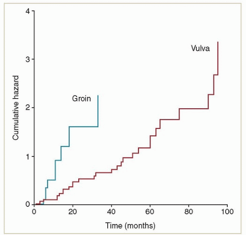
The outcome of salvage surgery for local vulvar recurrence has been the subject of several studies. Woolderink et al.8 reported a 5-year local recurrence rate of 30% after wide radical excisions of vulvar cancers. The median time to local recurrence was long (40 months, Fig. 12.2) with a wide range
(3 to 141 months), suggesting that many of these recurrences represented new cancers in a high-risk field. Although recurrences were frequently localized to the vulva, 72% of the 29 patients who were treated for local recurrence had a second recurrence in the vulva. The authors of this study did not discuss the effect of local recurrence on survival.


FIGURE 12.2 The time course of 29 vulvar and groin recurrences in 125 patients treated with wide local excision and superficial groin dissection between 1985 and 1999. Most groin recurrences occurred during the first year after surgery. Late vulvar recurrences likely represented new primary lesions associated with lichen sclerosis or HPV. The groin recurrence rate in this series was high, reflecting efforts during the 1990s to reduce the radicality of groin dissection to decrease the rates of perioperative and late side effects. This approach was subsequently abandoned because of the high rate of groin recurrence and death. (Woolderink JM, de Bock GH, de Hullu JA, et al. Patterns and frequency of recurrences of squamous cell carcinoma of the vulva. Gynecol Oncol. 2006;103:293-299.)

To summarize, the studies quoted above can be used to suggest some groups that appear to be at particularly high or low risk for local recurrence. The available data are insufficient to provide precise estimates, so the percentages should be considered to be only rough estimates of the risk of recurrence for patients treated with excision without adjuvant RT.

Very high risk (>70%)


  • Positive resection margins


  • Local recurrence <1 to 2 years after primary resection, particularly if other high-risk features are present.

High risk (about 50% to 60%)


  • Margins <5 mm, particularly with tumor >3 cm, deep invasion or positive lymph nodes


  • Multifocal disease or local recurrence more than 2 years after resection with other high-risk features

Moderately high risk (about 30% to 40%)


  • Margins 5 to 8 mm, particularly with tumor >3 cm, deep invasion or positive lymph nodes

Intermediate risk (<30%)


  • Margins >8 mm but tumor >3 cm, deep invasion or positive nodes

Very low risk (<5% to 10%)


  • Margins >1 cm, tumor <3 cm, superficially invasive (<2 to 3 mm), negative nodes

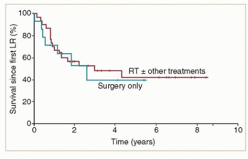
Although local recurrences can often be cured with re-excision, the prognosis is not uniformly good. In a review of the outcomes of 47 patients who had isolated local recurrences after initial surgical treatment, Faul et al.13 reported a 5-year overall survival rate of only 40% (Fig. 12.3). In their study, factors associated with a poor outcome after recurrence included short disease-free interval, tumor size >3 cm at the time of recurrence, high initial stage, and lymph-vascular space invasion (LVSI).

As will be discussed later, the risk of postexcision vulvar recurrence is only one factor that must be considered during the formulation of an overall treatment strategy for patients with vulvar cancer.


Predictors of Lymph Node Involvement

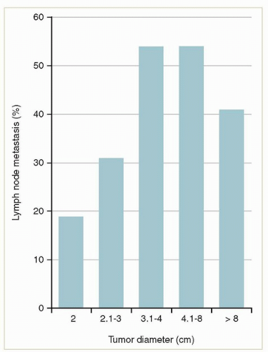
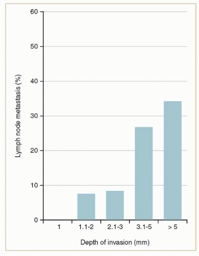
The vulva has a rich network of lymphatics that drain primarily to the inguinofemoral nodes (Chapter 5). The rate of lymph node metastasis is strongly correlated with tumor diameter5,14 (Fig. 12.4) and the depth of stromal invasion (as measured from the most superficial dermal papilla adjacent
to the tumor to the deepest focus of invasion) (Fig. 12.5).2 Only small tumors that invade <1 mm (stage IA) can be considered to have a negligible risk of lymph node metastasis. The risk rises steeply as the depth extends beyond 1 mm.


FIGURE 12.3 Survival from the time of first vulvar recurrence for 45 patients who had isolated vulvar recurrences after vulvectomy. Recurrences were treated with re-excision only (31 patients) or with RT with or without chemotherapy or excision (14 patients). The benefits of the two treatment approaches cannot be directly compared because patients who had treatment involving RT had significantly larger recurrent tumors than those treated with excision only. Although many recurrences appear to have been salvaged, the overall survival rate is only about 40%, even after exclusion of patients who had multiple sites of recurrence. (Faul CM, Miramow D, Gerzten K, et al. Isolated local recurrence in carcinoma of the vulva: prognosis and implications for treatment. Int J Gynecol Cancer. 1998;8:409-414.)


FIGURE 12.4 The relationship between tumor diameter and the incidence of lymph node metastases in 588 patients treated with vulvectomy and lymphadenectomy. (Homesley HD, Bundy BN, Sedlis A, et al. Radiation therapy versus pelvic node resection for carcinoma of the vulva with positive groin nodes. Obstet Gynecol. 1986;68:733-740.)


FIGURE 12.5 The relationship between the depth of stromal invasion and the incidence of lymph node metastases. (Hacker NF, Eifel PJ. Vulvar cancer. In: Berek JS, Hacker NF, eds. Gynecologic Oncology. Philadelphia, PA: Wolters Kluwer; 2015:560-607.)

Other factors that have been associated with lymph node metastasis are high tumor grade5,14 and the presence of LVSI14 in the primary tumor.


Predictors of Disease-Related Death

Local or regional recurrence plays a role in most deaths from vulvar cancer; however, distant metastases to lung, bone, or other sites may also occur in isolation or combined with locoregional failure. A number of clinical features can be used to predict outcome.


Inguinal Node Involvement

The most powerful and consistent predictor of outcome is lymph node involvement. The number of positive nodes and the size of the largest focus of intranodal disease are strongly associated with recurrence and death from disease.14,15,16,17 For patients who have a single microscopic nodal metastasis <5 mm, the rate of groin recurrence is <1% if a radical inguinofemoral lymphadenectomy has been performed. Patients who have more extensive node involvement or who have had less than a complete lymphadenectomy require adjuvant RT to prevent an unacceptably high rate of groin recurrence.1,18 For patients who receive adjuvant radiotherapy, the presence of extranodal extension has been consistently found to be an independent predictor of groin recurrence and death from disease.14,15,17

The prognosis of patients who have a groin recurrence is very poor. For those whose initial treatment was surgery alone, the survival rate after groin recurrence is <20% in most series, although the doses and techniques used for salvage radiation are rarely mentioned and most reports antedate modern image-based treatment methods.19 Patients who have a groin recurrence after initial treatment with surgery and radiation can rarely, if ever, be cured.


Pelvic Node Involvement

Historically, pelvic node metastases were associated with poor prognosis with reported survival rates of <20%1; patients who have pelvic node metastases are still categorized as having International Federation of Gynecology and Obstetrics (FIGO) stage IVB disease. However, most of the outcome data predate the standard use of adjuvant inguinal and pelvic radiation for node-positive vulvar cancer. In a 2015 review of 20 patients with grossly involved pelvic nodes who were treated with definitive RT, the 5-year overall survival rate was 43%, similar to the rates typically reported for stages IIIB and IIIC disease.20


Other Predictors

Most factors that have been associated with an increased risk of vulvar recurrence or lymph node involvement are also correlated with the rates of disease-specific and overall survival. These include tumor size,5,15,17,21 depth of invasion,5,15 histologic grade,5,17,21 and LVSI.17


FIGO Stage

The most recent (2009) version of the FIGO staging system incorporates the most powerful predictors of local recurrence, regional involvement, and survival (Table 12.1). As such, it will undoubtedly prove to be a strong predictor of outcome. However, it has a few limitations.


  • In 1994, FIGO moved to a surgical staging system with categories based on radical local excision and, after 2009, complete inguinal lymphadenectomy. This makes it difficult to categorize patients who are treated with lesser surgical procedures combined with radiotherapy or with radiotherapy alone.


  • The grouping of pelvic node metastases and hematogenous metastases together in the stage IVB category underestimates the curability of patients who have pelvic node disease.20


  • Although the 1994 and 2009 classifications improved the predictive power of the FIGO classifications, the three systems are so radically different that they cannot be readily translated from one to the other. This has made it extremely difficult to interpret stage designations in retrospective studies of vulvar cancer, most of which cross these historical divides (Chapter 2).


TABLE 12.1 Evolution of the FIGO/AJCC Staging System for Vulvar Cancer


AJCC Stage


1969-1993


1994-2008


2009-


FIGO Stage (2009)


(Based on clinical findings)


(Based on operative findings)


Primary tumor


T1


Tumor confined to the vulva ≤2 cm in diameter


T1a Confined to the vulva or perineuma; ≤2 cm diameter and ≤1 mm stromal invasion


IA


T1b Confined to the vulva or perineuma; ≤2 cm diameter and >1 mm stromal invasion


IB


T2


Tumor confined to the vulva >2 cm in diameter


Tumor confined to the vulva or perineum,a >2 cm in diameter


Tumor any size and growing into the anus, lower one-third of the vagina or urethrab


II


T3


Tumor any size and growing into the anus, perineum,a lower urethra, or vagina


Tumor any size and growing into the anus, lower urethra, or vagina


Tumor any size, growing into the upper urethra, bladder or rectum, upper two-thirds of vagina, or into pubic boneb


IVA


T4


Tumor any size, growing into the upper urethra, bladder or rectum, or into pubic bone


Nodes


N0


No nodes palpable


No lymph node metastasis


No regional lymph node metastasis


N1


Nodes palpable in either groin, not enlarged, mobile (not clinically suspect for neoplasm)


Unilateral regional lymph node metastasis


N1a one or two lymph node metastases, each <5 mm


IIIA (i)


N1b one lymph node metastasis ≥5 mm


IIIA (ii)


N2


Nodes palpable in either or both groins, enlarged, firm, and mobile (clinically suspect for neoplasm)


Bilateral regional lymph node metastasis


N2a three or more lymph node metastases, each <5 mm


IIIB (i)


N2b two or more lymph node metastases ≥ 5 mm


IIIB (ii)


N2c Regional lymph node metastasis with extracapsular spread


IIIC


N3


Fixed or ulcerated nodes


Fixed or ulcerated regional lymph node metastasis


IVA


Distant disease


M0


No clinical metastases


No distant metastasis


M1


Palpable deep pelvic lymph nodes


Any distant metastasis including pelvic lymph nodes


IVB


M2


Other distant metastases


a In 1994, tumors located on the perineum were moved out of the AJCC T3 category and were thereafter grouped with other vulvar cancers according to operative findings.

b In 2009, tumors involving the anus, distal vagina, or urethra were moved from the AJCC category T3 to T2 and from FIGO stage III to stage II; at the same time, tumors involving the proximal urethra, bladder, rectum, upper vagina, or pubic bone were moved from AJCC T4 to T3 but remained in the FIGO stage IVA category.


DEVELOPING A MULTIDISCIPLINARY TREATMENT STRATEGY

Locoregionally advanced vulvar cancer requires some of the closest multidisciplinary collaboration of any gynecologic cancer treated with RT. The decision whether to treat with surgery, RT, or a combination of the two treatments requires careful analysis of the role each treatment can play in controlling disease and preserving function. Whenever possible, these uncommon, complex cases should be treated in a center that has an experienced, subspecialized, and closely collaborative multidisciplinary team (Chapter 3).

Once the pretreatment evaluation has been completed, an initial treatment strategy should be developed after considering:


  • The risks and benefits of various treatment options for the primary site.


  • The risks and benefits of various treatment options for the nodes.


  • The combination of approaches to the primary site and groin that will achieve the best overall chance of cure with the least impact on quality of life.


Management of the Primary Site


Surgical Management

A traditional radical vulvectomy involves en bloc removal of the entire vulva, including the clitoris, the inguinofemoral nodes, and a “bridge” of intervening tissue. This extensive procedure, which was the standard treatment for most vulvar cancers until at least the late 1980s, had severe psychosexual and physical consequences and is rarely performed today. Vulvectomy and inguinal lymphadenectomy performed through separate incisions is better tolerated than the en bloc procedure and is equally effective.19 Recurrences in the skin bridge are rare after this staged procedure.19

Today, most invasive vulvar cancers are treated with an even more limited procedure called a “wide local excision” or “radical local excision.” With this procedure, the gross lesion is removed with, whenever possible, a skin margin of 1 to 2 cm; at depth, the dissection is usually carried down to the fascia of the urogenital diaphragm or pubic fascia. If necessary, a portion of the distal urethra can be removed, usually without compromising urethral function. Tumors that involve the perineum are frequently difficult to remove with an optimal margin because of proximity to the anus or rectum. Also, tumors that involve the vagina usually cannot be radically resected without compromising the urethra or rectum.

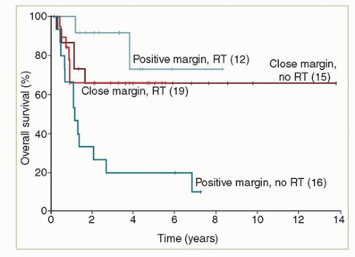
If preoperative evaluation suggests that organ-sparing surgery is likely to result in a positive resection margin, initial treatment with RT is usually preferred because (1) the dose required to prevent recurrence of disease after a positive resection margin is similar to that required to control gross disease; (2) postoperative wound complications can delay the start of RT, providing an opportunity for disease progression; and (3) the side effects of combined surgery and high-dose radiation tend to be greater than those of radiation alone. In some cases, however, resection with a close margin followed by planned postoperative RT may be preferred over treatment with radiation alone if the surgeon believes negative margins can be achieved and if the estimated risk of postresection wound complications is low. The goal should always be to achieve the best chance of local control with the least morbidity (Chapter 3). Close cooperation and early consultation between the surgeon and radiation oncologist are key to achieving the best result.


Postoperative Radiation Therapy

Predictors of postvulvectomy local recurrence are discussed earlier in this chapter. The most common indications for postoperative vulvar radiation are a close or positive margin. However, other risk factors should be considered and, as will be discussed later in this chapter, the presence of lymph node metastases may influence the decision whether to treat the vulva.

Only gold members can continue reading. Log In or Register to continue

Stay updated, free articles. Join our Telegram channel

Sep 29, 2018 | Posted by in ONCOLOGY | Comments Off on Vulvar Cancer

Full access? Get Clinical Tree
Get Clinical Tree app for offline access