Vaccines


Vaccines


Ennio De Gregorio

Ugo D’Oro

Sylvie Bertholet

Rino Rappuoli


INTRODUCTION

Over the last century, vaccination has been the most effective medical practice to control infectious diseases. Smallpox has been eradicated worldwide, and polio has been almost eliminated. Most viral and bacterial diseases traditionally affecting children worldwide are now preventable with vaccines (see www.who.int/vaccines-documents). Vaccination is estimated to save two to three million lives per year. Traditional vaccines designed following the basic Pasteur principle (isolate, inactivate, and inject) have been extremely successful in preventing infections by pathogens expressing relatively conserved antigens mainly through antibody-mediated mechanisms. However, there is still a long list of diseases that are not preventable by vaccination, and infectious diseases are still a major cause of death and disability worldwide. Some of these diseases are caused by pathogens that have a high degree of antigen variability and cannot be controlled only by antibodies but require a mix of humoral and cellular immune responses. As an example, the world is still waiting for effective vaccines against the three big killers: acquired immunodeficiency syndrome (AIDS), tuberculosis (TB), and malaria, which collectively kill almost five million people per year. Therefore, the development of new vaccines can still have a tremendous impact in reducing the mortality caused by infectious diseases throughout the world. Novel technologies for antigen identification, structural design, and formulation now allow for the development of safer vaccines that can better cope with pathogen diversity. In addition, progress in the field of innate immunity is translating into the development of novel classes of vaccine adjuvants that can promote, better than in the past, protective humoral and cellular immune responses. Significant research efforts are also being applied toward the development of therapeutic vaccines against cancer, allergy, autoimmunity, and other inflammatory disorders that could have a major impact on human health across the world. However, in this context, we will focus mainly on preventive vaccines against infectious diseases, although therapeutic vaccines against cancer will not be covered here. This chapter will give a short overview of the history of vaccine development and will describe most of the existing licensed vaccines for the prevention of infectious diseases. In addition, we will give details on novel approaches to develop effective vaccines against human immunodeficiency virus (HIV), malaria, and TB. One paragraph is dedicated to licensed and experimental vaccine adjuvants, with special focus on their immunologic mechanism of action. Other sections of this chapter will analyze new technologies that can either improve existing vaccines or allow the development of novel effective vaccines where conventional technologies have failed. Another section will concentrate on novel needle-free vaccine delivery systems. Finally, the last paragraph will review the major challenges pertaining to vaccines in the 21st century society.


HISTORICAL PERSPECTIVES


Smallpox: From Variolation to Vaccination

Smallpox, one of the most severe human viral diseases, is transmitted by inhalation of orthopoxvirus Variola. Before vaccination, smallpox outbreaks where quite frequent, killing more than 30% of infected people. It is estimated that in the late 18th century in Europe, 400,000 people died annually of smallpox. A method for preventing naturally acquired smallpox was discovered in India before AD 1000 and spread to China and western Asia. This method, called “variolation,” was introduced in Europe in 1721 by Lady Mary Wortley Montagu after her return to England from Constantinople were she observed the use of the technique. Variolation consisted in the inoculation of pustule material from patients infected with smallpox in the skin or in the nose of noninfected people to protect them from the outbreaks. Of course, the practice was not safe, with a mortality rate ranging from 1% to 2%; however, it generally produced an illness that was less severe than natural Variola infection by inhalation. A variolation campaign was introduced in Siena, Italy in 1758 as reported in a dedicated publication by the local Academy of Science in 1760 (Fig. 43.1). The symptoms experienced by a child in the days following variolation are described in the following translated excerpt.


In the year 1756, on the 8th of June, day on which the weather was very hot, Gherardo, son of Mr. Giovanni Pavolotti, was inoculated with smallpox…. A small cut was performed on each arm, and a wad of cotton wool soaked in the pus of a smallpox pustule of his brother, who had naturally acquired the disease, was applied. His arms were wrapped and the bandages were removed for the first time on the second day to treat the wounds. The cuts were almost dry and showed no change in color or other signs on the surrounding skin. The child showed not even the slightest discomfort; he went about the house enjoying his childish babble as usual; his pulse was steady, normal, the same as before the operation….

Fourth day. The cuts started to hurt occasionally and to a small degree, they were red and started to fester…

Sixth day. All became more serious. He complained a lot about the cuts; they were more open, festering,
swollen, and quite uneven around the edges. At midday he felt cold, then he became hot, feverish. In the evening, he vomited bitter, biliary fluids. His eyes were teary, light did not bother him very much. The cuts had small blisters in the middle and around the uneven edges. They were fetid. Moderate fever, no thirst. He was kept under watch overnight. Moderate urination with dark yellow sediment.

Seventh day. The cuts were more swollen, more festered, more fetid, the blisters were white, some were open. He complained about shooting pain in the armpits. At midday he became very hot, the fever rose, he was thirsty, somewhat shivering, frightened, spoke senselessly…

Eighth day. At sunrise he slept at length. All was very pleasant. The cuts were very festered. The blisters that had appeared on the sixth day were open. There were some smallpox pustules around the cuts…

Ninth day. No fever at all…

Tenth day. The rash stopped. The blisters started to fester. The child got up from bed and walked about the house.

Sixteenth day. The blisters were dry and the scabs started falling. The cuts were red, there was less pus, which was not so fetid…

Twenty-fourth day. The cuts had cicatrized. On that day, the child went outdoors for a walk and the blister marks were hardly visible.


FIG. 43.1. Front page of the publication by the Academy of Science of Siena reporting the history of the variolation campaign made in Siena between 1758 and 1760. Reproduction by courtesy of the Accademia de’ Fisiocritici, Siena, Italy.

A real vaccination practice was introduced when Edward Jenner replaced smallpox-infected material with pustule material from humans infected by cowpox. Cows can be infected by a virus similar to Variola, called Vaccinia virus, and develop a smallpox-like diseases called cowpox that can be transmitted to humans. It was quite frequent that people in close contact with cows, such as milkmaids, would contract cowpox and develop sores on fingers and hands. In the 18th century, several physicians noticed that people exposed to cowpox were protected from smallpox and did not react to variolation. In 1796, Edward Jenner demonstrated for the first time that pustule material taken from a milkmaid infected by cowpox when inoculated into the skin of another person produced a localized self-limiting infection, which, a few months afterward, was able to protect from smallpox challenge (by variolation). He also demonstrated that the same individual was still protected from smallpox variolation 5 years later, confirming the persistence of immunity. Jenner called the material used for the inoculum Vaccinia (from “vacca,” the Latin name of cow) and the process “vaccination.” For many years, vaccination became a standard practice in several countries and replaced variolation. At the beginning, vaccination was still relying on the arm-to-arm inoculation of human infected material. This process led to a progressive attenuation of the inoculum and was often associated with the transmission of other human diseases from the donor. Therefore, by 1890, human infected material was replaced by the use of
vesicle fluid from infected cows. In 1940, Collier developed a modern commercial process to produce a stable freeze-dried vaccine for large-scale smallpox prevention. Because Variola virus is transmitted from human to human and does not have any animal reservoir, it was hypothesized that eradication of smallpox through vaccination was possible. In 1959, the World Health Organization (WHO) set global smallpox eradication as a goal, but it was only in 1966 that a Smallpox Eradication Unit was formed. The global eradication program started in 1967. It is estimated that at that time, there were still 10 to 15 million cases of smallpox worldwide. The vaccination campaign lasted about 10 years and was extremely successful. The last case of naturally contracted smallpox was recorded in 1977 in Somalia, and in 1979, the Global Commission for Smallpox Eradication certified that global smallpox eradication had been achieved.


Nineteenth Century Vaccines

The first successful vaccine was discovered by Jenner without any understanding of its mechanism of action. The microbial origin of infectious diseases was discovered many years later by Louis Pasteur and Robert Koch. Both scientists gave a dramatic impulse to vaccine research. Pasteur discovered that bacteria grown in vitro for a sufficient amount of time lose their virulence and become attenuated. He proposed that these live-attenuated microorganisms could be used to inoculate individuals similarly to what Jenner had done in the case of cowpox. In honor of Jenner, Pasteur extended the word “vaccine” to all preparations used for immunization. Initially, Pasteur worked on animal models demonstrating that live-attenuated chicken cholera provides immunity against an experimental challenge with virulent organisms. Later, he showed that attenuated anthrax bacilli protected sheep from infection. After demonstrating the vaccination principle in animals, Pasteur worked on an attenuated rabies preparation that was first administered in humans in 1885. This vaccine was prepared by infecting rabbits with rabiesinfected material for several passages and then recovering rabbit spinal cord. The virulence of spinal cord material was further attenuated by exposing it to dry air. Pasteur’s rabies vaccine was administered for the first time subcutaneously in 13 doses over a 10-day period to Joseph Meister, a boy who had been bitten by a rabid dog 60 hours before and had no chance to survive. Thanks to vaccination, the patient resisted the development of the disease. At that time, Pasteur did not know that rabies was caused by a virus belonging to the family of Rhabdoviridae that takes several days from the time of infection to reach the central nervous system, making postexposure prophylaxis possible. Today, several antirabies vaccines exist and are all made of chemically inactivated virus grown in duck embryos or in cell culture.

Pasteur thought that only live-inactivated pathogens could be used for effective vaccination. The concept of killed vaccines was introduced in 1886. After Robert Koch discovered Vibrio cholerae, Daniel Elmer Salomon and Theobald Smith showed that a killed suspension of V. cholerae from hogs protected pigeons from the disease. Similar data were presented by a group working at the Pasteur Institute. These findings paved the way for the development of human killed whole-organism vaccines against typhoid fever (1896), cholera (1896), and plague (1897) (Table 43.1).


TABLE 43.1 Development of Human Vaccines


Live Attenuated


Killed Whole Organism


Toxoid/Protein


Polysaccharide


Glycoconjugate


Recombinant


Century


Smallpox


18th


Rabies


Typhoid


Cholera


Plague


19th


Tuberculosis (BCG)


Yellow fever


Polio (OPV)


Measles


Mumps


Rubella


Typhoid


Varicella


Rotavirus


Cholera


Pertussis


Influenza


Typhus


Polio (IPV)


Rabies


JE


TBE


HAV


Diphtheria


Tetanus


Acellular


Pertussis


Anthrax


Influenza


subunit


Pneumococcus


Meningococcus


Hib


Typhoid (Vi)


Hib


HBV


Lyme disease


Cholera toxin B


20th


Cold-adapted influenza


Rotavirus reassortants


Zoster


Pneumococcus


MenACWY


HPV


21st


BCG, Bacille Calmette-Guérin; HAV, hepatitis A virus; HBV, hepatitis B virus; Hib, Haemophilus influenzae type b; IPV, inactivated polio vaccine; JE, Japanese encephalitis; Men, meningococcus; OPV, oral polio vaccine; TBE, tick-borne encephalitis.


Adapted from Plotkin S, Orenstein WA, Offit P. Vaccines. 5th ed. New York, NY: Saunders-Elsevier; 2008, with permission.


Twentieth Century Vaccines

At the end of the 19th century, the basic principle acknowledged in vaccinology was to isolate the causative agent of disease, attenuate or kill the agent, and immunize. This principle allowed for the development of many live-attenuated and killed vaccines. The first important innovation in vaccinology in the first half of the 20th century was the discovery of vaccines based on inactivated toxins, also called toxoids. Bacterial toxins had already been identified in the 19th century. In 1888, Roux and Yersin identified diphtheria toxin. Two years later, Emil von Behring and Shibasaburo Kitasato discovered the presence of antitoxins in serum. In 1891, passive immunization using sera of animals immunized with low doses of diphtheria and tetanus toxins became available. However, toxins were not used as vaccines because they were too dangerous. At the beginning of the 20th century, Theobald Smith demonstrated that vaccines made from chemically inactivated toxins protected guinea pigs from infection. In 1923, Alexander Glenny showed that diphtheria toxin can be chemically inactivated by formalin, leading to the first human toxoid vaccine against diphtheria. In 1926, another formalin-inactivated toxoid vaccine against tetanus was introduced by Gaston Ramon and Christian Zoeller.

During the same period, the application of Pasteur’s principle led to the development of two new live-attenuated vaccines against TB and yellow fever. The TB vaccine was developed by Albert Calmette and Camille Guérin at the Pasteur Institute. They started from a Mycobacterium bovis strain that was attenuated for 13 years by 230 passages in culture media. The resulting Bacille Calmette-Guérin (BCG) strain became available for human vaccination in 1927. The first yellow fever vaccine was developed by attenuating the virus in a culture medium made of mouse brain tissue and was introduced in 1932. A few years later, the introduction of embryonated eggs as a medium for growing viruses allowed for the development of a new, safer, more attenuated vaccine to prevent yellow fever infection from a strain called 17D. Embryonated eggs were also used to develop the first two human vaccines against type A influenza. In the same year (1936), a live-attenuated influenza vaccine was introduced by Wilson Smith while a killed whole virus vaccine was developed by Thomas Francis and Thomas Magill. In the first half of the 20th century, two additional whole-killed bacterial vaccines against typhus and pertussis were developed, and in 1948, the first combination vaccine against diphtheria, tetanus, and pertussis became available.

The second half of the 20th century is considered the golden age of vaccine development. Probably, the most important technologies introduced at that time are related to the production of human viruses in controlled cell culture conditions, which allowed for the development of several new virus vaccines. Among them, the two vaccines against poliovirus are perhaps the ones that gained the greatest public interest. Before vaccination, more than 20,000 cases of polio were reported annually in the United States. A formalin-inactivated polio vaccine (IPV), developed by Jonas Salk, was licensed in 1955. A few years later (1960), a liveattenuated polio vaccine for oral administration (oral polio vaccine [OPV]) was developed by Albert Sabin. Owing to global vaccination programs, polio has been eradicated in most countries and remains endemic only in some areas of Afghanistan, India, Pakistan, and Nigeria. Other liveattenuated virus vaccines licensed between 1950 and 1970 are against measles, mumps, rubella, and varicella. More recently (1998), an oral live-attenuated vaccine against rotavirus infection was licensed in the United States, Europe, and other countries. In addition to live-attenuated vaccines, new whole-killed virus vaccines have also been licensed, including a hepatitis A virus (HAV) vaccine in 1979 (Table 43.1).

In the second part of the 20th century, several bacterial vaccines were developed using purified polysaccharides. Monovalent vaccines to prevent meningococcus (Men) group A was first licensed in 1974 followed by a MenC and a bivalent MenAC vaccine in 1975, and a quadrivalent MenACWY later in 1982. A 14-valent pneumococcus vaccine was licensed in 1977 followed by a 23-valent vaccine in 1983, whereas a vaccine against Haemophilus influenzae type b (Hib) was licensed in 1985. However, polysaccharide vaccines are not protective in children younger than 18 months, who are at most risk of contracting the disease. This problem was solved when it was shown that the immunogenicity of polysaccharides could be enhanced by linking them to a carrier protein such as diphtheria toxoid (DT) or tetanus toxoid (TT). This new class of vaccines, called glycoconjugates, is immunogenic in all age groups, including infants. The first glycoconjugate to be licensed was a Hib vaccine developed by Robbins and Schneerson (1987).1

Another important innovation in vaccinology introduced in the 20th century has been the use of a genetically modified toxin as a vaccine first applied to pertussis.2 Finally, the use of recombinant proteins instead of the antigens purified directly from the pathogens has been a major breakthrough. A vaccine made of hepatitis B surface antigen (HBsAg) expressed in yeast and adsorbed to alum was the first recombinant protein vaccine for human use licensed in 1987. The vaccine takes advantage of the property of HBsAg to self-assemble in a viral-like particle (VLP), resulting in an increased immunogenicity. A second recombinant protein vaccine based on the OspA antigen from Borrelia burgdorferi expressed in Escherichia coli was licensed in 1999 to prevent Lyme disease. However, the vaccine was withdrawn from the market in 2002 due to a lack of demand.


Twenty-first Century Vaccines

Several novel vaccines were licensed in the first decade of the 21st century. Three of them (cold-adapted influenza, novel rotavirus, and zoster) are based on live-attenuated pathogens. Others, such as 7-valent, 10-valent, and 13-valent pneumococcal conjugate vaccines or quadrivalent meningococcal vaccines, are based on the glycoconjugate technology. Finally, two vaccines to prevent human papillomavirus (HPV) infection are based on recombinant antigens (Table 43.1). Most of the vaccines listed in this paragraph will be described in more detail in the section dedicated to licensed vaccines. Another paragraph will describe new experimental vaccines that have not been licensed yet, such as vaccines to prevent AIDS, TB, and malaria.


CLASSIFICATION OF HUMAN VACCINES

To be efficacious, a vaccine needs to generate an immunologic status that is sufficient to block infection by the pathogen or at least to inhibit the establishment of disease. Depending on the type of infection to be prevented, an effective vaccine may require the induction of different humoral and cellular immune effector mechanisms. Protective humoral responses include neutralizing antibodies, opsonizing antibodies, or antibodies able to induce complement-mediated killing. Protective cellular mechanisms include different classes of cluster of differentiation (CD)4+ T cells and CD8+ cytotoxic T cells (CTLs). Based on the type of material from which they are made, licensed vaccines can be broadly classified into three general categories: live-attenuated vaccines, whole-killed vaccines, and subunit vaccines. Subunit vaccines can be further divided into polysaccharide, glycoconjugate, toxoid, or protein subunit and recombinant subunit vaccines. Different types of vaccines target diverse adaptive immune responses (Fig. 43.2). For example, glycoconjugate vaccines elicit high-avidity bactericidal antibodies but do not trigger any antigen-specific effector T-cell responses. Proteinbased vaccines can induce bactericidal and neutralizing antibodies and can also trigger robust CD4+ T-cell responses; however, they are not efficient in inducing CTLs. Finally, live-attenuated vaccines are able to generate all humoral and cellular responses, including CTLs. This section will describe all classes of licensed vaccines with a special focus on their immunologic correlates of protection. In addition, some promising technologies for vaccine design such as viral vectored and nucleic acid-based vaccines will be presented, even if they have not been applied yet to vaccine licensed for use in humans.


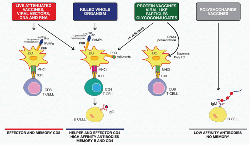
FIG. 43.2. Mode of Action of Different Classes of Vaccines. Live attenuated, viral vectors and nucleic acid vaccines induce both major histocompatibility complex (MHC) class I and MHC class II antigen presentation and activate pattern recognition receptors (PRRs) expressed by dendritic cells (DCs) through several pathogen-associated molecular patterns (PAMPs) including peptidoglycan, lipopolysaccharide, and bacterial or viral ribonucleic acid and deoxyribonucleic acid. They generally induce robust cluster of differentiation (CD)8 and CD4 T-cell responses, high antibody titers, and good memory. Killed whole-organism vaccines activate DCs through the same PAMPs and induce good CD4 and B-cell responses. Protein vaccines, viral-like particles, and glycoconjugate vaccines may need an adjuvant for optimal DC activation and CD4 T-cell priming. Some adjuvants such as polyinosinic:polycytidylic acid or saponins can also induce CD8 T-cell responses to subunit vaccines through cross-presentation mechanisms. Finally, polysaccharide vaccines induce a T-cell-independent antibody response.


Live-Attenuated Vaccines

As described in the previous paragraph on the history of vaccination, the very first vaccine of the modern era, Jenner’s vaccine against smallpox, was a live-attenuated viral vaccine, prepared from a natural strain of the cowpox or Vaccinia virus, which is not pathogenic for humans, to protect humans against Variola virus infection. Indeed, one type of live-attenuated vaccine is prepared from a microorganism that normally infects other animal species and does not cause disease in humans but is similar enough to the human pathogen to trigger a specific immune response that protects against the human infectious organism.


For most pathogens, however, a related animal pathogen that is inoffensive to humans does not exist, but luckily, a live-attenuated vaccine can also be obtained using a mutated strain of the infectious microorganism that has a reduced pathogenicity and therefore is able to infect the human being without inducing disease. It is not uncommon for attenuated strains of a pathogen to spontaneously develop during natural infection of the human population. These attenuated strains can therefore be isolated in order to develop a vaccine.3 However, attenuated viral strains can also be obtained by culturing the virus in vitro in nonhuman cells and selecting for mutant viruses that are adapted to this environment but are less fit to grow in the human cells. Most of the vaccines against viral infections in use today belong to this group of vaccines (eg, mumps, rubella, measles, varicella, and rotavirus vaccines). Although more difficult to obtain, and thus more rare, live-attenuated vaccines may also be developed against bacterial infections. Examples of live-attenuated bacterial vaccines among the currently licensed products are the vaccine against TB and an oral vaccine against typhoid fever. As the attenuated microorganism is capable of replication in the host mimicking a natural infection, this type of vaccine is generally able to induce strong and persistent antigen-specific immune responses in the vaccinees, resulting in a significant clinical efficacy. On the other hand, these vaccines may cause clinical manifestations associated with the infection, especially in immunocompromised subjects. Another potential risk associated with the use of attenuated strains is that they could revert to virulent form, therefore causing pathology in the vaccinated subject.


Whole-Killed or Inactivated Vaccines

Vaccines in this class contain the entire infectious agent that has been made harmless and incapable of replication by either chemical treatment (eg, formaldehyde) or a physical treatment like heating or radiations. Similar to live-attenuated vaccines, whole-killed or inactivated vaccines target bacterial and viral infections. Although this approach has been widely exploited in the past, only a few of the vaccines in use today are based on whole-killed microorganisms. In particular, only one whole-inactivated bacterial vaccine is still extensively used around the world, the whole-cell pertussis vaccine, whereas an oral vaccine against cholera, containing both heat-killed and formalin-inactivated strains of V. cholerae, is currently administered only to specific populations, such as travelers to an area where the infection is endemic. Examples of efficient vaccines prepared with whole-killed viruses are Salk’s IPV, rabies vaccine, and HAV vaccine. Inactivated vaccines are not able to cause infection or reproduce in the host, and, therefore they generally induce a lower antigenic stimulation compared to live-attenuated vaccines, requiring multiple immunizations to achieve efficacy. In addition, whole-killed vaccines can be reactogenic because they generally include microbe-derived proinflammatory molecules, also known as pathogen-associated molecular patterns (PAMPs). The high reactogenicity associated with some vaccines containing killed microorganisms has led to a progressive reduction of their use in favor of more purified vaccine preparations represented by the subunit vaccines.


Subunit Vaccines

A vaccine can be made of single or multiple antigenic components of a microorganism that are capable of stimulating a specific immune response sufficient to protect from the relevant pathogen infection or from the clinical manifestation of the disease. Depending on the molecular composition of the purified antigen used to prepare the vaccine, and on the techniques applied to obtain the final material used as a vaccine, different types of subunit vaccines can be defined.


Toxoids

Toxoids are toxins that are inactivated by chemical or physical treatment, but that nonetheless preserve their antigenic structure. This approach allows the formation of a protein that has completely lost toxic activity but is still capable of inducing the production of antibodies that will recognize and block the native toxin. This approach can be applied in case of infections in which the pathology exclusively depends on the damage induced by a single toxin produced by a pathogen, and, therefore, production of antibodies neutralizing the activity of the toxin or rapidly causing its elimination will be enough to avoid any disease. Vaccines against tetanus and diphtheria are based on toxoids; as in both cases, the pathology is caused entirely by a single toxic protein secreted by Clostridium tetani or Corynebacterium diphtheriae, respectively. Another commonly used toxoid is the inactivated pertussis toxin from Bordetella pertussis, which is one of the components of the acellular pertussis vaccine.


Protein Vaccines

In case immunization with a single protein or a combination of proteins from a pathogen is sufficient to stimulate a protective immune response against that particular microorganism, the approach of a protein-based vaccine is appropriate. Proteins can be purified from in vitro cultures of a pathogenic microorganism. The resulting vaccine preparations contain different amounts of contaminants depending on the efficiency of the purification process. Licensed acellular pertussis vaccines currently available contain from two to four different proteins purified from B. pertussis and are able to confer protection against whooping cough comparable to that obtained with the whole cell vaccine. One of the most widely used subunit protein vaccines is the influenza vaccine composed of hemagglutinin (HA) and neuraminidase (NA) purified from the inactivated influenza virus.


Recombinant Protein Vaccines

Development of the recombinant deoxyribonucleic acid (DNA) technology has made possible the expression of protective protein antigens in heterologous expression systems such as E. coli, yeast, mammalian cells, or baculovirus. This technology avoids the problems related to growing and manipulating large amounts of a pathogen from which the antigen is purified. Moreover, recombinant proteins are generally better purified from cultured microorganisms resulting in cleaner vaccine preparations with a better safety
profile. A drawback of a clean vaccine preparation containing pure recombinant protein(s) is their reduced immunogenicity that may require the addition of an adjuvant to achieve enhanced efficacy.


Viral-Like Particles

VLPs are supramolecular protein structures formed by viral proteins in vitro in the absence of a viral genome, which are structurally similar to the natural viral infectious particles while they are not infectious. One example of a VLP is the vaccine used to prevent hepatitis B virus (HBV). HBsAg spontaneously assembles into spherical particles of 22 nm diameter when expressed in yeast or mammalian cells.4,5,6 Many theoretical and practical aspects of VLP technology make this approach attractive to vaccine design. The viral antigens expressed in VLPs usually exhibit a conformational structure that is similar to what they present in the real virus, and this is usually associated with conservation of critical conformational epitopes that are targets of a neutralizing immune response. Another important characteristic of VLPs is that they express the antigenic epitopes in multiple repetitive structures in one single particle, which potentially can induce an efficient engagement of the antigen receptor on the surface of B cells, resulting in an improved antibody response compared to what is obtained with soluble protein antigens. As VLPs are not infectious, there are no issues related to manipulation of high amounts of infectious material. Moreover, no inactivation procedures, which may result in damage of important immune relevant epitopes, are required. VLPs are at the base of the two HPV licensed vaccines. The VLP approach has been applied to many vaccine candidates, some of which are currently being tested in clinical trials.7,8 This approach has also been used for antigens that do not naturally form VLPs by developing chimeric VLPs using two different methods. In one case using DNA recombinant techniques, a candidate vaccine antigen is genetically fused with a protein that is able to form VLPs, resulting in its expression in the context of the VLP structure. Alternatively, an antigen is chemically linked to heterologous VLPs that will provide the multimeric structure able to induce an appropriate immune stimulation. When these chimeric particles are administered, they are able to elicit a strong immune response to all components in the VLPs, including the epitopes from the fused candidate vaccine antigen. The chimeric VLP approach has been evaluated for development of vaccines against viral infections such as hepatitis C virus (HCV)9 and HIV,10 or against malaria,11 as well as of therapeutic vaccines for cancer or for addictive substances such as nicotine.12 However, thus far, none of these experimental chimeric VLP vaccines have progressed beyond phase I/II clinical trials.


Polysaccharide-Based Vaccines

The surface of many pathogenic bacteria is covered by a capsular shell that is mainly assembled from ordered polymeric glycans. This extensive polysaccharide coat entirely shields the bacteria outer membrane, preventing other surface bacterial components from becoming a target of the host immune response. Nevertheless, antibodies to bacterial surface polysaccharides can clear the bacteria from the host by different mechanisms, such as complement-mediated killing and opsonophagocytosis. Hence, stimulation of an antibody response against the surface polysaccharide of pathogenic bacteria is a valid strategy for the development of vaccines against capsulated bacteria. Experiments using the capsular polysaccharide isolated from Streptococcus pneumoniae demonstrated for the first time the immunogenicity of a bacterial carbohydrate and a protective effect of a polysaccharide-based vaccination.13 The chemical structure of capsular polysaccharides varies not only between bacteria of different species but also between different strains within a single species, which are usually differentiated and typed based on their capsular polysaccharides. As a consequence, a limitation of polysaccharide-based vaccine is that the immune responses they elicit are often serotype specific. Thus, if disease burden is caused by multiple polysaccharide types, a vaccine would optimally contain multiple polysaccharides in order to confer broad protection. In addition to S. pneumoniae, for which a vaccine against 23 serotypes is available, polysaccharide-based vaccines have been developed for MenACWY, Hib, and Salmonella typhimurium.


Glycoconjugate-Based Vaccines

Purified polysaccharides are able to stimulate only B-cell responses because they cannot enter the major histocompatibility complex (MHC) cavity (which evolved to bind peptides) and cannot be presented to T cells. For this reason, they are classically known as “T-independent” antigens. Thus, the antibody response generated by a polysaccharide vaccine is a typical T-cell-independent response that presents two major disadvantages from a vaccine point of view. First, the response does not take place in germinal centers, and it is therefore not associated with affinity maturation of the antibodies or the generation of memory B cells. As a consequence, a polysaccharide vaccine will induce low- to intermediate-affinity antibody responses that is not followed by immunologic memory, resulting in the need for booster administration to maintain immunity. Moreover, a T-cell-independent antibody response is deficient or absent in young children (younger than 18 months of age) and in the elderly, two populations at risk of infection with capsulated bacteria such as pneumococcus or meningococcus, and for which a polysaccharide-based vaccine is not efficacious. A way to circumvent the limitations of these vaccines is to conjugate a polysaccharide to a carrier protein, such as TT or DT, in order to induce a carrier-specific T-cell response. Following vaccination with glycoconjugate vaccines, T follicular helper cells promote differentiation of the polysaccharide-specific B cells in the germinal center, where somatic mutation and affinity maturation take place, resulting in higher affinity antibody response and generation of long-lived memory B cells conferring long-term protection. The first demonstration that conjugation to a protein increases immunogenicity of a capsular polysaccharide was obtained in 1929; however, it took 50 years for this strategy to be applied to vaccine design, when a glycoconjugated vaccine for Hib was first tested in animals.1 Compared to polysaccharide vaccines, glycoconjugated vaccines are indeed
more efficacious, being able to induce a long-lasting antibody response not only in adults but also in infants, young children, and the elderly. Similarly to the polysaccharidebased vaccines, the glycoconjugate vaccines are also serotype specific, and, therefore, multivalent formulations are required to achieve protection against multiple serotypes.


Combination Vaccine

In clinical practice, vaccines for different pathogens or for different strains of a particular pathogen are combined in one single formulation, allowing a reduction in the number of injections administered, with a concomitant reduction in costs and distress for the individuals. This last advantage is particularly important for childhood vaccinations that are frequently associated with psychological distress for parents, which may affect compliance with the vaccination schedule and be accompanied by possible negative effects on vaccination coverage. Indeed, it has been reported that the use of combination vaccines has improved both coverage rates and timeliness of vaccination.14,15 A combination of diphtheria and tetanus toxoids with the whole-killed pertussis vaccine (DTwP) was the first multipathogen vaccine to be licensed. Years later, a similar combination containing the acellular pertussis component (DTaP) has been used as the basis to build new expanded combinations that may include one or more components such as IPV, HBsAg, and Hib glycoconjugate. Another commonly used pediatric combination is a mix of measles, mumps, and rubella vaccine, although a quadrivalent vaccine containing also the varicella vaccine became available only recently after solving problems related to compatibility and stability of the different components. Together with the continuous development of new vaccines, demands for additional combinations will be increasing with the aim of reducing costs and simplifying the vaccination schedule, in particular in the infant population. However, the increase in complexity of combination vaccines will raise the risk of a possible interference on antigen stability and immunogenicity.


Novel Technologies for Vaccine Design

Novel technologies and new strategies are continuously tested in preclinical research and applied to the development of new candidate vaccines. Novel types of vaccines can therefore be identified among the experimental vaccines currently evaluated in both preclinical and clinical research, some of which may become established products in the future.


Viral Vector-Based Vaccines

Priming of CTL responses requires presentation of the antigen by the MHC class I molecules, a pathway usually followed by endogenously synthesized proteins (eg, viral antigens) but not by exogenously administered antigens. For this reason, inactivated or subunit vaccines are not per se able to stimulate a CTL response, which is instead induced by live-attenuated viral vaccines. The observation that a gene encoding an external antigen could be inserted in the genome of the Vaccinia virus and expressed in the virus-infected cells has prompted the idea of genetically manipulating an attenuated or nonpathogenic virus to express an exogenous antigen from a pathogenic virus against which vaccines need to be developed. Because these antigens are synthesized by the host cells, they undergo all posttranslational modifications and acquire a correct conformation and should be able to stimulate a strong neutralizing antibody response. Moreover, antigens are presented in the context of the MHC class I pathway generating therefore a CTL response. Examples of attenuated strains of Vaccinia virus are the modified Vaccinia Ankara (MVA) strain obtained by repetitive passages through in vitro cell cultures and the New York Vaccinia strain obtained by genetic manipulation of the viral genome. These strains have been evaluated in both nonhuman primates and humans as vector vaccines for HIV, malaria, influenza, TB, and other infectious diseases.16,17,18 Other poxviruses tested as vector vaccines include fowlpox and canarypox.19 Despite extensive testing of these vectors as vaccine candidates, so far, no successful vaccine has been developed using this approach.

Because of their immunogenicity and efficacy in the induction of a robust cellular response, replication-incompetent recombinant adenoviral vectors (rAds) have been successfully tested in many animal models as vaccine vectors for many pathogens such as Ebola, HIV, anthrax, influenza, and malaria.20,21,22,23 Adenoviral vaccine vectors have also been evaluated in many human clinical trials, particularly for HIV and cancer. On the other hand, natural adenovirus circulates in the human population, and, therefore, preexisting immunity, in particular neutralizing antibodies, may be present in the subject who will receive the vaccine, compromising efficacy of these vaccines.24 It is important to note that tens of adenovirus serotypes exist but most of the clinical studies have used a vector derived from a single serotype (Ad5). Serotype 5 is one of the most common serotypes spread in the human population, and a high frequency of subjects possesses anti-Ad5 antibodies. In order to overcome potential problems related to preexisting anti-Ad5 antibodies, new vectors based on different human serotypes25 or on adenovirus from other species26,27,28 were developed. At the present, the most promising are the chimpanzee-derived adenoviruses.27

Alphaviruses are positive single-stranded ribonucleic acid (RNA) viruses in which the genome can be manipulated to obtain replication-defective infectious particles that express an exogenous antigen. Besides this favorable safety feature, upon entering the target cell, alphavirus RNA self-replicates, producing many copies of the coding RNA, which can then direct the synthesis of a very high amount of the antigen, making it a promising platform for a vector-based vaccine.29 Alphavirus-based vaccines have been tested in animal models for several pathogens and have demonstrated excellent immunogenicity.30,31,32,33 Some clinical trials have been conducted, but the results have not been published.

The prime-boost vaccination strategy using viral vectors has received wide evaluation in the vaccine field. Heterologous prime-boost regimens mainly use a viral vector or a DNA vaccine (see subsequent discussion) at priming
followed by a boost with a protein-based vaccine, although other options have also been used. This unconventional immunization schedule usually results in the induction of a strong cellular immune response, with generation of both CD4+ and CD8+ antigen-specific T cells. This is in turn associated with a higher and more specific antibody response against the vaccine target compared to homologous immunization with either DNA or viral vector. The immunologic mechanisms responsible for these improved responses are not entirely known. It is hypothesized that while DNA or viral vector priming is required to induce a strong T-cell response, boosting with only the target protein focuses the B-cell response on a single protein antigen for which high-affinity antibodies are desirable and at the same time avoiding interference by antivector immunity.34


Nucleic Acid-Based Vaccines

At the beginning of the 1990s, experiments designed to evaluate new approaches for gene therapy were performed to test if intramuscular injection of a liposome formulation containing a plasmid DNA or a messenger RNA coding for a reporter protein could result in in vivo expression of the protein. Surprisingly, animals in control groups that were injected with pure DNA or RNA showed a significant and prolonged expression of the reporter protein in muscle cells.35 This observation inspired a series of studies that demonstrated that mouse immunization with an antigen encoded by DNA could induce a strong antigen-specific immune response characterized by antibody production and generation of both T-helper and CTL responses.36,37,38 One of these studies also established the protective effect of a DNA-mediated immune response, showing that mice injected with a plasmid encoding the nucleoprotein from a H1N1 strain of influenza A virus were completely protected from challenge with a virulent H3N2 strain.38 Based on these promising results, a huge amount of experimental work has been performed in the last two decades to apply DNA vaccination to many disease targets with both prophylactic and therapeutic approaches. As with vector-based vaccines, the advantage of a DNA-encoded vaccine is that the antigen is synthesized within host cells, facilitating correct folding of conformational neutralizing epitopes and MHC class I antigen presentation, allowing generation of CTL responses. An additional advantage of DNA vaccines compared to viral vectors is that the former contains only DNA, therefore nullifying any effects of preexisting vector immunity. DNAbased vaccines are composed of plasmid DNA containing an antigen gene sequence. To be efficiently expressed in the host mammalian cells, the antigen sequence has to be under the control of a strong eukaryotic promoter, such as those present in many viral genomes (eg, the early promoter of the human cytomegalovirus [HCMV]). Classical plasmids used for DNA vaccination also contain antibiotic resistance genes and prokaryotic origin of replication sequences, both required for selection and replication in bacteria. These prokaryotic sequences may contain unmethylated CpG motifs that are recognized by toll-like receptor (TLR)9 and were originally shown to play an immunostimulatory role during DNA vaccination.39 However, subsequent investigations have excluded a role for TLR9 and CpG motifs in the immunogenicity of DNA vaccines and have suggested that the immunostimulatory activity might be due to the double-stranded structure of DNA and its capacity to activate other innate immune receptors.40,41,42,43 After intramuscular injection, DNA is primarily localized within myocytes where the encoded antigens are expressed, although expression can also be observed in antigen-presenting cells (APCs).44,45 Indeed, several studies have demonstrated that expression of antigen by myocytes is critical for induction of a CTL response, although they do not directly present the antigen to T cells. Experiments using parental bone marrow transplantation into F1 mice established that generation of antigen-specific CTL responses by DNA immunization depends on a cross-priming mechanism. Transfected myocytes transfer the antigen to professional APC, driving its processing and presentation by MHC class I molecules, which then leads to triggering of a CD8+ T-cell response.46,47 Manufacturing of DNA vaccines has several benefits over all other type of vaccines: 1) design of DNA construct is straightforward; 2) production of DNA plasmids is uncomplicated and cheap and does not require manipulation of infectious materials; and 3) plasmids are stable even at room temperature, reducing the complexity and the cost of distribution. Because of the potential advantages of a DNAbased vaccine, this approach has been extensively tested for many pathogen such as rabies, HBV, and HIV in different animal models with promising immunogenicity results.48,49,50 Furthermore, the approach has also been evaluated in humans in several clinical trials.51,52 The human studies have demonstrated that DNA vaccination is generally safe and able to stimulate an immune response; however, immunogenicity is usually lower than expected based on the results of animal studies, resulting in a diminished confidence in this approach. The initial failure of DNA vaccine in the clinical setting led to the exploration of a series of techniques to improve their immunogenicity. To improve expression in vivo, the antigen-coding sequences were modified in order to contain only codons that match transfer RNAs with high frequency in mammalian cells, whereas stronger promoters were included in the plasmids. In an attempt to increase stability of the administered plasmid, different formulations and delivery systems, such as liposomes or microparticles, have also been used.53 In addition, several new injection techniques have been tested to improve in vivo transfection efficiency. A forced delivery of DNA into the skin can be obtained by the use of gold microparticles in gene gun techniques54 or by a high-pressure liquid stream created by the Biojector device (Bioject Medical Technologies, Inc., Tigard, OR, U.S.A.),55 whereas electroporation can greatly increase efficiency of intramuscular injection of DNA.56


Peptide-Based Vaccines

Although B cells can recognize linear epitopes within a protein, most of the epitopes recognized by a protective antibody response during a natural infection are conformational. Linear epitopes are usually not efficient at inducing a good neutralizing response. The goal of peptide vaccines is therefore to create T-cell epitopes capable of stimulating
T-cell responses and in particular CTLs, in cases of viral infections57 or cancer,58 where these type of immune responses are required to obtain a complete protection. Although the peptide approach is able to induce protection from viral infections and cancer in the mouse model, polymorphism of the MHC system has limited the application of peptides to human preventive vaccines. The use of peptides has been mostly restricted to therapeutic vaccines against cancer.59


VACCINE ADJUVANTS

Live-attenuated and inactivated vaccines have been successful in preventing and, in some cases, eradicating infectious diseases in the last century. However, there is an increasing demand for subunit vaccines composed of highly purified antigens because they are generally better tolerated compared to vaccines made of inactivated or live-attenuated pathogens. The limitation of subunit vaccines is that they are in general poorly immunogenic and often require the addition of an adjuvant to achieve protective immune responses. The term vaccine adjuvant has been used to define several compounds that enhance the immunogenicity of a coadministered antigen in vivo. As a consequence of this functional and empirical definition, the vaccine adjuvants group is composed of diverse classes of molecules such as microbial products, emulsions, mineral salts, small molecules, microparticles, and liposomes that have different mechanisms of action.60 In general, adjuvants are believed to boost vaccine response by increasing the persistency of the antigen in vivo or by targeting innate immune pathways normally associated with response to infection60,61 (Box 43.1).

Several studies have linked the mechanism of action of known adjuvants to the upregulation of major histocompatibility complex proteins, costimulatory molecules, and cytokines in dendritic cells (DCs). All these events increase the potential of DCs to prime naive T cells, a central step in the activation of both humoral and cellular adaptive responses. Interestingly, DC activation following adjuvant injection can occur either directly, through the stimulation of innate immune receptors, or indirectly, through the stimulation of blood cells or stromal cells at the injection site.62 It has been shown that various cell types can react to adjuvant injection by producing cytokines or danger-associated molecular patterns, such as endogenous DNA and uric acid crystals that in turn can activate DCs.


Many independent studies have suggested that although activation of DCs is probably required for all vaccine formulations, it is not sufficient for optimal adjuvanticity, which is achieved when an adjuvant able to activate DC (generally called immunopotentiator) is formulated with a second adjuvant that enhance antigen uptake (also known as antigen delivery systems).63

Examples of immunopotentiators are microbial compounds, small molecules, and nucleic acids that are able to activate pattern recognition receptors (PRRs) such as TLRs, nucleotide oligomerization domain-like receptors (NLRs), and C-type lectins receptors (CLRs); cytokines such as granulocyte macrophage-colony stimulating factor, interleukin (IL)-12, IL-2, and interferon (IFN)-α saponins and microbial compounds targeting invariant natural killer T cells (αGal-Cer and derivatives).

Examples of antigen delivery systems are mineral salts (alum), emulsions, microparticles (polylactide coglycolide), liposomes, and monoclonal antibodies targeting DC internalization receptors such as DEC-205. All these compounds are able to enhance antigen uptake by APCs in vitro. However, alum and oil-in-water emulsions can also induce the expression of cytokines and other innate immune mediators at the site of injection that may contribute to DC activation (described in detail in following paragraphs).

It has been shown that codelivery of antigen and an immunopotentiator in the same APC can greatly enhance adaptive responses to vaccination. In most studies, codelivery has been achieved by adsorbing the antigens (generally recombinant proteins or purified subunits) and immunopotentiators to the same antigen delivery system. However, codelivery can also be achieved by linking directly immunopotentiators such as flagellin, oligonucleotides, lipopeptides, and small molecules to the vaccine antigen.

As a consequence of the innate immune stimulation, adjuvants can have a strong impact on adaptive responses. In preclinical and clinical studies, it has been demonstrated that adjuvants can increase the amount, avidity, and the cross-reactivity of antigen-specific antibodies, potentiate T-cell responses, and modulate the immune response toward a targeted CD4+ T-cell phenotype adapted against defined pathogens. Finally, adjuvants can improve adaptive responses to vaccination in immunologically hyporesponsive populations including the elderly and infants and can allow for antigen dose sparing. Despite the importance of adjuvants, only very few compounds selected in preclinical studies have been licensed for human use (Table 43.2). Among them, alum has been widely used for more than 70 years; oil-in-water emulsions have been licensed for
adjuvanted influenza vaccines; and AS04, a combination adjuvant composed of the TLR4 agonist monophosphoryl lipid A (MPL) adsorbed to alum, has been approved for HBV and HPV vaccines. The identification and development of new adjuvants is necessary because the limited number of currently approved vaccine adjuvants do not always elicit the desired immune responses capable of preventing the targeted infection. In particular, there is a need for new adjuvants eliciting potent CD8+ T-cell and Th1 responses. A large number of compounds have demonstrated vaccine adjuvant activities in preclinical models, and it would be difficult to review all of them in this chapter. Therefore, we will focus only on the adjuvants that have been either licensed (Table 43.2) or already tested in human (see Table 43.3), with a special focus on their use with subunit vaccines targeting infectious diseases.


TABLE 43.2 Licensed Human Vaccine Adjuvants


Name


Class


Components


Vaccine


Reference


Aluma


Mineral salts


Aluminum phosphate, aluminum hydroxide


Diphtheria, tetanus, pneumococcus, HAV, HBV, anthrax, tick-borne encephalitis, MenC, HPV


64


MF59


Oil-in-water emulsion


Squalene, Tween 80, Span 85


Seasonal and pandemic influenza


84,85,88


AS03


Oil-in-water emulsion


Squalene, Tween 80, α-tocopherol


Pandemic influenza


89


AF03b


Oil-in-water emulsion


Squalene, Montane 80, Eumulgin B1PH


Pandemic influenza


86


Virosomes


Liposomes


Phospholipids, cholesterol, HA


Seasonal influenza, HAV


115,360


AS04a


Alum-adsorbed TLR4 agonist


Aluminum hydroxide, MPL


HBV, HPV


111,361


RC-529c


Alum-adsorbed TLR4 agonist


Aluminum hydroxide, synthetic MPL


HBV


114


HAV, hepatitis A virus; HBV, hepatitis B virus; HPV, human papillomavirus; Men, meningococcus, MPL, monophosphoryl lipid A; TLR, toll-like receptor.


a Adjuvants approved by the U.S. Food and Drug Administration.


b Approved in Europe but not marketed.


c Licensed only in Argentina.


Licensed Vaccine Adjuvants


Alum

Numerous subunit vaccines in use today, including those against HAV, HBV, HPV, anthrax, diphtheria, tetanus, MenC, tick-borne encephalitis, and pneumococcal conjugate vaccines, are adsorbed to aluminum salts.64 The most used aluminum salts for licensed vaccines are aluminum hydroxide and aluminum phosphate, and they are generically called “alum.” Alum adsorption enhances the antibody responses to the coadministered antigens and, in some cases, can also increase antigen stability allowing for more extended vaccine shelf life. Only recently has the mechanism of action of alum been deeply investigated.65,66,67 Early work suggested that alum is able to form a depot with the antigen at injection site increasing antigen persistency and availability. However, in the last 20 years, the depot effect of alum has been challenged by several studies that reported a similar half-life of antigen formulated in the presence or absence of alum.68,69,70 Alum absorption increases antigen uptake by DCs in vitro, suggesting that it acts as an antigen delivery system.71 More recently, it has been shown that the interaction between alum and DCs requires membrane sphingomyelin and cholesterol.72 Moreover, alum injection results in cell recruitment to the injection site,73,74 especially local recruitment of monocytes, which migrate to the draining lymph nodes and differentiate into inflammatory DC capable of priming T cells.75 Altogether, these data suggest that alum has a dual function in promoting antigen uptake and a local proinflammatory environment at injection site. In vitro experiments conducted on mouse and human blood cells led to the identification of at least one of the molecular targets of alum inflammatory activity. Alum activates a cytoplasmic NLR protein called NLRP3, which associates with the adaptor protein ASC and the caspase-1 protease to form a protein complex called inflammasome.76,77,78 Activation of NLRP3 ultimately results in the processing of pro-caspase-1 into its active form, which cleaves several proinflammatory cytokines including IL-1β and IL-18. It is still not clear how alum activates NLRP3; however, it has been proposed that phagosomal destabilization induced by aluminum and by other crystal structures (silica crystals) following phagocytosis plays an important role.79

In vivo studies conducted in NLRP3-deficient mice have shown that direct NLRP3 activation contributes to alum adjuvanticity.75,77,80 However, the requirement of NALP3 for the adjuvant activity of alum has been challenged by other studies using alternative antigens or different immunization routes.76,81,82 Probably, other proinflammatory pathways triggered by alum exist and are independent from inflammasome activation. Recently, it has been proposed that host DNA released from dying cells acts as a danger signal that mediates alum adjuvanticity through the activation of several innate immune pathways including interferon regulatory factor 3 (IRF3). In addition, it has been demonstrated that (IRF3) is required for immunoglobulin (Ig)E antibody response to alum adjuvanted vaccines.83


Oil-in-Water Emulsions

Oil-in-water emulsions are liquid dispersions of oil droplets stabilized by one or more surfactants. The first emulsion to be licensed for human use was MF59 composed of squalene
oil, emulsified with two surfactants (Tween 80 and Span 85). MF59 has been licensed in Europe since 1997 in an adjuvanted seasonal influenza vaccine for its capacity to increase flu immunogenicity in the elderly.84 More recently, MF59 and a second squalene-based oil-in-water emulsion called AS03, composed of squalene, Tween 80 and α-tocopherol, have been licensed in Europe for pandemic influenza vaccines and have been widely used for the 2009 H1N1 pandemic flu campaign.85 A third squalene-based oil-in-water emulsion called AF03 has been approved for pandemic influenza in Europe but has not been marketed yet.86 Clinical trials demonstrated that avian H5 pandemic influenza vaccines containing oil-in-water emulsions are superior to alum-adjuvanted or nonadjuvanted H5 vaccines in the induction of protective antibody titers.87 Both AS03 and MF59 allowed for antigen dose sparing and increased seroconversion and cross-protection.88,89 It is important to notice that in the case of H5 avian flu, for which most of the population has not been previously exposed, nonadjuvanted vaccines did not reach the antibody titers considered to be the serologic correlate of protection, whereas emulsion-adjuvanted vaccines were able to elicit protective antibody titers after two doses. A phage display library approach demonstrated that MF59 can enhance the breadth and the affinity of the antibody responses to H1 and H5 pandemic influenza vaccines.90,91,92 In particular, the addition of MF59 enhanced the recognition of the HA globular head region that contains most of the neutralizing epitopes. Two additional clinical trials have been conducted on individuals vaccinated several years before with H5 vaccines either nonadjuvanted or mixed with MF59 or AS03 by administering a new dose of oil-in-water emulsion-adjuvanted H5 vaccine (boost). These studies demonstrated that priming with avian H5 vaccines mixed with MF59 or AS03 allows for better and more rapid responses to a booster dose compared to individuals primed with nonadjuvanted vaccines.93,94 Priming with adjuvanted vaccines was effective even when H5 antigens from different influenza clades were used for priming and boosting, inducing a rapid increase of cross-neutralizing antibody titers. These studies suggest that adjuvanted influenza vaccines may be used for a prepandemic vaccination strategy. Another recent study highlighting the strong impact that adjuvants can have on vaccine efficacy was recently performed on children between 6 and 72 months of age. The addition of MF59 to seasonal flu subunit vaccine increased the efficacy of the vaccine from 43% to 86% against polymerase chain reaction confirmed influenza-like illness.95 Interestingly, in the age subgroup 6 to 24 months, nonadjuvanted influenza vaccine was unable to protect, whereas the efficacy of MF59-adjuvanted vaccine was 77%.

Several studies using the mouse model and human blood cells have addressed the mechanism of action of oil-in-water emulsion. In contrast to alum, which physically binds antigens through an adsorption process, oil-in-water emulsions are simply mixed with antigens. MF59 neither increases antigen persistence at the injection site nor induce any depot effect.96 However, MF59 can enhance antigen uptake by activated DCs in mouse muscle97 and phagocytosis in vitro in human peripheral blood mononuclear cells.98 These data suggest that MF59 can act as an antigen delivery system; however, other studies have shown that it has also immunostimulating properties. In vitro analysis of human blood cells showed that MF59 induces the differentiation of monocytes toward DCs and can stimulate chemokine secretion (CCL2, CCL3, CCL4, and CXCL8) in human macrophages, monocytes, and granulocytes.98 Intramuscular administration of MF59 in mice induced the recruitment of mononuclear cells partially dependent on chemokine receptor 2 (CR2).99 The local effect of MF59 at an injection site has been evaluated in detail by combining DNA microarray and immunofluorescence techniques. Transcriptome analysis showed that the injection of MF59 in mouse muscle induced the activation of several innate immune genes, including Ccr2 and its ligands (CCL2, CCL7, and CCL8), further supporting the data described previously on CR2-dependent cell recruitment.100 In addition, MF59 promoted the upregulation of many proinflammatory cytokines and other innate immunity genes and genes involved in leukocyte migration.100 Two of the early genes modulated by MF59, JunB and pentraxin 3, were upregulated in the muscle, suggesting that the activation of muscle cells by MF59 might play some role in its adjuvant activity.

Intramuscular injection of fluorescently labeled MF59 and antigen revealed that MF59 colocalizes with the antigen in APCs in the muscle and enhances the number of antigenpositive DCs and B cells in the draining lymph node.101

Similarly to MF59, AS03 induces transient cytokine activation at the injection site and triggers the secretion of cytokines in human monocytes and macrophages.102 In addition, AS03 induces cytokine expression in the draining lymph node in a mechanism that is at least partially dependent on the presence of image-tocopherol in the formulation.

In summary, the data described previously suggest that squalene-based oil-in-water emulsion function by enhancing antigen uptake and migration of antigen-positive APCs in the draining lymph nodes and by inducing an immunostimulatory environment at the injection site. However, the innate immune pathways required for the mechanism of action of emulsions are not well characterized. Unlike alum, MF59 does not activate NLRP3 in vitro and is not dependent on NLRP3 or caspase-1 in mouse immunization studies.82,103 Despite the fact that MF59 adjuvanticity is inflammasome independent, adaptive responses induced by an H5 influenza subunit vaccine adjuvanted with MF59 largely depend on the inflammasome component ASC. Therefore, it has been proposed that ASC plays an inflammasome-independent function required for MF59 adjuvanticity.103 Surprisingly, MF59 adjuvanticity to a recombinant MenB subunit vaccine requires another adapter protein called MyD88, which plays an essential role in TLR and IL-1R signaling. However, MF59 does not have any TLR agonist activity and the MyD88-dependent pathway required for its adjuvanticity is still unknown.82


Monophosphoryl Lipid A and Derivatives

TLRs are key pathogen sensors that modulate the host immune system and play a fundamental role in response to microbial infection.104 TLRs are PRRs expressed on innate immune cells including DCs that sense PAMPs. Upon pathogen recognition, DCs are activated and provide essential
signals for naive T-cell priming and development of an adaptive immune response. In addition to pathogen infection, microbial products from live-attenuated or heat-killed vaccines can also stimulate TLR pathways providing adjuvant activities.105,106 For example, the yellow fever vaccine activates DCs by signaling through TLR2, 7, 8, and 9.107 Many preclinical and clinical studies using purified TLR agonists have been conducted and demonstrated that all TLRs examined can be exploited to enhance adaptive responses to vaccination. However, despite the large amount of work performed, so far, only the TLR4 agonist 3-O-desacyl-4′-MPL, a component of lipopolysaccharide (LPS) purified from Salmonella minnesota has been licensed for human use. MPL is approved for two vaccines against HPV and HBV. In both vaccines, MPL has been adsorbed to alum to form a combination adjuvant called AS04. The HBV vaccine formulated with AS04 was developed for patients with renal insufficiency and was tested in several immunocompromised individuals including the elderly and patients on hemodialysis. The incorporation of MPL allowed for a more rapid increase of antibody titers and enhanced seroprotection rates using fewer vaccine doses.108 HPV vaccine formulated in AS04 induced higher neutralizing antibody titers and memory B-cell responses compared to the same vaccine formulated only with alum.109 A head-to-head trial was conducted to compare the alum-MPL adjuvanted HPV vaccine versus another HPV licensed vaccine that is adjuvanted only by alum.110,111 Both vaccines exploit VLP technologies for the production of antigens and are highly efficacious in preventing HPV16/18-associated cervical intraepithelial neoplasia in women. However, the AS04-adjuvanted vaccine produced significantly higher neutralizing antibody titers to HPV16 and HPV18, and higher frequencies of memory B cells compared to the other vaccine.

The molecular mechanism of action of AS04 is well understood. The MPL component activates directly the LPS sensor TLR4 that is expressed by human innate immune cells including DCs and macrophages. Adsorption of MPL to alum does not interfere with its ability to activate human immune cells including DC.112 AS04 injection in the mouse muscle lead to a transient production of interferon and proinflammatory cytokines at injection site and to an increased number of antigen-loaded DCs in the draining lymph node.112 TLR4 stimulation on the surface of conventional DCs induces the production of IL-12 and activates the expression of MHC class II and costimulatory molecules leading to efficient CD4+ T-cell priming and to strong Th1 adaptive responses.

Synthetic TLR4 agonists have the advantage over MPL to be more pure and less expensive. Several synthetic TLR4 agonists such as RC-529 and GLA have been developed and tested as vaccine adjuvants.113 RC-529, a fully synthetic monosaccharide mimetic of MPL, is the most advanced and has been approved in Argentina for a vaccine against HBV.114


Virosomes and Liposomes

Virosomes were licensed for an influenza vaccine in 1994 and are composed of a mixture of phospholipids and cholesterol that are assembled in vitro with purified influenza HA and NA to form synthetic influenza viral-like structure. Influenza virosomes can be also used as adjuvants to incorporate additional antigens from other viruses or other pathogens in general. One formulation composed of H1N1 virosomes and inactivated HAV was licensed in 1997 to target HAV. The mechanism of action of virosomes is mainly linked to antigen delivery functions. Virosomes increase antigen uptake by APCs and display the antigen in draining lymph nodes in a multimeric form, facilitating B-cell-receptor engagement. The presence of preexisting immunity against influenza further enhance the adjuvant effect of virosomes probably through the formation of immune complexes that are rapidly internalized by APCs.115

Other liposome adjuvants made of phospholipid bilayers have been tested in preclinical models and in clinical trials in combination with various antigens and in some cases with other adjuvants, as described in the following paragraphs.


Vaccine Adjuvants in Clinical Development Phase


New Toll-Like Receptor Agonist Adjuvants

There is a great interest worldwide in exploiting TLRs as targets for vaccine adjuvants. Besides MPL, several additional TLR agonists are known to be effective adjuvants in preclinical studies.116 Among them, agonists of TLR2 and TLR9 and novel TLR4 agonists have been extensively tested in human as vaccine adjuvants (Table 43.3). Clinical data are also available for vaccine formulations including TLR5 and TLR7 agonists. Human data on TLR3 agonists are not yet published; however, clinical trials have already started.

Toll-Like Receptor 2. Bacterial lipoproteins targeting TLR2/1 or TLR2/6 dimers are validated vaccine adjuvants in humans and multiple TLR2 ligands have undergone clinical evaluation. The most advanced TLR2 agonist is the lipopeptide moiety of OspA, the surface protein of B. burgdorferi, the major component of a vaccine preventing Lyme disease.117 The vaccine was efficacious and licensed by the U.S. Food and Drug Administration (FDA) in 1998. However, despite the fact that the vaccine was safe and well tolerated, it was withdrawn by the manufacturer 3 years later following media coverage of possible autoimmune side effects.

Toll-Like Receptor 3. Agonists of the double-stranded RNA sensor TLR3 have not been extensively tested in human yet. However, a synthetic analogue of double-stranded RNA called polyinosinic:polycytidylic acid (Poly I:C) has been tested in multiple animal models with various antigens. Poly I:C can activate TLR3 and a second double-stranded RNA sensor called MDA-5, both leading to the production of type I IFN. In mice, Poly I:C was as effective as other TLR agonists in boosting antibody responses to HIV gag antigen and more efficient than any other TLR agonist tested in inducing specific IFN-γ+ CD4+ T cells.118 The same study showed that the adjuvanticity of Poly I:C depends on type I IFN. A study conducted with a Plasmodium falciparum antigen has shown that Poly I:C is effective in inducing specific antibodies and promoting multifunctional CD4+ T-cell responses in nonhuman primates.119


TABLE 43.3 Vaccine Adjuvants in Clinical Development


Name


Class


Components


Vaccine


Reference


CpG 7909


TLR9 agonist


CpG ODN


HBV, malaria, influenza, anthrax, cancer


116


CpG 1018


TLR9 agonist


CpG ODN


HBV, cancer


126


Imiquimod


TLR7 agonist


Small molecule formulated in a topical cream


Cancer


124


Pam3Cysa


TLR2 agonist


Lipopeptide


Lyme disease


117


Flagellin


TLR5 agonist


Bacterial flagellin fused to antigen


Influenza


121


Iscomatrix


Combination


Saponin, cholesterol, dipalmitoylphosphatidylcholine


HCV, influenza, HPV, cancer


133


AS01


Combination


Liposome, MPL, saponin (QS21)


Malaria


140,141


AS02


Combination


Oil-in-water emulsion, MPL, saponin (QS21)


Malaria, TB, cancer


131,139


IC31


TLR9 agonists


ODN, cationic peptides


TB


129


Montanide ISA 51


Water-in-oil emulsion


Mineral oil, surfactants


Malaria, HIV, cancer


23


Montanide ISA 720


Water-in-oil emulsion


Squalene, surfactants


Malaria, HIV, cancer


23


LT


Bacterial toxins


A subunit + B pentamer of Escherichia coli heat-labile enterotoxin


Influenza (intranasal), ETEC, pandemic influenza (Patch)


149,154,155


LTK63


Bacterial toxins


Genetically modified A subunit +B pentamer of E. coli heat-labile enterotoxin


Influenza, TB, HIV (intranasal)


152,153


ETEC, enterotoxigenic Escherichia coli; HBV, hepatitis B virus; HCV, hepatitis C virus; HIV, human immunodeficiency virus; HPV, human papillomavirus; MPL, monophosphoryl lipid A; ODN, oligodeoxynucleotide; TB, tuberculosis; TLR, toll-like receptor.


a Licensed and then withdrawn from market.


Toll-Like Receptor 5. TLR5 recognizes bacterial flagellin and is a validated adjuvant target in animal models. A vaccine composed of a recombinant fusion protein consisting of flagellin and four tandem copies of the ectodomain of the conserved influenza matrix protein M2e protects mice from a lethal challenge with influenza A virus.120 Furthermore, a recombinant fusion protein made of S. typhimurium flagellin type 2 and the globular head of influenza HA could increase HA inhibition (HAI) titers by 12-fold over baseline in subjects over 65 years old.121 However, seasonal influenza vaccines are known to be immunogenic even in the absence of a coadministered adjuvant, and the absence of a control group in this vaccine trial makes it impossible to assess the real contribution of flagellin in HA responses.

Toll-Like Receptor 7/8. Preclinical data in mice and in nonhuman primates showed that small molecules agonist of the single-stranded RNA sensors TLR7 and TLR8 can improve the immunogenicity of various vaccine antigens if adequately formulated or directly conjugated to protein antigens.122,123 Agonists of TLR7/8 have not been used as adjuvant in human vaccine formulations. However, a cream formulation of the TLR7 agonist imiquimod, which is licensed for dermatologic diseases, has been applied at the site of injection of several tumor antigens.116 The application of imiquimod-containing cream at the site of injection of recombinant human NYESO-1 protein antigen in melanoma patients induced measurable CD4+ T-cell and antibody responses in a subset of subjects. Unfortunately, the trial was designed without a control group that did not receive the adjuvant, and it is not possible to estimate the impact of imiquimod on the adaptive immune responses directed against NY-ESO-1.124

Toll-Like Receptor 9. DNA oligodeoxynucleotides (ODNs) targeting TLR9 are in advanced development stage.125 Two synthetic CpG-containing ODNs called 1018 and 7909 have been incorporated in various vaccine formulations and tested in humans. Both ODNs belongs to the B type CpG class; therefore, they are strong activators of B cells and induce large amounts of type I IFN by plasmacytoid DCs. ODN 1018 has been evaluated as an adjuvant to HBsAg vaccine and induced more rapid and sustained seroprotection in healthy adults and vaccine hyporesponsive populations.126 The ODN 7909 has been tested with HBV, malaria, influenza, anthrax, and various cancer antigens.116 Clinical data demonstrated that 7909 enhances antibody responses to vaccines directed against infectious diseases. In addition, in some vaccine trials targeting cancer, 7909 had also a significant impact on CD8+ T-cell responses.116 Another class of TLR9 agonist adjuvant called IC31 consists of a cationic peptide KLKL(5)KLK complexed with the immunostimulatory ODN1a sequence.127 IC31 was found to promote highly efficient Th1 responses through a MyD88- and TLR9-dependent manner.128 The complex between the cationic peptide and the immunostimulatory ODN results in a depot effect at the injection site. Furthermore, the presence of the peptide reduces the amount of ODN needed to elicit an immune response. IC31 has been combined to the
Mycobacterium tuberculosis (Mtb) antigens Ag85B-ESAT-6 and tested in a phase I clinical trial.129 IC31 significantly boosted antigen-specific IFN-γ-producing T cells.


Saponins

Saponins are natural products extracted from plants, which have been known to exert adjuvant activities for more than 80 years. Most of the saponins used as vaccine adjuvants today are extracted from the bark of Quillaja saponaria. The QS21 is a fraction of Quillaia saponaria extract purified by reverse-phase chromatography.130 QS21 is known to promote cross-presentation and stimulate specific CD8+ T-cell responses to subunit vaccines in preclinical models and has been formulated with liposomes, emulsions, MPL, and CpG to obtain various combination adjuvants that have been tested in human.131 Two of these combinations, AS01 and AS02, have been successfully adopted for a vaccine preventing malaria transmission.

Saponins have also been formulated with cholesterol and phospholipids to obtain a particulate adjuvant called Iscomatrix® (CSL, Victoria, Australia).132 Iscomatrix® has been formulated with several vaccine antigens and tested in various animal species including mice, guinea pigs, cattle, and nonhuman primates.132 In addition, experimental vaccines containing Iscomatrix® targeting HCV, influenza, and HPV16 have been tested in humans.133 More recently, a subunit vaccine composed of a recombinant NY-ESO-1 testis cancer antigen formulated with Iscomatrix was tested in a phase I trial.134 In animal models, Iscomatrix® is an efficient adjuvant in boosting antibody and both CD4+ and CD8+ T-cell responses even to recombinant antigens.133 Mechanism of action studies showed that Iscomatrix® increases antigen translocation from the endosome to the cytosol of DCs in vitro and can promote cross-presentation in both proteasome-dependent and tripeptidyl peptidase II-dependent pathways.135 Accordingly, when Iscomatrix® was combined to ovalbumin and injected in mice, it potently enhanced the number of DCs in draining LN able to activate OT-I CD8+ T cells ex vivo.136 More recently, it was shown that Iscomatrix® injection in mice induced cytokine expression and antigen-independent cell recruitment in draining lymph nodes and enhanced antigen uptake by CD8+ and CD8- DCs; however, CD8+ DCs were more efficient in cross-presenting ovalbumin peptides to CD8+ T cells.137 Unfortunately, CD8+ T-cell responses induced in human by subunit vaccines are in general less strong compared to those induced in animal models, suggesting that cross-presentation in humans is more difficult to achieve. For example, the experimental vaccine using HCV core protein antigen formulated in Iscomatrix® induced high antibody titers in all patients and specific CD4+ T-cell responses in seven out of eight patients; however, in only two of eight patients with HCV could core-specific CD8+ T cells be detected.138


Water-in-Oil Emulsions

Water-in-oil emulsions were developed by Freund in 1937 and are still largely used in preclinical models. Incomplete Freund adjuvant is composed of mineral oil emulsified with mannide monooleate and is a potent adjuvant with a large number of antigens. However, when incomplete Freund adjuvant was tested in humans, it produced several local adverse reactions including sterile abscesses. Later, novel water-in-oil formulations were developed by using less reactogenic oils and surfactants. The most advanced are Montanide ISA 51 and ISA 720 (Seppic, Paris, France) that have been used in malaria, HIV, and cancer vaccine trials.139 Montanide ISA 51 is composed of mineral oil and surfactants, whereas ISA 720 is made of squalene oil emulsified in mannide monooleate to obtain droplets of 1 µm of diameter. The different oil composition affects the reactogenicity profiles of this class of adjuvants: Mineral oils persist at injection site for a long time, whereas squalene is more rapidly metabolized. The mechanism of action of water-inoil surfactants has been linked to their ability to 1) increase the persistence of the antigens that are slowly released at the injection site (depot effect), 2) increase antigen uptake by APCs like other antigen delivery systems, and 3) induce local inflammation that promotes blood cell recruitment at injection site. In a clinical trial, Montanide enhanced antibody and cellular adaptive responses to coadministered antigen. Cellular responses could be further enhanced by the inclusion of CpG ODNs.


Novel Adjuvant Combinations

In some cases, a single adjuvant may not be sufficient to achieve a protective immune response. For this reason, several combinations composed of various classes of adjuvants have been tested in preclinical and clinical settings. The most successful example of adjuvant combination is AS02 composed of MPL, QS21 saponin and an oil-in-water emulsion. When P. falciparum recombinant antigen RTS,S was formulated with AS02, a 37% reduction of malaria infection was observed in a clinical trial.140 Recently, the same vaccine has been optimized with the adjuvant combination AS01, in which the oil-in-water emulsion is substituted with liposomes, further increasing the protection rate.141 The AS02 combination has been also used for the non-small cell lung cancer antigen MAGE-A3. The vaccine gave a significant antibody and CD4+ T-cell responses. However, only a small subset of patients developed antigen-specific CD8+ T-cell responses.142 In the future, adjuvant combinations may be further improved by using different TLR agonists. In vitro data on human cells showed that TLR3 and TLR4 agonists can synergize with TLR7, TLR8, and TLR9 inducers for cytokine production and DC activation.143 Accordingly, preclinical data in mice showed that combining TLR3 with TLR9 or TLR2 agonists resulted in a synergistic effect on the development of antigen-specific CD8+ T cells.144 Furthermore, the small molecule TLR7 agonist imiquimod/R837 synergized with the TLR4 agonist MPL in mice inducing more persistent germinal centers, increased antibody titers, and increased frequencies of antigen-specific CD4+ and CD8+ T cells and plasma cells.145


Mucosal Adjuvants LT and LTK63

Mucosal vaccines elicit protective immune responses at the site of pathogen infection and have the additional advantage to be needle free, thereby improving accessibility and
cost-effectiveness. Out of five licensed mucosal vaccines, four are live attenuated (intranasal flu, oral rotavirus, OPV, and typhoid) and one is made of killed V. cholerae given with cholera toxin (CT) B antigen. Notably, no subunit vaccine has been licensed for mucosal administration due mainly to the absence of safe and effective human mucosal adjuvants. In fact, the administration of antigens in the absence of adjuvants in respiratory or gastrointestinal tracts can induce tolerance. Mucosal adjuvants are required to 1) protect antigens from degradation by mucosal proteases, 2) enhance internalization of vaccine antigen by intraepithelial DCs or M cells, and 3) overcome regulatory mechanisms that prevent the development of effector B- and T-cell responses in the mucosal-associated lymphoid tissue. Mucosal immunity can protect from pathogen infection by various mechanisms including local production of IgA, IgG, and IgD antibodies by mucosal B cells; activation of mucosal CD4+ T cells producing IL-17 (Th17); and activation of mucosal CD8+ T cells.146 A large number of animal studies showed that some TLR agonists such as CpG ODNs, MPL, and flagellin can act as mucosal adjuvants; however, they have not been tested in humans yet.147 The most advanced mucosal adjuvants are CT and heat-labile toxin (LT) from E. coli. Both are adenosine diphosphate (ADP)-ribosyl tranferase enterotoxins composed of a pentameric B subunit and a catalytic A subunit. The B pentameric subunit binds the ganglioside GM1 expressed on the surface of several host cells including mucosal M cells. The binding of the B pentamer promotes internalization of LT and CT into target cells followed by processing of the complex and release of the A subunit into the cytosol, where it catalyzes the ADP-ribosylation of trimeric G proteins.148 Both CT and LT induce potent mucosal responses to coadministered antigens when administered intranasally in mice. However, the use of wild-type toxins as adjuvants in human has been prevented by a nonfavorable safety profile. LT was included in an inactivated virosome influenza intranasal vaccine licensed in Switzerland. After licensure, the vaccine was associated with a 19-fold greater risk of facial nerve paralysis (Bell palsy) compared to unvaccinated controls and therefore was withdrawn by the manufacturer.149 In order to improve the safety profile of CT and LT, several nontoxic mutants have been designed. LTK63 and CTK63 have a single amino acid substitution in the nicotinamide adenine dinucleotide-binding site of the subunit A of LT and CT, respectively, which results in a complete loss of enzymatic activity. Both protein complexes assemble into a holotoxin and induce mucosal immune responses.150,151 LTK63 was superior to CTK63 in inducing mucosal responses in mice and was selected for further investigation in human clinical trials. In a phase I clinical trial, LTK63 was tested as mucosal adjuvant with an intranasal trivalent inactivated influenza vaccine. LTK63 was simply coadministered with influenza HA antigens or formulated in a nanoparticle called biovector made of polysaccharide, phospholipids, and cholesterol. Both intranasal formulations were safe and enhanced IgA mucosal responses to HA in nasal secretions. The presence of the biovector enhanced both IgA responses and HAI titers.152 Later, LTK63 was also administered intranasally with Mtb Ag85B-ESAT6 fusion protein or with HIV glycoprotein 140 in two phase I clinical trials. In both trials, one subject developed a transient form of Bell palsy, questioning further development of LTK63 in intranasal administration.153 It is important to report that LT has been used also for transdermal immunization using a skin patch system. In a first approach, the LT patch has been used to induce anti-LT immune responses and protect from enterotoxigenic E. coli infection. In a phase II placebocontrolled field trial, the LT patch protected 75% from moderate-to-severe diarrhea and 84% from severe diarrhea.154 The same patch has been used as an adjuvant to enhance immune responses to a coadministered intramuscular H5 influenza vaccine. The application of the LT patch to the vaccine injection site increased HAI titers and seroconversion.155 The mechanism of action of CT and LT as mucosal adjuvants is not well characterized. However, the data on genetically modified toxins showed that the enzymatic activity is not required. Experiments conducted in mouse lungs demonstrated that LTK63 activated the expression of several innate immune genes that may be involved in adjuvanticity by activating mucosal APCs.156 The same study demonstrated that LTK63 induced recruitment of DCs and activation of alveolar macrophages in the airways. These events may increase antigen capture and presentation in the lung. Recently, one study demonstrated that LT and LTK63 can have direct effects on DC in vitro. Mouse bone marrow -derived DC stimulated ex vivo with LT or LTK63, and antigen have an increased capacity to stimulate specific IL-17, IL-10, or IFN-γ-positive CD4+ T cells in recipient mice. The same study showed that LT and LTK63 can stimulate the production of MIP-2 by mouse DCs and synergize with TLR agonists for the production of other cytokines including IL-1α and IL-1β.157 Similarly to what has been previously observed for alum (see previous paragraph), LT and LTK63 increased IL-1β production in vitro by activating the NLRP3 inflammasome complex. In agreement with in vitro data, IL-1R has been implicated in IL-17 and IFN-γ CD4+ T-cell responses elicited by parenteral immunization with LT, LTK63, and keyhole limpet hemocyanin antigens. It remains to be demonstrated if the same pathway is involved in mucosal adjuvanticity.

Only gold members can continue reading. Log In or Register to continue

Stay updated, free articles. Join our Telegram channel

Aug 29, 2016 | Posted by in IMMUNOLOGY | Comments Off on Vaccines

Full access? Get Clinical Tree

Get Clinical Tree app for offline access