Gastroesophageal Reflux Disease in the Elderly
Gastroesophageal reflux disease (GERD) is defined by symptoms and/or histopathological alterations (esophagitis) caused by reflux of gastric contents into the esophagus. Manifestations of GERD range from mild episodes of heartburn and acid regurgitation, without esophagitis, to chronic mucosal inflammation with erosive esophagitis and ulceration, complicated in severe cases by stricture and bleeding. While it is still unclear whether the incidence and prevalence of GERD symptoms increase with aging, several studies suggest that frequency of esophagitis is significantly higher in the elderly than in adult or young subjects. Indeed, older age was found to be a significant risk factor in the development of severe forms of GERD in both epidemiological and clinical studies from the United States, Japan, and Europe.
Pathophysiological changes in esophageal functions that occur with aging may be responsible, at least in part, for the high prevalence of GERD in old age. These include (1) a shorter intra-abdominal segment of the lower esophageal sphincter (LES), (2) a reduction of secondary peristalsis, (3) an increase in the prevalence of tertiary contractions, (4) alterations in salivary secretion, (5) a reduction in gastric emptying, (6) a decreased esophageal mucosal resistance resulting from impaired epithelial cell regeneration, and (7) duodenogastroesophageal reflux of bile salts (Table 91-1). Elderly subjects have a high prevalence of other risk factors that predispose the aging esophagus to lesions (Table 91-1): (1) difficulty in maintaining an upright position after meals; (2) hiatus hernia associated with both repeated episodes of acid reflux and with more severe diseases sucs as Barrett esophagus; (3) increased drug use, including those that may have a directly damaging effect on esophageal mucosa or an indirect effect on reducing LES pressure; and (4) delayed esophageal transit time of many drugs, creating a potentially dangerous situation when it coexists with acid reflux, as reported for alendronate and nonsteroidal anti-inflammatory drugs (NSAIDs) (Table 91-2).
FUNCTIONAL CAUSES | ANATOMICAL CAUSES |
|---|---|
Impaired motility of the esophagus | Hiatus hernia |
Reduced LES pressure and length | Difficulty in maintaining an upright position |
Normal gastric acid secretion | |
Delayed gastric emptying transit time | |
Reduced salivary secretion | |
Decreased tissue resistance as a result of impaired epithelial cell regeneration | |
Duodenogastroesophageal reflux of bile salts |
DIRECT EFFECT ON ESOPHAGEAL MUCOSA | REDUCTION IN LES PRESSURE |
|---|---|
Aspirin | Theophylline |
NSAIDs | Nitroderivates |
Potassium salts | Calcium channel-blockers |
Ferrous sulfate | Benzodiazepines |
Corticosteroids | Dopaminergics |
Alendronate | Tricyclic antidepressants |
Anticholinergics |
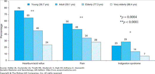
Particular attention has been given to the clinical presentation of GERD in the elderly since important differences between young and adult patients have been reported. Indeed, with advancing age, a significantly lower prevalence of typical symptoms, i.e., heartburn, acid regurgitation, and epigastric pain was reported by patients (Figure 91-1). Atypical symptoms also are relatively rare in the elderly (Table 91-3). In contrast, the prevalence of nonspecific symptoms, i.e., vomiting, anorexia, weight loss, and anemia significantly increased with age (Figure 91-2). Reflux esophagitis in the elderly may thus be missed, and a substantial number of patients may suffer subclinical relapses of the disease. The cause of such a different clinical expression of the disease in the elderly is not clear, but a diminished sensitivity to visceral pain has been documented in the elderly. Moreover, 24-hour esophageal pH monitoring and endoscopy examinations demonstrated an age-related reduction in acid chemosensitivity and a reduced symptom severity despite a tendency toward increased severity of esophageal mucosal injury.
TYPICAL SYMPTOMS | ATYPICAL SYMPTOMS |
|---|---|
Heartburn | Pulmonary symptoms: |
Acid regurgitation | Bronchial asthma |
Epigastric pain | Bronchiectasis |
Chronic bronchitis | |
Chronic cough | |
Otorhinolaryngeal symptoms: | |
Hoarseness | |
Chronic laryngitis | |
Odynophagia | |
Noncardiac chest pain |
Figure 91-2.
Prevalence of nonspecific symptoms in 840 subjects with reflux esophagitis divided according to age. (Adapted from Pilotto A, Franceschi M, Leandro G, et al. Clinical features of reflux esophagitis in older people: a study of 840 consecutive patients. J Am Geriatr Soc. 2006;54:1537–1542.)
Since the presentation of GERD is often with nonspecific symptoms in the elderly, endoscopy should be undertaken early as the initial diagnostic test in all elderly patients suspected of having GERD. Early endoscopy is very useful in diagnosing the presence and the grade of severity of esophagitis (Table 91-4) and/or hiatus hernia, which are important prognostic factors to be considered in the long-term treatment of patients. Endoscopy will also identify GERD complications, especially esophageal stricture and Barrett esophagus, and concomitant gastroduodenal diseases, that is, gastric or duodenal ulcers and/or Helicobacter pylori (H pylori) infection. Barium radiography of the esophagus is a useful test to establish the presence of a hiatus hernia and is indicated as part of the evaluation of the patient with suspected motility abnormalities or peptic stricture. A barium study may also identify rings and webs or other obstructive lesions. The barium swallow test is also a key test in studying elderly patients with dysphagia, and it should be performed in conjunction with endoscopy in all elderly patients with this symptomatology. Barium studies are widely available and usually well tolerated by older people. Esophageal 24-hour pH testing is helpful before antireflux surgery and in those patients not responsive to medical treatment. In the endoscopy-negative patient, an abnormal esophageal pH test may suggest the need for more aggressive drug therapy, whereas a normal test may indicate the presence of a functional disorder. In the patient with persistent esophagitis, a normal test could differentiate pathophysiological mechanisms, that is, a drug-induced esophagitis from acid reflux disease. Esophageal manometry is useful in identifying abnormalities of LES pressure or esophageal motility. In elderly patients, its major use is reserved for the localization of LES before pH testing and for obtaining preoperative information on esophageal peristalsis.
GRADE | DESCRIPTION | |
|---|---|---|
Grade A | One or more mucosal breaks no longer than 5 mm, none of which extends between the tops of the mucosal folds |  |
Grade B | One or more mucosal breaks more than 5 mm long, none of which extends between the tops of two mucosal folds |  |
Grade C | Mucosal breaks that extend between the tops of two or more mucosal folds, but which involve less than 75% of the esophageal circumference |  |
Grade D | Mucosal breaks that involve at least 75% of the esophageal circumference |  |
A therapeutic trial with proton pump inhibitors (PPI-test) has been suggested as a useful diagnostic test in patients with GERD. Meta-analyses of clinical studies demonstrated that the PPI test has 78% sensitivity and 54% specificity in confirming the diagnosis of GERD when compared with endoscopy or 24-hour esophageal pH monitoring. In patients with noncardiac chest pain, sensitivity and specificity are reported to be 80% and 74%, respectively. Since typical (heartburn, acid regurgitation) and extraesophageal symptoms (asthma, chronic cough, noncardiac chest pain) of GERD are often absent in elderly patients, the patient’s history is less reliable and high prevalences of severe esophagitis are common despite mild symptoms, a trial with PPIs should be used with great caution in older patients, possibly only after endoscopy as a first diagnostic test.
The objectives of GERD treatment are (1) relief of symptoms, (2) healing of the esophagitis, (3) prevention of the relapses of the disease, and (4) prevention of the complications.
Recommendations for lifestyle modifications are based on the presumption that certain foods, body position, tobacco, alcohol, and obesity contribute to a dysfunction in the body’s antireflux defense system. Although there is clinical and physiologic evidence that smoking, alcohol, chocolate, peppermint, coffee, fatty or citrus intake may adversely affect symptoms or esophageal pH, there is little evidence that cessation of these agents will improve GERD variables. Elevation of the head of the bed and weight loss, however, have been associated with improvement in GERD variables in case–control studies. Medications that decrease LES pressure and promote gastroesophageal reflux as well as drugs that may cause direct esophageal injury (Table 91-2) should be avoided when possible or used with caution in older patients with GERD. In any case, these drugs must be taken while maintaining an upright position and with a full glass of water.
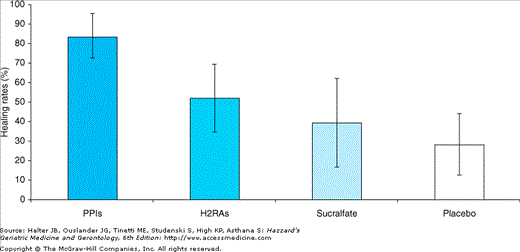
Antacids and alginic acid provide symptomatic relief in mild nonerosive esophagitis. Prokinetic drugs, either alone or in combination with antisecretory drugs, are only moderately effective in GERD. Possible side effects of antacids include salt overload, constipation, hypercalcemia, and interference with absorption of other drugs, particularly antibiotics such as tetracycline, azithromycin, and quinolones. Currently, prokinetic drugs include metoclopramide, clebopride, domperidone, and partially levosulpiride. Unfortunately, no controlled clinical trials have evaluated the role of these drugs in the treatment of GERD in the elderly. Antireflux therapy is focused largely on suppressing gastric acid secretion with H2-blockers and proton pump inhibitors (PPI). Meta-analysis of 43 articles including more than 7600 patients aged 18 to 89 years with grade II to IV esophagitis treated for ≤12 weeks reported higher healing rates with PPIs (83.6%±11.4) than H2-blockers (51.9%±17.1), sucralfate (39.2%±22.4), or placebo (28.2%±15.6). Moreover, PPIs provided faster and more complete heartburn relief than H2-blockers (Figure 91-3). Currently, five PPIs are available: omeprazole, lansoprazole, rabeprazole, pantoprazole, and esomeprazole. Some age-associated differences in pharmacokinetics and pharmacodynamics of the PPIs have been reported. However, it is unknown if these differences are associated with different clinical effects, particularly in older patients. Indeed, a series of meta-analyses evaluating acute therapy of esophagitis reported that the PPIs were superior to ranitidine and placebo in healing erosive esophagitis, without significant differences in efficacy between omeprazole 20 mg daily and lansoprazole 30 mg daily or pantoprazole 40 mg daily or rabeprazole 20 mg daily. Recently, a systematic review of randomized controlled trials comparing standard dose PPIs reported a lower 8-week healing rate of esophagitis with omeprazole 20 mg or lansoprazole 30 mg daily, but not with pantoprazole 40 mg daily, compared to esomeprazole 40 mg daily (Table 91-5). The recent finding that omeprazole has considerable potential for drug interactions, since it has higher affinity for the cytochrome CYP2C19 than other PPIs, may explain these differences in efficacy, especially in elderly patients who are treated with concomitant therapies.
Figure 91-3.
Percentage of patients that experience complete healing after treatment for grade II to IV reflux esophagitis with the indicated therapies. PPIs, proton pump inhibitors; H2RAs, histamine type 2 receptor antagonists. (Adapted from Chiba N, De Gara CJ, Wilkinson JM, et al. Speed of healing and symptom relief in grade II to IV gastroesophageal reflux disease: a meta-analysis. Gastroenterology. 1997;112:1798–1810.)
TOTAL NUMBER OF PATIENTS | ESOPHAGITIS HEALED | % | p | |
|---|---|---|---|---|
Lansoprazole | 3262 | 2698 | 82.7 | 0.0001 |
Esomeprazole | 3264 | 2807 | 85.9 | |
Omeprazole | 2431 | 1998 | 82.2 | 0.0001 |
Esomeprazole | 2446 | 2167 | 88.6 | |
Pantoprazole | 1709 | 1507 | 88.2 | ns |
Esomeprazole | 1688 | 1523 | 90.2 |
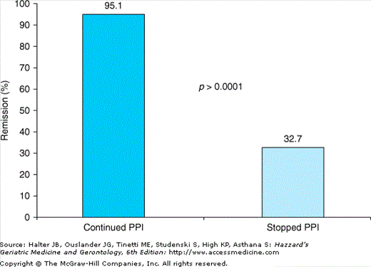
GERD is a chronic disease with a 70% to 90% annual relapse rate after the interruption of an effective antisecretory therapy. Risk factors for relapse of the disease are shown in Table 91-6. Maintenance therapy with antisecretory drugs is a significant protective factor in reducing the occurrence of relapse of GERD in elderly patients. Several studies have shown higher efficacy of PPIs over H2-blockers and prokinetics in maintaining a healed esophagitis. There are two main approaches to drug therapy for GERD: step-up and step-down. In the step-up approach, therapy is initiated with weak inhibition of gastric acid (e.g., an H2-blocker or half dosage of a PPI), and progresses to a higher degree of acid inhibition (standard and then escalating doses of PPI), until adequate symptom control is obtained. The step-down approach involves starting with the most effective regimen (full dosage of a PPI) and switching to lower doses of PPI for maintenance therapy once symptoms are under control. This latter approach is perhaps more rational, based on evidence showing superior efficacy of PPIs over H2-antagonists across all grades of severity of GERD. Withdrawal of maintenance therapy with PPI after 6 months significantly reduces the percentage of remission of esophagitis at 1 year from 95% to 33% in patients older than 65 years (Figure 91-4). Presently, no comparative studies have been carried out to evaluate which strategy (step-down vs. step-up) is more cost-effective in older subjects.
RISK FACTORS |
|---|
Patient’s characteristics |
Old age |
High body mass index (BMI) |
Clinical features |
Long history of reflux symptoms |
Latency prolonged between appearance of symptoms and therapy |
Persistence of symptoms after the healing of esophagitis |
Endoscopic characteristics |
Severity of the esophagitis at baseline |
Presence of a large hiatus hernia |
Pathophysiology features |
Reduced pressure of the lower esophageal sphincter (manometry) |
Modified curve at the 24-h pH-metry |
Figure 91-4.
Remission at 12 months in elderly patients with esophagitis who continued versus stopped PPI maintenance treatment after 6 months. (Data from Pilotto A, Leandro G, Franceschi M, et al. Short- and long-term therapy for reflux oesophagitis in the elderly: a multi-centre, placebo-controlled study with pantoprazole. Aliment Pharmacol Ther. 2003;17:1399–1406.)
A study explored the long-term safety of omeprazole for up to 11 years in patients (including some elderly) with refractory reflux esophagitis. Annual incidence of gastric corpus mucosal atrophy was 4.7% in Hpylori-positive individuals and 0.7% in Hpylori-negative patients. Moreover, mucosal atrophy was mainly observed in elderly patients who had moderate or severe gastritis at entry into the study. Corpus intestinal metaplasia was rare and no dysplasia or neoplasms were observed. Long-term therapy with a PPI does not appear to have marked effects on protein and carbohydrate digestion. Antisecretory drugs, however, may interfere with calcium absorption through induction of hypochlorydria; PPIs, moreover, may reduce bone resorption through inhibition of the osteoclastic vacuolar proton pump. Indeed, a long-term PPI therapy has been associated with an increased risk of hip fractures. Inhibition of acid secretion may induce an increase of the bacterial flora at the gastrointestinal level with potential to increase the incidence of systemic infections particularly in immunodepressed elderly subjects. Indeed, the use of antisecretory drugs has been associated with an increased risk of infections of the lower gastrointestinal tract, mainly because of Salmonella spp. and Clostridium difficile. Recent studies, moreover, reported that the use of PPIs may be associated with an increased risk of community-acquired pneumonia.
Since PPIs are mainly metabolized by the cytochrome P450 (CYP), particularly the subfamily CYP2C19, clinically relevant drug interactions may occur in case of concomitant administrations of CYP-2C19-metabolized drugs. For these reasons a thorough surveillance of the long-term antisecretory therapy is suggested in elderly patients who frequently present with infections, malabsorption and/or diarrhea, osteoporosis, and/or are treated with CYP450-metabolized medications.
The role of surgery in the treatment of GERD is controversial. Laparoscopic fundoplication has greatly reduced the morbidity and mortality of antireflux surgery, also in the elderly. Thus, the indications for surgery seem to be evolving. At present, evidence suggests that surgery may be indicated in elderly patients who (1) are medical treatment failures; (2) have severe complications, i.e., strictures not treatable by endoscopy; (3) have severe dysphagia, aspiration, or atypical symptoms associated with a large hiatal hernia; and/or (4) have preneoplastic lesions, that is, Barrett esophagus with high-grade dysplasia. Randomized clinical studies are needed to compare the outcome of antireflux surgery with that of medical therapy in the elderly. Furthermore, surgery for GERD should be centralized to units specialized in these techniques to reduce surgical complications and improve successful clinical outcomes.
Peptic Ulcer Disease
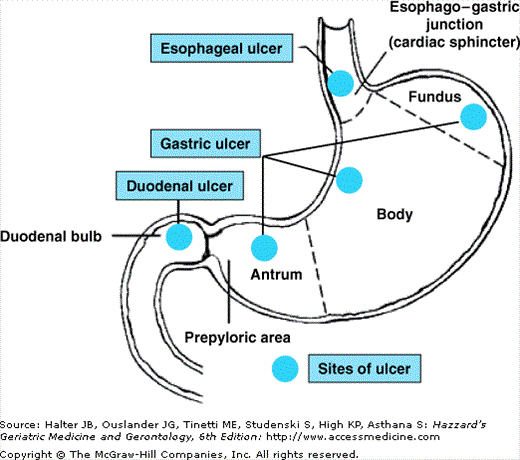
Peptic ulcer is a break of the mucosa lining the stomach or the duodenum. According to their anatomical location, peptic ulcers are divided into gastric ulcers, i.e., peptic ulcers of the gastric fundus, body or antrum, prepyloric and pylori ulcers, i.e., located within 3 cm from the pyloric ring and in the pyloric ring respectively, and duodenal ulcers, i.e., located into the bulb or in the second portion of the duodenum (Figure 91-5).
The worldwide peptic ulcer prevalence differs, with duodenal ulcers dominating in western populations and gastric ulcers being more frequent in Asia. Although the incidence of peptic ulcer disease in western countries has declined over the past 100 years, almost 1 in 10 Americans is still affected. Even if the most recent epidemiological studies indicate that the prevalence and the incidence of the peptic ulcer show a constant decline in the general population, the incidence of complicated gastric and duodenal ulcer hospitalization (Figure 91-6) and mortality (Figure 91-7) remain very high in older patients.

