Tumor Necrosis Factor-Related Cytokines in Immunity


Tumor Necrosis Factor-Related Cytokines in Immunity


Carl F. Ware


INTRODUCTION

Lymphocytes are highly mobile, transiting the vascular and lymphatic circulatory systems with limited stopovers in organized lymphoid tissues (lymph nodes, spleen, and Peyer’s patches) in which they commune to initiate immune responses. Cytokines, both secreted and membrane-anchored proteins, serve as the communication media of the immune system. Cytokines in the tumor necrosis factor (TNF) superfamily (TNFSF) help orchestrate the development, homeostasis, and effector actions of cells in the immune system. The diversification of the TNFSF is evidenced by its roles in regulating cells of neuronal, skeletal, and ectodermal origin. This chapter’s focus is on members in the TNFSF that are primarily involved in regulating immunity.

The ligands belonging to the TNFSF initiate intercellular communication by binding specific receptors on the surface of the receiving cell. The TNF-related ligands are membrane-bound and require cell-cell contact to initiate signaling, although some ligands may be secreted in soluble form influencing cellular responses at locations distant from the source of production. The TNF receptors (TNFRs) form a corresponding superfamily of cognate membrane proteins that initiate intracellular signaling pathways that influence cellular growth, differentiation, and survival. Each ligandreceptor pair forms a “system” with over 40 distinct ligandreceptor systems. Many of the ligands and receptors engage more than one cognate, thereby forming “circuits” of signaling systems, which often function together as coordinated or integrated networks with other cytokines to regulate a specific cellular process. The signals delivered to the cytosol activate transcription factors such as NF-&kgr;B and AP1, which in turn initiate the expression of hundreds of genes that alter cellular differentiation. Additionally, some TNFRs activate cell death pathways, both apoptotic and necrotic, terminating cellular life. Genetic mutations have revealed the importance of individual TNFSF members in human immune responses and development. Moreover, the beneficial effect of TNF antagonists in patients with certain autoimmune diseases brings the need to understand the complexities of the TNFSF into sharp focus.

The nomenclature of the TNFSF is a bit of a morass for students new to the field, although introduction of a numerical system standardizing gene names (www.genenames.org/) helps in accessing genomic databases. However, the use of more common, often colorful, acronyms continues. The name, rank, and genomic identification of each ligand (Table 27.1) and receptor (Table 27.2) is tabulated along with additional pertinent information for human and mouse genes.


TUMOR NECROSIS FACTOR LIGAND FAMILY

The TNF-related cytokines are type II transmembrane proteins (intracellular N-terminus) with a short cytoplasmic tail (15 to 25 residues in length) and a larger extracellular region (˜150 amino acids) containing the signature TNF homology domain where the receptor binding sites are located (Fig. 27.1). The TNF homology domain assembles into trimers, the functional unit of the ligand. Atomic analysis of several members of the family1,2,3,4,5 revealed the ligands have a highly conserved tertiary structure folding into a &bgr; sheet sandwich, yet amino acid sequence conservation is limited to < 35% among the family members. The conserved residues defining this superfamily are primarily located within the internal &bgr; strands that form the molecular scaffold, which promote assembly into trimers. The residues in the loops between the external &bgr;-strands are variable and in specific loops make contact with the receptor. Although most of the TNF ligands self-assemble into homotrimers, heterotrimers can also form between LT&agr; and LT&bgr;6 and APRIL and BAFF.7 The stoichiometry of the LT heterotrimer is 1:2 (LT&agr;&bgr;2), which imparts its distinct receptor specificity from the LT&agr; homotrimer. Interestingly, complement component C1q and several related proteins are structurally related to the TNF family, containing a TNF homology domain, a rather surprising finding given the apparent functional divergence between the complement and TNF systems.8 Alternate splicing can also generate distinct ligands, such as the splice form joining TWEAK and APRIL (TWE-PRIL),9 the alternate ligands for ectodysplasin receptor,10 and a cytosolic form of LIGHT.11

The genetic organization of TNF ligands is highly conserved and typically encoded in three or four exons, with the fourth exon encoding most of the extracellular TNF homology domain. The genes encoding TNF, LT&bgr;, and LT&agr; reside adjacent to each other in a compact loci in the class III region of the major histocompatibility complex (MHC) (in humans at chromosome 6p21) sandwiched between the antigenic-peptide presenting MHC proteins encoded by MHC class I and II genes.6,12 Three other genetic clusters of TNF-related cytokines are found within the corresponding MHC paralogous regions, located on chromosomes 1, 9, and 19. These genetic clusters share a remarkably conserved gene structure and transcriptional orientation, and a similar function linked to regulating cellular immune responses.11 The evolutionary pressures retaining this genetic configuration of the MHC in general is encompassed in the paralogy theory of genome
evolution.13 Conservation of gene structure of the TNF ligands outside these MHC paralogs is limited. An evolutionarily conserved pathway in Drosophila melanogaster, Eiger-Wengen system, is structurally and functionally related to the TNFSF-related signaling pathway, although this system is predominantly expressed in the nervous system in invertebrates.14,15,16


TABLE 27.1 Tumor Necrosis Factor Superfamily Chromosomal Locations


Chromosomal Location


Gene Name/Alias


Human


Mouse


Ligand Symbol


TNF


6p21.3


ch17 (19.06 cm)


TNFSF1A


LT&agr;


6p21.3


ch17 (19.06 cm)


TNFSF1B


LT&bgr;


6p21.3


ch17 (19.06 cm)


TNFSF3


OX40-L


1q25


ch1 (84.90 cM)


TNFSF4


CD40-L, CD154


Xq26


chX (18.0 cM)


TNFSF5


Fas-L


1q23


ch1 (85.0 cM)


TNFSF6


CD27-L, CD70


19p13


ch17 (20.0 cM)


TNFSF7


CD30-L, CD153


9q33


ch4 (32.20 cM)


TNFSF8


4-1BB-L


19p13


ch17 (20.0 cM)


TNFSF9


TRAIL


3q26


ch3


TNFSF10


RANK-L, TRANCE


13q14


ch14 (45.0 cM)


TNFSF11


TWEAK


17p13


ch11


TNFSF12


APRIL/TALL2


17P13.1


ch13


TNFSF13


BAFF, BLYS, TALL1


13q32-q34


ch8 (3 cM)


TNFSF13B


LIGHT


19p13.3


ch17 (D-E1)


TNFSF14


TL1A


9q33


ch4 (31.80 cM)


TNFSF15


G1TRL, A1TRL


1q23


Unknown


TNFSF18


EDA1


Xq12-q13.1


chX (37.0 cM)


EDA1


EDA2


Xq12-q13.1


cX (37.0 cM)


EDA2


CD, cluster of differentiation; LT, lymphotoxin; TNF, tumor necrosis factor; TNFSF, tumor necrosis factor superfamily; TRAIL, TMF-related apoptosis inducing ligand.


From Ware CF. The TNF superfamily-2008. Cytokine Growth Factor Rev. 2008;19:183-186.


TUMOR NECROSIS FACTOR RECEPTOR FAMILY

Members of the TNF receptor superfamily (TNFRSF) include proteins of vertebrate and viral origin. Most of the signaling receptors in the TNFRSF are type I transmembrane glycoproteins (N-terminus exterior to the cell). However, several TNFRSF members lack a membrane-anchor domain, are proteolytically cleaved from the surface, or are anchored via glycolipid linkage (eg, TNF-related apoptosis inducing ligand [TRAIL]R3). These soluble receptors, termed “decoy receptors,” retain their ligand-binding properties and compete with cellular receptors for the specific ligands, thus earning the title of decoy. The structural motifs in the cytoplasmic domains further categorize the TNFR into two groups based on their signaling properties: those contain a death domain (DD) and others that engage TNFR-associated factors (TRAFs).


TABLE 27.2 Tumor Necrosis Factor Receptor Superfamily


Chromosomal Location


Gene Name/Aliases


Human


Mouse


Gene Symbol


TNFR-1, p55-60


12p13.2


ch6 (60.55 cM)


TNFRSF1A


TNFR2, p75-80


1p36.3-36.2


ch4 (75.5 cM)


TNFRSF1B


LT&bgr;R


12p13


ch6 (60.4 cM)


LT&bgr;R


OX40


1p36


ch4 (79.4 cM)


TNFRSF4


CD40


20q12-q13.2


ch2 (97.0 cM)


CD40


FAS, CD95


10q24.1


ch19 (23.0 cM)


TNFRSF6


DcR3


20q13


unknown


TNFRSF6B


CD27


12p13


ch6 (60.35)


TNFRSF7


CD30


1p36


ch4 (75.5 cM)


TNFRSF8


4-1BB


1p36


ch4 (75.5 cM)


TNFRSF9


TRAILR-1, DR4


8p21


Unknown


TNRSF10A


TRAIL-R2, DR5


8p22-p21


ch14 (D1)


TNFRSF10B


TRAILR3, DcR1


8p22-p21


ch7 (69.6 cM)


TNFRSF10C


TRAILR4, DcR2


8p21


ch7 (69.6 cM)


TNFRSF10D


RANK, TRANCE-R


18q22.1


ch1


TNFRSF11A


OPG, TR1


8q24


ch15


TNFRSF11B


FN14


16p13.3


ch17


TNFRSF12A


TRAMP, DR3, LARD


1p36.3


ch4 (E1)


TNFRSF25


TACI


17p11.2


ch11


TNFRSF13B


BAFFR


22q13.1-q13.31


ch15


TNFRSF13C


HVEM, HveA, ATAR


1p36.3-p36.3


ch4


TNFRSF14


P75NTR, NGFR


17q12-q22


ch11 (55.6 cM)


TNFRSF16


BCMA


16p13.1


ch16 (B3)


TNFRSF17


AITR, GITR


1p36.3


ch4 (E)


TNFRSF18


RELT


11q13.2


unknown


TNFRSF19L


TROY, TAJ


13q12.11-q12.3


ch14


TNFRSF19


EDAR


2q11-q13


ch10


EDAR1


EDA2R


Xq11.1


ch X


EDA2R


DR6


6P12.2-21.1


ch17


TNFRSF21


IGFLR1, Tmem149


19q13.12


ch7


IGFLR1


CD, cluster of differentiation; HVEM, herpesvirus entry mediator; LT, lymphotoxin; OPG, osteoprotegerin; TNFR, tumor necrosis factor receptor; TNFRSF, tumor necrosis factor receptor superfamily; TRAIL, TMF-related apoptosis inducing ligand.


From Ware CF. The TNF superfamily-2008. Cytokine Growth Factor Rev. 2008;19:183-186.


The cysteine-rich domain (CRD) in the extracellular, ligand-binding region defines membership in the TNFRSF (see Fig. 27.1). Each CRD typically contains six cysteine residues forming three disulfide bonds. The CRD is pseudorepeated in different members ranging from one to six. Based upon the crystal structures solved for several TNFRSF members, the CRD confers an elongated shape and sidedness to the ectodomain.17,18 The crystal structure of the complex between TNFR1 and one of its ligands, the LT&agr; homotrimer, revealed
that residues in CRD2 and 3 of TNFR1 contact the ligand. Variation in binding interactions has been identified; for example, the receptors for BAFF have one functional CRD.2,19


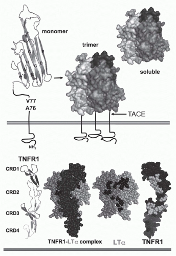
FIG. 27.1. Structure of Tumor Necrosis Factor (TNF) and TNF Receptors (TNFRs). Top: TNF. The &bgr;-sandwich of the TNF monomer (1A8M.pdf) (shown as a ribbon diagram) contains two stacked &bgr;-pleated sheets each formed by five antiparallel &bgr; strands (wide ribbons given letter designations, A-G) that fold into a Greek key or “jelly-roll” topology.219 The inner &bgr;-sheet (strands A, A′, H, C, and F) is involved in contacts between adjacent subunits, which promotes assembly into a trimer. The trimer is formed such that one edge of each subunit is packed against the inner sheet of its neighbor. The outer &bgr; sheet (strands B, B′, D, E, and G) is surface exposed. The trimeric structure is characteristic of all TNF-related ligands. The type II configuration of TNF (N-terminus inside the cell) anchors TNF to the membrane. The TNF trimer is ˜60 Å in height with a relatively flat base residing close to the membrane, resembling a bell-shape (shown as surface representation with different shades of gray used for each subunit of the trimer). The surface exposed loops between A-A′ and D-E strands are involved in receptor binding. TNF is cleaved by TNF&agr;-converting enzyme (TACE), a member of the ADAM family of metalloproteinases (ADAM17) involved in processing of many cell surface proteins. TACE is a type 1 transmembrane protein that cleaves TNF between residues Val77 and Ala76, when all three sites in the trimer are cleaved TNF is released from the membrane. Bottom: TNFR and ligand complex. The ectodomain of TNFR1 forms an elongated molecule with CRD1 proximal to the N-terminus (ribbon diagram). The face of TNFR1 on the left engages LT&agr;. In the ligand-receptor complex, the elongated receptor (dark) lies along the cleft formed between adjacent subunits. Shown (space-filling) is a single TNFR1 in complex with two subunits of LT&agr; (lighter shades); the receptor N-terminus points upward, closest to the base of the ligand (transorientation). In the exploded view, TNFR1 is removed from in front of the ligand revealing the contact residues in the ligand, which are primarily located in the D-E and the A-A″ &bgr;-strands (dark shade). TNFR1 is rotated 180 degrees, exposing the contact residues in the receptor (light shade). Structures from 1TRN.pdf17 as visualized with MacPyMOL (www.pymol.org).


RECEPTOR-LIGAND COMPLEX

The binding specificity of the various members of the TNF ligand and receptor superfamilies show monotypic interaction, yet several members interact with multiple partners20 (Figs. 27.2 and 27.3). The binding interactions between TNF-related ligands and TNFR are typically high affinity, with equilibrium binding constants measured in the high pM to low nM range. The membrane position of the ligands further enhances the binding interaction with their receptors. In the membrane anchored position, the ligands and receptors must be in trans to form a complex. In the LT&agr;-TNFR1 complex, the surface loops between A-A″ and D-E &bgr; strands contain many of the amino acid residues that make contact with the receptor, with the receptor binding site formed as a composite of adjoining subunits in the trimeric ligand17 (see Fig. 27.1).

The trimeric architecture of the TNF ligands, containing three equivalent receptor-binding sites, provides the basis for initiating signaling through aggregation or clustering of receptors. This concept is supported by the finding that receptor-specific bivalent antibodies can act as agonists mimicking the signaling activity of the natural ligand.21 Indeed, antibodies or peptide mimetic to TNFR can function as antagonists, blocking the ability of the natural ligand to bind the receptor while simultaneously activating the receptor as an agonist.22 Some ligands such as FasL, TRAIL, and BAFF form higher ordered oligomers of the basic trimer. These higher ordered oligomers promote supraaggregation of receptors, enhancing or sustaining signaling pathways in the receptor-bearing cell.23,24 Overexpression of TNFR in cells can also lead to ligand-independent signaling, a
feature bestowed in part by the propensity of the cytosolic tails to self-associate.25 In the physiologic setting, the expression level and compartmentalization of these receptors are tightly controlled. A subregion in the first CRD of TNFR1 known as the preligand assembly domain may restrict the orientation of the unligated receptor to prevent spontaneous activation.26 It is not known if this mechanism applies to all TNFRSF. Interestingly, some patients with periodic fever syndrome have mutant TNFR1 that form abnormal disulfide-linked oligomers that are retained intracellullarly and provoke misfolded protein response.27


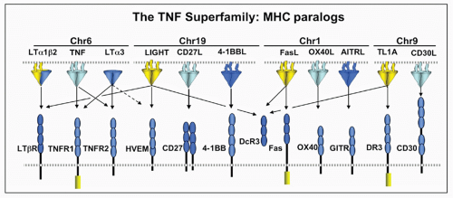
FIG. 27.2. The Tumor Necrosis Factor (TNF) Superfamily-Major Histocompatibility Complex (MHC) Paralogs. Members of the TNF ligand superfamily (above) and their corresponding receptors (below) are identified by connecting arrows. The ligands are grouped according to their chromosomal locations in the MHC paralogous regions. The number of cysteine-rich domains are depicted for each TNF receptor, and TNF receptors containing a death domain are identified by cylinder in the cytoplasmic tail. (Modified from Ware336).


Alternate Ligands

There is significant divergence in the ligands and the mechanisms of ligand binding by the TNFR family. A major branch point is exemplified by the ligands for p75 neurotrophin receptor (nerve growth factor and the other neurotrophins), which are structurally unrelated to TNF ligand family. Molecular contacts between NGF and p75NTR occur through two spatially separated binding regions located at the first and second CRD and the junction between the CRD3 and CRD4.28 The p75NTR functions in complex with two other proteins, Nogo66 and LINGO, to engage myelin-associated inhibitory factors. Taj/TROY can supplant p75NTR in this complex.29 Like p75 NTR, DR6, ILGF1R, and TROY/Taj do not bind any of the known TNF ligands but do engage other ligands. The pathways activated by p75NTR and TROY/Taj systems show both positive and inhibitory regulation of axonal regeneration.29,30

The herpesvirus entry mediator (HVEM; TNFRSF14) provides an example of a TNFR system that binds alternate ligands. Although HVEM engages two TNF-related ligands, LIGHT and LT&agr;, it also engages two members of immunoglobulin (Ig) superfamily, B- and T-lymphocyte attenuator (BTLA)31 and cluster of differentiation (CD)160.32 BTLA binding to HVEM occurs in CRD1, on the opposite face of where LIGHT/LT&agr; bind in CRD2 and 3. The BTLA binding site in CRD1 of HVEM is a region also targeted by herpesviruses.33,34,35 Recent evidence indicates the neurotrophin and chondrocyte growth factor-like protein progranulin interacts with TNFR1 and TNFR2 and competes with TNF for binding.36

An insulin growth factor-like protein was recently identified as a ligand for a TNFR-like type 1 transmembrane protein (insulin growth factor-like R, formerly TMEM149).37 Insulin growth factor-like R has conserved positioning of cysteines delineating a CRD1, but atypical CRD2/3. Insulin growth factor-like messenger ribonucleic acid (mRNA) is expressed in psoriatic skin lesions and the receptor is detected in T cells suggesting a possible role in skin inflammation.

Death receptor-6 (DR6) engages the growth factor-like domain in &bgr;-amyloid precursor protein38,39 and thus may function as a negative regulator more like p75 neurotrophin receptor.40 Emerging evidence suggests DR6 has a role in neuroimmune function. In a mouse experimental autoimmune encephalomyelitis (EAE) model, DR6-deficiency was shown to protect against central nervous system demyelination and leukocyte infiltration, but also enhanced overall CD4+ T-cell proliferation and TH2 differentiation, underscoring a role for DR6 in mediating TH1-specific immune responses in EAE progression.41 Taj/TROY and RELT also do not bind any of the known TNFSF ligands; however, recent evidence indicates that Taj/TROY functions more like p75NTR, capable of binding myelin inhibitory factors.29,30 A role for Taj/TROY and RELT in immune function is presently unclear.
The engagement of ligands distinct from TNFSF members implicates a higher level of integration with other signaling pathways.


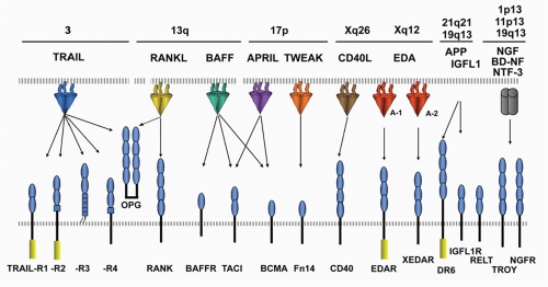
FIG. 27.3. The Tumor Necrosis Factor (TNF) Superfamily—Part 2. Members of the TNF ligand superfamily (above) and their corresponding receptors (below) are identified by connecting arrows. The ligands are grouped according to their chromosomal locations. The number of cysteine-rich domains are depicted for each TNF receptor, and TNF receptors containing a death domain are identified by cylinder in the cytoplasmic tail. (Modified from Ware336).


Viral Orthologs

TNFR-like proteins are found in the genomes of several viral pathogens representing captured cellular genes that have evolved as part of that pathogen’s immune evasion strategy (Table 27.3). Poxviruses were the first pathogens identified harboring a version of a cellular TNFR.43 Poxvirus TNFR displays significant sequence homology to TNFR2 and binds TNF and LT&agr;. The rabbit poxvirus protein T2 is secreted by virally infected cells and contributes to the virulence of infection.44,45 Smallpox virus, the former scourge of mankind, also harbored viral versions of TNFR2.46


TABLE 27.3 Viral Orthologs and Modulators of the Tumor Necrosis Factor Superfamily


Virus


Name/ORF


Orthologa


Mechanism


Poxvirus


Myxoma


T2


Soluble TNFR2


TNF and LT&agr; decoy


Variola


crm B (G2R)


Soluble TNFR2


TNF and LT&agr; decoy


Cowpox


vCD30


Soluble CD30


CD30 ligand inhibitor


Herpesvirus


HSV1&2


Glycoprotein D


BTLA


Entry; HVEM blockade


HCMV


UL144


HVEM


BTLA activation


EBV


LMP1


CD40 intracellular


TRAF activation


&ggr;HV


vFLIP


FLIP


Caspase 8 blockade


Adenovirus


E3-10.4, 14.5, 6.7


?


Fas and TRAILR downmodulation


Retrovirus


EIAV


Envelope gp90


?


HVEM entry factor


FIV


Envelope gp95


?


Ox40 entry factor


ASLV


Envelope gp85


?


TRAILR entry factor


Rabies virus


Envelope RVG


?


NTRp75 entry factor


ASLV, avian sarcosis and leukosis virus; BTLA, B- and T-lymphocyte attenuator; CD, cluster of differentiation; EBV, Epstein-Barr virus; HCMV, human cytomegalovirus; HSV, herpes simplex virus; &ggr;HV, equine gamma herpesvirus; EIAV, equine infectious anemia virus; FIV, feline immunodeficiency virus; TNFR, tumor necrosis factor receptor; TRAF, TNFR-associated factor; TRAIL, TMF-related apoptosis inducing ligand;?, no homology recognized.


a Relationship determined by sequence or structural homology to the indicated TNF superfamily member.


SIGNALING PATHWAYS AND CELLULAR RESPONSES


Regulation

The cellular response activated by a TNF-related cytokine depends on several factors including the temporal patterns of expression of the ligands and receptors on the interacting cells, and the cellular context (the state of differentiation of the responding cell). Regulation is achieved at the level of transcriptional and translational controls, and by modulating the availability of the ligand or receptors (Fig. 27.4). For some ligands, transcriptional activation is a critical feature controlling the duration of mRNA expression. Some ligands exhibit inducible, transient expression of mRNA following signals from antigen receptors or innate receptor recognition systems. The half-life of mRNA
for TNF is short, controlled by an adenylate-uridylate-rich element in the 3′ untranslated region.47 Deletion of the adenylate-uridylate-rich element in TNF mRNA in mice leads to a profound inflammatory disease.47,48 TNF mRNA is inducible in macrophages by multiple pathways, particularly innate activation pathways such as toll-like receptor signaling, whereas other ligands like 41BBL and OX40L are constitutively expressed in differentiated antigen-presenting dendritic cells (DCs). T- and B-lymphocytes require activation prior to expression of TNF. In these cells, signals via the antigen receptor utilizing nuclear factor of activated T cells (NFAT) transcription factors are needed for the induction of TNF transcription. The inducible or constitutive patterns of expression are also observed with some receptors as well.


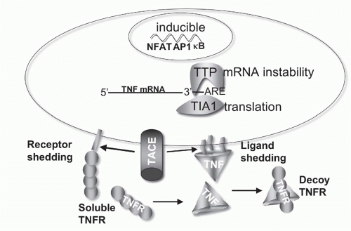
FIG. 27.4. Regulation of Tumor Necrosis Factor (TNF) Bioavailability. The expression of TNF is regulated at the transcriptional and translational levels, and bioavailability by altering its physical location and cellular receptors. TNF transcription is regulated by the action of multiple transcription factors including nuclear factors of activated T cells (NFAT), activated protein-1 (AP1), and NF-&kgr;B. NFAT is a predominant acting transcription factor regulating TNF transcription in T- and B-lymphocytes, and NF-&kgr;B and AP1 are important in myeloid lineage cells following activation via innate pattern recognition receptors, such as the toll-like receptors. TNF messenger ribonucleic acid (mRNA) stability is controlled by an adenylate-uridylate-rich element (ARE) in the 3′ untranslated region present in many transiently expressed inflammatory genes.337 Stability of TNF mRNA is decreased by the action of tristetraprolin, and T-cell intracellular antigen silences translation of TNF mRNA through the ARE.338 TACE proteolytically controls TNF at the membrane, generating the soluble form of TNF. TACE also cleaves TNFR1 and 2,339 downregulating cell surface receptors and releasing soluble receptors that retain TNF binding activity. Soluble TNFR can stabilize the TNF trimer at sub saturating concentrations, and at higher, saturating concentrations act as decoys competing for TNF binding to cellular receptors.340

Posttranslational regulation of signaling is achieved by proteolytic cleavage of the ligand or receptor from the cell’s surface, which places the protein into the soluble phase, where its half-life may be dramatically shortened. TNF and Fas ligand, for example, are shed from the surface by membrane proteases. ADAM17 (also known as TNF&agr;-converting enzyme [TACE]), the enzyme that processes TNF into a soluble form, is involved in cleaving multiple cell surface proteins including transforming growth factor &agr;, L-selectin, and TNFR1 and 2. Production of soluble TNFR1 and 2 may be important in regulating TNF bioavailability (see Fig. 27.4).

Most of the TNF-related ligands are expressed by DCs, activated lymphocytes, and myeloid cells, particularly macrophages, but can also be produced by nonlymphoid cells. TNF is an example of a ligand expressed by many cell types depending on the stimulus. Expression of TNFR is widespread. TNFR1 is expressed on most cells, while TNFR2 is limited to cells of hematopoietic origin and is expressed following activation of B or T cells. Macrophages are a primary source of TNF in response to toll-like receptor signaling, and T cells produce TNF when activated by antigenic stimuli. Fibroblasts also produce TNF in response to virus infection, and nonlymphoid tumor cells may ectopically express TNF. FasL is another example of a ligand with a varied cellular expression pattern. FasL is expressed by effector T cells and natural killer (NK) cells as a component of their cell lytic activity, yet FasL mRNA is also detected in reproductive organs and epithelium of the eye, which may use this TNFSF member to kill organ infiltrating inflammatory cells as a mechanism to dampen inflammation.


Signal Transduction Pathways

TNF receptors initiate signaling pathways that alter gene expression patterns, changing the differentiation status of a cell, as well as apoptotic pathways that terminate cellular life. The propagation of signals from receptor to enzymatically active proteins is mediated by two distinct types of signaling motifs in the cytosolic domain of the TNFR: DDs and TRAF-binding motifs. TNFR that contain a DD include Fas, TNFR1, DR3, and TRAILR1 and 2. Other TNFRs have TRAF recruitment motifs. Three basic schemes are used by TNF receptors to connect to enzymatically-driven signaling pathways (Fig. 27.5A). Adaptor molecules are required to establish the signaling connections between the receptor and signaling enzymes. The DD connects TNFR to cytosolic proteins containing a Death Effector Domain (DED), which in turn link to caspases (cysteine based, aspartic acid specific proteinases). Alternately, the TRAF proteins connect to the cytosolic domain of the TNFR, altering ubiquitin-dependent pathways that regulate key serine kinases that activate NF&kgr;B and AP1 transcription factors. The third scheme involves an indirect link between the death domain and TRAFs via the adaptor TNFR-associated death domain (TRADD).

The apoptotic and NF-&kgr;B pathways activated by the TNFR family help regulate cellular homeostasis by controlling cell death and survival. In the immune system, apoptosis is essential for homeostasis and for eliminating antigen-bearing cells from the host. Many TNFR can induce activation of survival or death pathways depending in part on the differentiated state of that cell. In all nucleated cells, apoptosis is the default pathway; that is to say, all of the constituents of the pathway are expressed and ready to be activated (Fig. 27.6), whereas cellular survival requires transcriptional control of genes that encode regulatory proteins that suppress the progression of the apoptotic pathway.


Apoptosis

Ligation of TNFR promotes assemble of the death-inducing signaling complex (DISC) that promotes dimerization of procaspase 8 forming an active enzyme complex.49 Activated caspase 8 acts directly to cleave procaspase 3 and 7, which are known as the executioner caspases as they directly cleave critical cellular substrates leading to apoptotic death. Caspase 8 also cleaves BID, a crucial connector to the mitochondria-associated death mechanism, which greatly amplifies the apoptotic process.50 A cell must be capable of actively transcribing and translating genes to resist apoptosis signaling. A variety of genes can inhibit apoptosis and cell death pathways. For example, the cellular inhibitor of apoptosis (XIAP) is a direct caspase inhibitor that is regulated at the level of gene expression by transcription factor NF-&kgr;B. Another regulator, FLIP (FLICE inhibitory protein), is also an NF-&kgr;B-regulated gene that contains a DED and a pseudocaspase domain that attenuates the apoptotic pathway by blocking conversion of procaspase 8 to the active form.51 Many viral pathogens parasitize the transcriptional capabilities of the cell and prevent the cell from making new survival proteins, allowing apoptosis to proceed. As expected, viruses have evolved many different strategies to alter proapoptotic pathways (eg, viral orthologs of XIAP and FLIP prevent premature death of the cell) (see Fig. 27.6).


Cell Survival Signaling

Activation of transcription factors by TNFR utilizes the TRAF adaptors. There are seven members of the TRAF family (given numerical designations) that play key roles in regulating TNFR signaling and activation of host defenses. Each TRAF appears to play different roles in modulating signaling. Each TRAF protein contains a TRAF homology domain that binds TNFR and a RING and/or zinc finger domain characteristic of E3 ubiquitin ligases.52,53,54 Ubiquitination plays an essential role in regulating signal transduction by TNFR55,56 and in the pathogen recognition receptors as part of innate host defenses.57

Clustering of the TNFR promotes the recruitment of TRAFs into a complex with the receptor through two different mechanisms. TRAF2, 3, and 5 bind directly to the receptor’s cytosolic domain recognizing a consensus PXQS/T motif,58 whereas TNFR1 uses TRADD to couple to TRAF2. TRAF3 and TRAF6 are preassociated with different proteins, including protein kinases involved in multiple signaling pathways including the pathogen recognition and interferon (IFN) pathways.53,59 TRAF adaptors link TNFR directly to protein kinase cascades, which in turn lead to activation of transcription factors including NF-&kgr;B and AP1.

The NF-&kgr;B family of transcription factors control expression of genes critical for cell survival, inflammatory, and immune responses. In mammalian cells, the NF-&kgr;B family consists of five members: RelA, RelB, c-Rel, p50/NF&kgr;B B1, and p52/NF&kgr;B B2 (proteolytic processing of p105 and p100 yields the active forms p50 and p52, respectively).60,61 Activation of NF-&kgr;B releases inhibitors that restrict nuclear translocation.62 Homo- and heterodimers of NF-&kgr;B family members are held inactive in the cytosol by inhibitors of &kgr;B (I&kgr;B), such as I&kgr;B&agr;, that mask nuclear localization motifs on the NF-&kgr;B dimers. A complex consisting of the kinase catalytic subunits IKK&agr; and IKK&bgr; and the regulatory/scaffold subunit IKK&ggr; form the IKK complex that mediates phosphorylation and ubiquitination of I&kgr;B, leading to its degradation and release of the active transcription factor. I&kgr;B is the common target of a variety of signals that control the activation of the RelA NF-&kgr;B transcription factor, which in turn regulates expression of many proinflammatory genes within a signal responsive cell.63

A distinct pathway regulates the activation of RelB NF-&kgr;B through the NF-&kgr;B-inducing kinase (NIK) and IKK&agr; kinases. In unstimulated cells, NIK levels are maintained at vanishing low levels by ubiquitination and proteosome degradation through an E3 ligase complex comprised of TRAF3, TRAF2, and CIAP1/2.52 Receptor ligation releases the active form of NIK by competitively displacing TRAF3, preventing further ubiquitination and allowing NIK to accumulate.64 NIK phosphorylates IKK&agr;, which induces the proteosome dependent processing of p100 to p52, degrading the inhibitory domain of p100 and allowing the RelB/p52 complex to activate gene transcription.


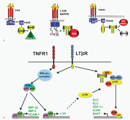
FIG. 27.5. Signaling Pathways and NF-&kgr;B. A: Adaptors link tumor necrosis factor receptors (TNFRs) to proteinases and kinases (upper panel). Three basic schemes link activated TNFRs to signaling pathways: the death inducing signaling complex formed with Fas is initiated by ligand clustering of Fas, promoting death domain (DD) interactions, which recruits Fas-associated death domain (FADD) (heterotypic interaction). The death effector domain of FADD links to the death effector domain of procaspase 8. The proximity of multiple procaspase 8 domains forms an active enzyme that can process other caspase 8 molecules. By contrast, TNFRs bind TNF-associated factor (TRAF) adaptors via short peptide motifs that release TRAF from associated kinases, such as NF-&kgr;B-inducing kinase (NIK). The third scheme is a combination of DD and TRAF recruitment. TNFR1 uses the DD protein TRADD to recruit RIP and TRAF2, promoting the activation of NF-&kgr;B. There are currently seven TRAF members with distinct interaction patterns with the TNFR family. TRAF proteins may function as regulators of key kinases. TRAF2, 3, and 6 function as modulators of several different kinases involved in toll-like receptor signaling, induction of type 1 interferon responses, and signaling by some TNFR family members. TRAF proteins contain an N-terminal RING finger motif, a coiled coil domain (isoleucine zipper), and the receptor association domain (TRAF) domain. The TRAF are trimers formed through their TRAF and coiled domains. The TRAF domain can bind to several different TNFR through a relatively short proline-anchored sequence that is responsible for binding directly to the mushroom head of various TRAF molecules.341 The zinc RING of TRAF6 functions together with Ubc13 and Uev1A as ubiquitin E3 ligase targeting proteins for proteosome degradation. TRAF2, 3 and cIAP form the ubiquitin E3 ligase that targets NIK. Modified with permission from Ware CF. Tumor necrosis factors. In: Bertino JR, ed. Encyclopedia of Cancer. San Diego, CA: Academic Press, Inc.: 2002;475-489. B: NF-&kgr;B activation. TNFR1 and LT&bgr;R induce distinct forms of the NF-&kgr;B family of transcription factors. TNFR1 signaling is a potent activator of RelA/p50 but does not activate the RelB pathway, whereas lymphotoxin &bgr;R can activate both. The RelA and RelB forms of the &kgr;B family control transcription of distinct sets of genes; however, the two pathways are interrelated through the control of p100 expression by the RelA/p50 complex. Modified from Dejardin et al.66


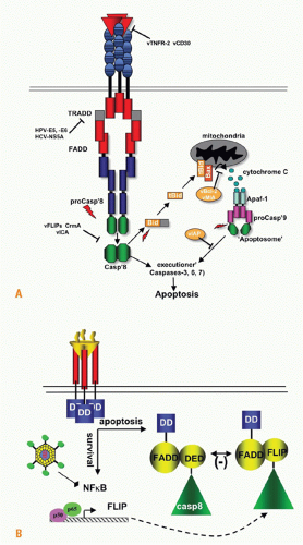
FIG. 27.6. Apoptosis and Survival Signaling. A: Ligation of tumor necrosis factor (TNF) receptor 1, Fas, or TNF-related apoptosis inducing ligand 1 and 2 activates apoptosis pathways. The nascent death-inducing signaling complex can directly cleave the executioner caspases 3, 6, and 7. Caspase 8 also cleaves BID to tBID, activating the mitochondrial regulated apoptotic pathway, dramatically accelerating cellular death. A number of viral proteins interfere with apoptosis induced through TNF pathway including human papilloma virus, hepatitis C virus, herpesvirus vFLIP, and virus orthologs of BCL2 and vMIA act on the mitochrondria. Adapted with permission.111 B: The components of the apoptotic pathway are preformed in the cytosol enabling the cell to respond rapidly to death signaling. In contrast, the key regulators of the apoptotic pathway, including Bcl2 and inhibitor of apoptosis, require new gene expression controlled by NF-&kgr;B. An important NF-&kgr;B-induced gene is cellular FLIP, which contains a pseudocaspase domain. The cFLIP protein interferes with caspase 8 activation, blocking the prodeath pathway. If the cell is transcriptionally inactive or unable to activate NF-&kgr;B, which often occurs in a pathogen-infected cell, then the proapoptotic pathway dominates the signals emanating from the TNF receptor 1 signaling complex leading to apoptosis and curtailing parasitism of the cell.

As examples, TNFR1 and LT&bgr;R activate separate yet related pathways that lead to distinct forms of NF-&kgr;B, the RelA/p50, and RelB/p52 complexes.65 Each form of NF-&kgr;B activates a large number of genes with distinct roles in physiology (see Fig. 27.5B). TNFR1 signaling rapidly mobilizes (within minutes) the RelA/p50 complex, which controls expression of many proinflammatory and survival genes. By contrast, the processing of p100 and accumulation of nuclear RelB/p52 takes several hours after the initial stimulus. RelB-dependent genes are often involved in lymphoid tissue organogenesis and homeostasis. NIK is also required for NF-&kgr;B RelB and RelA activation by CD27, CD40, and BAFF-R, but not by TNFRI, which is restricted to activating RelA/p50 complex.66,67 Components of the NF-&kgr;B pathway, including TRAF3, cIAP, NIK, and A20, are frequently mutated in cancer, leading to constitutive expression of survival factors such as the BCL2 family.68


THE IMMEDIATE LYMPHOTOXIN AND TUMOR NECROSIS FACTOR FAMILY

TNF (formerly TNF&agr; or TNFSF2) and lymphotoxin (LT)&agr; (formerly TNF&bgr; (or TNFSF1) were originally pursued and characterized as inducers of tumor cell death, holding promise as antitumor therapeutics. However, the potent inflammatory action of TNF, particularly in the cardiovasculature, was quickly realized by the response of cancer patients injected with recombinant protein. We now recognize that TNF and LT&agr; are two components of an interconnected network of “signaling circuits” that include LT-&bgr;, LIGHT (TNFSF14), and their specific receptors and regulatory proteins (Fig. 27.7). Each individual pathway has unique and cooperative signaling activities with other members of this immediate family. The immunologic processes controlled by this cytokine network are extensive, ranging from the development and homeostasis of lymphoid organs to the mobilization of innate defense systems to cosignaling activity promoting adaptive immune responses.69,70,71

TNF, LT&agr;, LT&bgr;, and LIGHT define the immediate group of TNF-related ligands that bind four cognate cell surface receptors with distinct but shared specificities. TNF and LT&agr; both bind two distinct receptors, TNFR1 (p55-60, TNFR1A) and TNFR2 (p75-80, TNFR1B). The heteromeric LT&agr;&bgr;2 complex binds the LT&bgr;R, which also binds LIGHT (TNFSF14). LIGHT also engages the HVEM (TNFRSF14), which acts as a ligand for BTLA and CD160,31 an Ig superfamily member. Two distinct human herpesviruses, herpes simplex virus and cytomegalovirus, target the HVEM-BTLA pathway using different mechanisms35,72 (see Fig. 27.7).

TNF mediates a diverse range of cellular and physiologic responses linked to acute and chronic inflammatory processes. The diversity of responses is due in part to the broad expression of TNFR1 and the release of TNF as a soluble mediator where it can act in a systemic fashion. TNF production is triggered by many of the innate recognition systems, such as the toll-like receptors and by T and B cells. In chronic inflammation, TNF production can lead to tissue damage and organ failure. Genes expressed in response to TNF signaling
coordinate the physiologic responses during inflammation (Table 27.4). The responses will reflect the characteristic of the inflamed organ, the local or systemic source of TNF, and the duration of TNF signaling. Acute inflammatory responses involve rapid changes in hemodynamics (plasma leakage and edema) and leukocyte adherence, extravasation, and organ infiltration induced by TNF. TNF production during chronic inflammation may contribute to systemic metabolic derangements and wasting (cachexia) or loss of organ structure and function (bone erosion in joints of patients with rheumatoid arthritis [RA]). In some models, TNF can promote an inflammatory environment that promotes tumor formation.73,74,75


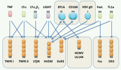
FIG. 27.7. The Immediate Tumor Necrosis Factor (TNF) Lymphotoxin (LT) Network. The cartoon depicts the signaling network formed between TNF, LT&agr;, LT&bgr;, and LIGHT, and their receptors. Each arrow indicates a ligand-receptor interaction. The network is defined by the extensive cross-utilization of ligand and receptors. Herpesvirus entry mediator (HVEM) forms a switch between positive cosignaling through LIGHT-HVEM interaction and inhibitory signaling through B- and T-lymphocyte attenuator (BTLA). LIGHT bound to HVEM activates TNF receptor-associated factor-dependent activation of NF-&kgr;B, whereas HVEM-BTLA acts through an immunoreceptor tyrosine-based inhibitory motif of BTLA to recruit the phosphatase SHP2, attenuating kinases activated by T-cell receptor signaling. The herpes simplex viron envelope protein gD attaches to HVEM acting as an entry step for infection. UL144 gene of human cytomegalovirus binds BTLA, but not LIGHT, selectively mimicking the inhibitory pathway of HVEM-BTLA. Fas Ligand and TL1A are included in this network through their interaction with decoy receptor-3.

Elucidation of the functions associated with this TNF/LT signaling network has been aided by studies with genetically modified mice engineered with null or transgene expression of the cytokine or receptor (Table 27.5). Deficiency in TNF or TNFR1, but not TNFR2, have similar phenotypes with alterations in host defense to intracellular bacterial pathogens, like Listeria monocytogenes and Mycobacterium tuberculosis, but surprisingly modest susceptibility to some viral pathogens. These results demonstrated a role for TNF in acute-phase response of the host defense system. In contrast, LT&agr;-deficient mice showed a failure in formation of peripheral lymphoid organs, a phenotype not observed in mice deficient in either TNFR or TNF, which implicated the LT&agr;&bgr; complex signaling through the LT&bgr;R as a key developmental pathway for lymphoid organogenesis.


Lymphotoxin&agr;&bgr;-Lymphotoxin&bgr;R, A Mammalian Organ Development Pathway

Gene-deficient mice sharing a common phenotype of no lymph nodes revealed the framework of a signaling pathway involved in mammalian organ development. LT&agr;-, LT&bgr;-, or LT&bgr;R-deficient mice fail to develop secondary lymphoid tissues.76,77 Several other knockout mice, including the transcriptional regulators Ikaros, ID2, and ROR&ggr;t, also lack lymph nodes,78,79,80,81,82 as do mice deficient in components of the NF-&kgr;B
activation pathway, including TRAF6, NIK, IKK&agr;, and Rel B, and their gene targets. Target genes including CXCR5, the receptor for CXCL13, show defective lymphoid organ structure, as do CCR7-/- mice (receptor for CCL19/CCL21). The developmental program of secondary lymphoid organ formation initiates at 9 days post coitus progressing in organized fashion from dorsal to lateral movement, with intestinal Peyer’s patches forming during the first postnatal week.83 The defect is irreversible in that transferring LT&agr;&bgr;-sufficient bone marrow into an adult LT-mutant mouse failed to induce lymph node formation. Accumulating studies indicate that the formation of lymphoid organs involves two distinct cell types, an embryonic LT&bgr;R-expressing mesenchymal stromal cell that responds to LT&agr;&bgr; expressed in a cell of hematopoietically derived lineage, termed the lymphoid tissue inducer cell. The lymphoid tissue inducer cell develops separately from lymphocytes and myeloid cells in a pathway dependent on ID2 and ROR&ggr;t and different cytokines including interleukin (IL)7, RANK ligand (TRANCE, TNFSF11), and TNF. These cytokines induce surface LT&agr;&bgr; in lymphoid tissue inducer cells that differentially engenders formation of lymph nodes and Peyer’s patches.84 Cells of the lymphoid tissue inducer lineage are maintained in the adult tissues at low levels (0.5%), presumably aiding in the homeostasis of lymphoid organs.85,86


TABLE 27.4 Physiologic Correlates of Tumor Necrosis Factor-Mediated Gene Induction


Induced Gene


Response


iNOS


Vasodilation, edema


VCAM-1


Leukocyte margination and extravasation


IL-8


Leukocyte chemotaxis


MHC-1


Antigen presentation


Caspase 8 activation


Apoptosis


LPL


Cachexia


Caspase 8, cysteine-dependent aspartic acid specific proteinase-8; IL-8, interleukin-8 (CXC chemokine); iNOS, inducible nitric oxide synthetase; LPL, lipoprotein lipase; MHC-1, major histocompatibility complex-1; VCAM-1, vascular cell adhesion molecule-1.


TABLE 27.5 Phenotypes in Mice Deficient in Lymphotoxin and Tumor Necrosis Factor Immediate Family


Phenotypes


Gene Deletion


LNa


PPb


Architecturec


NKd


NKTe


DCf


LT&agr;


Disrupted


Impaired


Impaired


CD8-DC


LT&bgr;


Disrupted


Impaired


Impaired


CD8-DC


LIGHT


+


+


+


+


+


+


LT&bgr;-Bg


+


+


Disrupted


nr


nr


nr


LT&bgr;-Tg


+


+


+


nr


nr


nr


TNFh


+


+


+


+


+


Maturation


LT&bgr;R


Disrupted


CD8-DC


TNFR1


+


Disrupted MZ


+


+


Maturation


TNFR2


+


+


+


+


+


+


-, absent;, +, normal; CD, cluster of differentiation; DC, dendritic cell; LN, lymph node; LT, lymphotoxin; MZ, marginal zone; NK, natural killer; NKT, natural killer T; nr, not reported; PP, Peyer’s patches; TNF, tumor necrosis factor; TNFR, tumor necrosis factor receptor.


a LT&bgr;-/- mice have -75% of mesenteric LN; LIGHT/LT&bgr;-/- mice have fewer mesenteric nodes than LT&bgr;-/- mice.76,342,343

b See Rutschmann et al.344 and Neumann et al.345

c Architecture of the splenic white pulp includes T- and B-zone segregation, MZ, germinal center and follicular DC network.

d NK-cell deficiency includes reduced cell numbers and enhanced tumor susceptibility.346

e NKT cells V&agr;14 subset.347

f CD8- DC subsets in spleen are diminished from failure to proliferate.95

g LT&bgr; conditionally deleted in B cells or T cells.348 LT&bgr;-B cells showed partial disruption in architecture; normal for LT&bgr;-T, but combined knockout in both B and T cells was worse than LT&bgr;-B.

h Normal architecture observed in TNF point mutant344; abnormal architecture in TNFR1-/- mice.345


Modified with permission from Ware.70


The microarchitecture of the white pulp in the spleen87 is disrupted in LT- and TNF-deficient mice.88 Multiple abnormal features of the architecture are observed in LT- and TNF-deficient mice, including missing macrophages in the marginal sinus and the loss of positional segregation of T and B cells into discrete zones. The segregation of T and B cells into discrete compartments depends on expression of the tissue organizing chemokines CCL19 and CCL21, which attract T cells, and CXCL13, which attracts B cells. CCL19 and CCL21 act through the chemokine receptor CCR7 expressed on T cells, and CXCL13 binds CXCR5 on B cells to promote localization in the follicles. An LT-chemokine circuit is formed by migration of B cells to LT&bgr;R+ stromal cells expressing CXCL13, which in turn induces expression of LT&agr;&bgr; on B cells.89,90,91 Circulating B cells lack surface LT&agr;&bgr;, but expression is regained upon reentry into the CXCL13 rich microenvironment.91 The formation of the splenic microarchitecture depends on B-cell expression of LT&agr;&bgr;, which induces differentiation of specialized stromal cells (eg, secretion CCL20 and CXCL13 chemokines) in the spleen during postnatal maturation. Remodeling of the microarchitecture of the secondary lymphoid organs occurs during immune responses, which requires both TNF and LT pathways signaling on fibroblastic reticular cells.92 A viral pathogen, cytomegalovirus, can induce specific changes in the splenic microenvironment through modulation of CCL21 expression.93

The LT&bgr;R and TNFR pathways facilitate lymphocyte entry into lymphoid tissues in part by modulating expression of adhesion molecules, such as peripheral node and mucosal addressins on high endothelial venules.94 LT&bgr;R signaling is necessary to maintain networks of follicular DCs involved in capturing antigen and immune complexes that aid in activating B cells. These cellular interactions are further enhanced by LT&bgr;R signals that provide growth signals for some conventional myeloid DC (CD8- subsets) within the lymphoid organ, whereas TNF plays a role in differentiation of DC progenitors in bone marrow.95

T and B cells can communicate with stromal and myeloid cells via the LT&agr;&bgr;-LT&bgr;R pathway and thus modify their immediate microenvironment during the course of an immune response. Nonlymphoid tissues suffering from chronic inflammation associated with autoimmune disease, graft
rejection, or microbial infection often contain organized accumulations of lymphocytes reminiscent of secondary lymphoid organs, called tertiary lymphoid organs. Activated T and B cells provide the source of LT&agr;&bgr; that helps drive the process of forming these structures, but as antigen is cleared, immune responsiveness subsides and these structures resolve. A gradation of features may be found in the tertiary lymphoid organs including presence of DCs, expression of chemokines, high endothelial venules, segregated regions of T and B cells, and germinal centers, but these structures typically lack the permanence of a lymph node. TNF also contributes to formation of granuloma that assists in walling off bacteria.96 Thus, the LT&agr;&bgr; and TNF pathways operative in embryonic life also play critical roles in the adult in the formation of tertiary lymphoid structures.


Influence of the Lymphotoxin&agr;&bgr; Pathway on Lymphocyte Development

Mounting evidence indicates the LT&agr;&bgr;-LT&bgr;R pathway contributes to the ontogeny of unconventional T cells, including &ggr;&dgr; T cells and invariant NK T cells, whereas conventional T cell subsets are normal in mice deficient in the TNF and LT&bgr;R pathways. The LT&bgr;R pathway seems to operate at distinct levels during thymic development.97 Double positive thymocytes regulate the differentiation of early thymocyte progenitors and &ggr;&dgr; T cells via the LT&bgr;R pathway,98 yet the LT&bgr;R is not expressed in thymocytes, suggesting an indirect mechanism. In this regard, LT&bgr;R signaling is required for the proper formation and function of the thymic stroma, which influences T-cell development.99 The thymic medulla appears to control the export of invariant NK T cells from the thymus.100,101 In addition, LT&bgr;R signaling in thymic stroma affects central tolerance to peripherally restricted antigens, which may be either dependent or independent upon the autoimmune regulator Aire.102 Thymic differentiation depends on the LT&bgr;R pathway to mediate the cellular communication between lymphoid and stromal compartments.


Autoimmunity

Dysregulated expression of several members of the TNFSF leads to autoimmune-like diseases in humans and animal models. For example, enforced expression of TNF or LIGHT, which overrides the normal transient expression, causes severe autoimmune and inflammatory processes in mice.103,104,105 LT&agr; or LT&agr;&bgr; transgenic expression in the pancreas leads to insulitis and formation of tertiary lymphoid structures.106 These types of results have implicated members of the TNF superfamily as immune regulators and support the notion that LT&agr;&bgr; and LIGHT pathways contribute to inflammation and tissue destructive processes.


Infectious Diseases

TNF is a major inflammatory cytokine required for the acutephase response to bacterial infection. For instance, lipopolysaccharide in gram-negative bacteria is a potent inducer of TNF secretion through the TLR4 innate recognition system. In mice, lipopolysaccharide induces a shock syndrome that is rapidly lethal owing to profound changes in blood circulation. However, mice survive lipopolysaccharide in the genetic absence of TNFR1 or if treated with an TNF-neutralizing antibody, indicating that host-derived TNF mediates pathogenesis.107,108,109 On the other hand, TNFR1 is essential for resistance to infection with a live organism, such as Listeria monocytogenes, through multiple processes including enhancement of phagocytosis and bacteriocidal destruction by macrophages. By contrast, T-cell immunity is not overtly impaired in TNFR1-/- mice. Humans treated with TNF inhibitors show some increase in susceptibility to selective pathogens, particularly Mycobacterium tuberculosis, reinforcing the role of TNF as a critical host defense system.110

In contrast, LT-deficient mice showed significant variability in susceptibility to individual pathogens (Table 27.6). Increased susceptibility resulted from either developmentally controlled aspects of lymphoid organ structure (eg, lymphocytic choriomeningitis virus, Leishmania) or a requirement for LT&agr;&bgr;-LT&bgr;R pathway as an effector system in innate and adaptive immune systems (eg, murine cytomegalovirus). Viewed from an evolutionary perspective, this variation in the requirement for LT signaling may reflect specific contributions from the pathogen used to evade the broader TNF- and LT-dependent pathways.35,111


TUMOR NECROSIS FACTOR SUPERFAMILY AND T-CELL COSIGNALING

Antigen recognition together with cooperating signaling “cosignaling” systems determine the quality of the adaptive immune responses. Lymphocyte responses to antigen are dynamic processes that start with the activation of naïve cells and transition through effector and memory phases. Cosignaling systems assist these phases by promoting more efficient engagement of antigen-binding TCR molecules to enhance initial cell activation and cell division (clonal expansion), augment cell survival (clonal contraction or memory cell differentiation), or induce effector functions such as cytokine secretion or killer function. Negative signals (inhibitory cosignaling) may also be delivered to T cells depending upon the particular system, which may prevent initial cellular activation or eliminate excess activated cells to dampen inflammation. Cosignaling can be quantitative, modifying thresholds of common signaling intermediates, or qualitative, involving signals distinct from other cosignaling systems or the TCR. Cosignaling receptors and ligands can be up- or downregulated at the transcriptional and protein levels depending on the stage of the T-cell response and the inflammatory milieu. In the absence of cosignaling, T cells may become unresponsive (anergic) or die.

The TNFRSR is one of two major families of cosignaling regulators that modulate T cells. The other cosignaling systems belong to the Ig superfamily, such as CD28,112 cytotoxic T-lymphocyte (CTL)A-4,113 ICOS,114 PD1,115 and BTLA.116,117 TNFRSF members involved in T-cell cosignaling include OX40, 41BB, DR3, CD27, CD30, and HVEM,118,119,120,121,122 whereas CD40 and BAFFR are more involved in cosignaling in B lymphocytes.123,124 However, considerable crossover of activities in both lymphocyte populations can be demonstrated. Death
receptors such as Fas and TNFR1 are thought to be involved in clonal contraction through apoptosis of activated effector cells, although TNF via TNFR2 also shows costimulatory action in naïve T cells. In tissue culture models, several of the TNF-related signaling pathways show costimulatory activities for T cells, which probably reflects the common induction of NF-&kgr;B-dependent survival genes, a common trait of TNFRSF members. However, analyses of physiologic models using genetically deficient mice reveal distinct roles for these molecules in the life cycle of T cells. Cosignaling systems are emerging as important targets to enhance immune responses to tumors or attenuate autoimmune diseases.114,125,126,127,

Only gold members can continue reading. Log In or Register to continue

Stay updated, free articles. Join our Telegram channel

May 30, 2016 | Posted by in IMMUNOLOGY | Comments Off on Tumor Necrosis Factor-Related Cytokines in Immunity

Full access? Get Clinical Tree

Get Clinical Tree app for offline access