Transplantation Immunology


Transplantation Immunology


Megan Sykes

Kathryn Wood

David H. Sachs


INTRODUCTION

The transplantation of organs and cells between individuals saves or prolongs thousands of lives each year. The growing list of organs transplanted includes corneas, kidneys, livers, hearts, lungs, small intestines, pancreata, and even hands and faces. Currently, clinical cellular transplantation includes islets of Langerhans and hematopoietic cells, but the list is likely to expand in the future to include other cell types, such as hepatocytes and myoblasts, which are currently under investigation in experimental models. Success of all types of transplants depends on the ability to avoid rejection due to a host-versus-graft immune response. Hematopoietic cell transplantation (HCT) is, in addition, associated with some special considerations, as the administration of a donor graft that contains mature T cells to a conditioned, and consequently immunoincompetent recipient, is associated with the risk of rejection in the graft-versus-host direction (ie, graft-versus-host disease [GVHD]). These transplants, performed between different members of the same species, are referred to as allotransplants. However, the success of organ transplantation has presented a new limitation, namely the insufficient supply of human organs. This organ shortage has led to consideration of alternative sources of organs, such as artificial organs, tissue grafts engineered from stem cells, and other species. The latter, referred to as xenografts, are a promising alternative, but present even greater immunologic challenges than allografts. This chapter presents an overview of the immunology of organ and cellular allotransplantation and xenotransplantation.

Allogeneic and xenogeneic responses differ from other immunologic responses in at least two fundamental ways. First, they exhibit extraordinary strength that includes multiple, redundant pathways. Second, they involve two different sets of antigen-presenting cells (APCs): those of the donor and those of the recipient. In this chapter, we will emphasize the uniqueness of and challenges presented by the immune response to transplantation.


ORIGINS OF TRANSPLANTATION IMMUNOLOGY


Early History

Although there were sporadic reports of tissue transplants in ancient times, skin grafting did not become an accepted practice until the late 1800s. Even then, however, many workers did not distinguish between autografts (where the donor and recipient are the same individual) and allografts (where the donor and recipient are of the same species), or even xenografts (where donor and recipient are of different species). The last of these formed the basis for an extensive practice known as zoografting in which patients were subjected to grafts from animals ranging from pigs to frogs. According to Billingham, no one apparently cared whether the grafted skin “took” or merely promoted healing of the wound.1 The results of these efforts led to a period of confusion in transplantation. Without any clear understanding of the processes involved, surgeons embarked on all sorts of transplants, and a series of operations were reported that we know, from our present understanding of the laws of transplantation, could not possibly have been successful. The transplantation of internal organs awaited the development of techniques for vascular anastomosis. In 1908, Carrel, one of the pioneers of vascular surgery, reported the results of en bloc allotransplantation of both kidneys in a series of nine cats.2 He was able to obtain up to 25 days of urine output in some cats, but ultimately all of them died. The first successful clinical renal transplant was not performed until 1952 in Boston, using the kidney of an identical twin.3

During this same period, the closely related field of tumor transplantation gained momentum. Although many of the experiments with tumor transplants provided information essential to an understanding of transplantation immunology, this was not clear at the time. Many workers were committed to the idea that they were studying an effect peculiar to tumor tissues. In his Harvey lecture, Medawar summed up the confusion neatly by the statement, “Nearly everyone who supposed that he was using transplantation to study tumors was in fact using tumors to study transplantation.” Medawar was largely responsible for establishing the immune basis of transplant reactions. Following a series of grafting experiments in rabbits and mice, he concluded in 1945 that rejection of skin “belongs to the general category of actively acquired immune reactions.”4


History, Principles, and Discoveries of Immunogenetics


Inbred Strains

Rodents have provided an invaluable model for the study of the genetic basis for graft rejection, largely due to the availability of a large number of inbred strains. Such strains consist of genetically identical animals that have been produced by sequential pedigreed brother-sister matings for at least 20 generations. They are, therefore, homozygous for all autosomal chromosomes and produce identical homozygous
progeny. The reason that sequential inbreeding leads to homozygosity is illustrated in Figure 46.1. In essence, the probability of fixation of a given autosomal locus at each broter-sister mating is 1 in 8, which is mathematically equivalent to stating that on average, 1 in 8 of all segregating loci are fixed at each generation. If the locus in question is not fixed during this random breeding, then the chances that it will be fixed at the next breeding are just a little less than 1 in 8 (ie, 1 in 8 of the remaining unfixed loci). Thus, as indicated in Figure 46.1, the probability of fixation is given by the following formula:


FIG. 46.1. Breeding Scheme for Inbred Strains.

Probability of fixation = 1- (7/8)n-1 which describes a curve that asymptotes toward 100% fixation (Fig. 46.2). For practical purposes, one considers a strain inbred after 20 such brother-sister matings, as at this point there is a very small chance that any locus will not have reached homozygosity. Hundreds of such well-characterized inbred strains are now available.

Inbred strains have also been produced in several other species, including rats, guinea pigs, and rabbits, although in much more limited numbers. Both space requirements and other genetic features, such as gestation times, age of sexual maturity, and litter size, make production of inbred strains in larger species much more difficult. Over the past 40 years, studies in one of our laboratories (D.H.S.) have produced highly inbred miniature swine.5,6 Swine were chosen for this purpose because they are one of the few large animal species in which breeding characteristics make genetic experiments possible. These characteristics include a large litter size, a short gestation time, early sexual maturity, and an estrous cycle every 3 weeks,5 and have made it possible to develop major histocompatibility complex (MHC) homozygous lines
of miniature swine in a relatively short time, to isolate and characterize new MHC recombinants, and to carry out experiments involving the segregation of genetic characteristics. The Massachusetts General Hospital (MGH) miniature swine thus represent the only large animal model in which MHC genetics can be reproducibly controlled, and they have been particularly useful in studying the role of MHC matching on rejection and/or tolerance induction.7


FIG. 46.2. Probablity of Fixation Curve.

At present, swine of three homozygous swine leukocyte antigen (SLA) haplotypes, SLAa, SLAc, SLAd, and five lines bearing intra-SLA recombinant haplotypes are maintained, as illustrated in Figure 46.3. All of these lines differ by minor histocompatibility loci, thus providing a model in which most of the transplantation combinations relevant to human transplantation can be mimicked. In addition, one subline of SLAdd animals was selected for further inbreeding in order to produce a fully inbred line of miniature swine. This subline has now reached a coefficient of inbreeding of > 94% and is now sufficiently inbred that reciprocal skin grafts among the offspring are not rejected.6 This histocompatibility makes these animals particularly appropriate for adoptive transfer studies for the first time in a large animal model.8


Genetic Principles Governing Tissue Transplantation: The “Laws of Transplantation”

In the early 1900s, it was noted by tumor biologists that tumors arising in inbred animals could frequently be transplanted successfully to other animals of the same line, while this was usually impossible in outbred animals. Little studied this phenomenon systematically and in the process produced and characterized a large number of inbred strains of mice.9 In summarizing the results of these studies of tumor grafting in mice, Little described what have since been called the five laws of transplantation (Table 46.1). Little’s remarkable insight was to reconcile these observations with the classical Mendelian principles by proposing that recipients would reject grafts if the donor expressed a product of any histocompatibility locus that was not expressed by the recipient. His explanation for the unusual inheritance pattern in Table 46.1 was to suggest, first, that there must be codominant expression of the histocompatibility genes, and second, that there must be a relatively large number of histocompatibility loci. Under these conditions, members of the F1 generation would express both parental alleles at all histocompatibility loci (and thus would fail to reject grafts from parental, F2, or subsequent generations) and members of the F2 generation would be unlikely to express all of the products of histocompatibility genes that are expressed by either parental generation (and thus would usually reject parental allografts).


FIG. 46.3. Origin of Miniature Swine Haplotypes.


TABLE 46.1 The Laws of Transplantation


1.


Transplants within inbred strains will succeed.


2.


Transplants between inbred strains will fail.


3.


Transplants from a member of an inbred parental strain to an F1 offspring will succeed but those in the reverse direction will fail.


4.


Transplants from F2 and all subsequent generations to F1 animals will succeed.


5.


Transplants from inbred parental strains to the F2 generation will usually, but not always, fail.


Adapted from Little.9


Estimating the Number of Histocompatibility Genes

One can experimentally determine the number of histocompatibility loci by which any two inbred strains differ by breeding a large F2 population between these strains and then transplanting tissues from one of the parental strains to all of the F2 offspring, measuring the fraction of grafts that survive. As illustrated in Figure 46.4, if the two strains were to differ at only one histocompatibility locus,
one would predict that three-quarters of the grafts would survive. If, however, the two strains differed by two independently segregating histocompatibility loci, then one would predict that (three-quarters)2 or nine-sixteenths of the grafts would survive. Similarly, if there were n loci by which these two strains differed, one would expect (threequarters)n to be the fraction of surviving grafts. When this equation has been solved for “n” experimentally, using skin grafts as the challenging transplant, numbers as high as 30 to 50 have been reported.10 Because there are only 20 chromosome pairs in the mouse genome, these larger numbers imply that many chromosomes carry more than one histocompatibility locus.


FIG. 46.4. Estimating the Number of Histocompatibility Loci.


Producing Congenic Strains: Identifying the Major Histocompatibility Complex

A process to generate strains differing from one another genetically at only a single one of these numerous histocompatibility loci was pursued by Snell at Jackson Laboratory and involved the production of congenic strains (inbred strains that differ from one another at only one independently segregating genetic locus) using the rejection of parental skin grafts as the trait used to select successive matings.11 It became apparent during this process that one histocompatibility locus could be distinguished from all the others by the speed with which it caused skin graft rejection. This is now called the MHC. All of the other 30 to 50 histocompatibility loci have since been called minor histocompatibility loci. There are now a very large number of H-2 congenic strains of mice available (Table 46.2), as well as some that isolate minor histocompatibility loci and some rat congenic strains.


TABLE 46.2 List of H-2 Congenic Resistant Strains


Strain


H-2 Haplotype


Origin of Background


MHC


A


a


A


A./BY


b


A


Brackyury


A./CA


f


A


Caracal


A./SW


s


A


Swiss


BALB/c


d


BALB/c


BALB/c


BALB.B


b


BALB/c


C57BL/10


BALB.K


k


BALB/c


C3H


B6.AKR-H-2k


k


C57BL/6


AKR


B6.SJL


s


C57BL/6


SJL


B10


b


C57BL/10


C57BL/10


B10.A


a


C57BL/10


A


B10.D2


d


C57BL/10


DBA/2


B10.M


f


C57BL/10


Outbred


B10.BR


k


C57BL/10


C57BR


B10.SM


v


C57BL/10


SM


B10.RIII


r


C57BL/10


RIII


B10.PL


u


C57BL/10


PL/J


C3H


k


C3H


C3H


C3H.SW


b


C3H


Swiss


C3H.JK


j


C3H


JK


C3H.NB


p


C3H


NB


D1.C


d


DBA/1


BALB/c


D1.LP


b


DBA/1


LP


LP.RIII


r


LP


RIII


MHC, major histocompatibility complex.


One of the most useful breeding schemes to produce congenic resistant (CR) lines is illustrated in Figure 46.5. Starting with two inbred strains, labeled Strain A and Strain B for simplicity, the objective is to obtain a strain that will share its entire genome with Strain A except for the major histocompatibility locus H-2, which will be derived from Strain B. The end product will be designated Strain A.B. Using the cross-intercross scheme illustrated in Figure 46.4, a skin graft or tumor graft from Strain A is placed onto all F2 offspring. Animals that reject the graft must be of genotype bb in at least one histocompatibility locus. Obviously, as there are many histocompatibility loci, most animals at this generation will reject the graft. However, if only animals rejecting vigorously are chosen, and if numerous such animals are selected, then one can be reasonably certain to have selected bb homozygotes at the H-2 locus by this procedure. Because mammalian genes are transferred as linked units in chromosomes, this process will always lead to the retention of a variable amount of bb genetic information at genes closely linked to the locus being selected. However, as described in the following, the occurrence of recombination during intercrossing generations leads also to fixation of the aa genotype at loci on the same chromosome as the MHC (chromosome 17 in mice) but at a variable distance from H-2. For practical purposes, animals that have been through at least nine cycles of such selected breeding are considered to be congenic.

As indicated in Table 46.2, H-2 congenic mouse strains are available on a variety of backgrounds. In general, the names of each of these strains follow the rule A.B, with Strain A being the background strain used in the production of the congenic and Strain B being the other parental strain from which the alternate allele at H-2 was selected. All of the early inbred mouse strains were assigned a small letter designation to represent the particular constellation of alleles that they possessed at genes in the MHC. This small letter designation is often called the haplotype designation, as indicated in Table 46.2. Thus, for example, Strain C57BL/10 (or B10) is assigned the haplotype designation H-2b and Strain DBA/2 the haplotype designation H-2d. Thus, the congenic strain B10.D2 represents a line in which the background is derived from the C57BL/10 and the MHC from the DBA/2. It thus resembles in almost every way the C57BL/10 congenic partner, except that it differs from this partner for all properties controlled by MHC-linked genes.


Intra-Major Histocompatibility Complex Recombinant Strains: Class I and II Antigens

As can be seen in Figure 46.5, every alternate generation in this mating scheme involves the crossing of animals heterozygous at H-2. Whenever heterozygotes are bred, there is always a possibility of recombination between autosomal chromosomes at meiosis. During the production of congenic lines, such recombination will tend to decrease the amount of linked genetic information carried into the congenic from the H-2 source. Therefore, the more backcrosses a particular congenic line has been subjected to, the closer
will be the boundaries on either side of H-2 at which the chromosome reverts to the background strain. Because it soon became apparent that the MHC was in fact made up of multiple loci, there was also the possibility for recombination within H-2 to occur during such crosses. Fortunately, mouse geneticists were aware of this possibility and saved numerous recombinants during the production of H-2 congenic lines. Indeed, it was through the detection and characterization of such recombinants that the linkage map of H-2 was constructed. Thus, for example, there are now a series of recombinants between strain C57BL/10(H-2b) and A/WySn(H-2a) that were isolated by Stimpfling during production of the B10.A CR line and which have provided a great deal of information on the genetic fine structure of the H-2 complex. Strains B10.A(2R) and B10.A(4R), for example, have been used to map a variety of immune response genes within the MHC. Table 46.3 presents a listing of many of the most useful congenic recombinant strains now available and their known or presumed points of recombination. Among the most important contributions that came from the study of intra-MHC recombinant strains was the progressive understanding that the loci within the MHC encoded two general types of MHC antigens, now referred to as class I and class II MHC antigens.


FIG. 46.5. Schematic Representation for Production of a Congenic Line.


TABLE 46.3 List of H-2 Recombinant Strains


Recombinant Interval Haplotypes


Parental Haplotypes


Haplotypes Designation


KAESD


Presence of Additional Recombinant Site


Strain-Bearing Recombinant


K-A


b/m


bq1


b/k k k q


Yes


B10.MBR


s/a1


t1


slk k k d


Yes


A.TL


A-E


a/b


h4


k klb b b


No


B10.A(4R)


b/a


i5


b blk d d


Yes


B10.A(5R)


b/a


i3


b blk d d


Yes


B10.A(3R)


E-S


k/d


a


k k kld d


No


A, B10.A


S-D


d/b


g


d d d dlb


No


HTG, B10.HTG


d/k


o2


d d d dlk


No


C3H.OH


a/b


h1,h2


k k k dlb


Yes


B10.A(2R)


k/q


m


k k k klq


No


AKR.M, B10.AKM


q/a


y2


q q q qld


No


B10.T(6R)


s/A


t2


s s s sld


No


A.TH


Congenic recombinant haplotypes available from The Jackson Laboratory.


Note that many of the recombinants involve at least one haplotype already containing a point of recombination. These are indicated by “Yes” and are listed only under the recombinant interval representing the most recent recombination in the haplotype’s history.


DONOR ANTIGENS RESPONSIBLE FOR GRAFT REJECTION


Major Histocompatibility Antigens

As discussed previously, the genetic analysis of graft rejection indicated that the antigens encoded within the MHC are of particular importance in graft rejection. Table 46.4 summarizes important aspects of the MHC antigens that are especially relevant for transplantation, while a much more detailed description of their structure and function can be found in Chapter 21.


Basic Features of Major Histocompatibility Complex Antigens

Polymorphism. The MHC antigens exhibit extraordinary polymorphism, providing the species with a broad capacity to present the peptides of, and thus respond to, a large number of pathogens. In the human leukocyte antigen (HLA) complex, for example, there are currently >600 named alleles at each of the HLA-A and B (class I) and >300 at the DRB1 (class II) locus.12 The high degree of polymorphism has important consequences for transplantation. Given that there are three class I loci (A, B, and C) and three to four class II loci (DQ, DP, DRB1, ± an expressed DRB 3, 4, or 5 locus present in some haplotypes) on each haplotype, the likelihood of achieving identity for MHC antigens in two unrelated humans is extremely small, though for individuals with two major conserved HLA haplotypes,13,14 the likelihood is increased.


TABLE 46.4 Summary of Features of Major Histocompatibility Complex


Class I antigens


Single polymorphic chain Three domains: alpha 1, 2, and 3 MW: 45,000


Associated with beta 2 microglobulin A, B, and C loci in humans


Expressed on all tissues and cells


Class II antigens


Two polymorphic chains: alpha and beta


Each with two domains: alpha 1 and 2, beta 1 and 2


MW: 33,000 and 28,000


DP, DQ, and DR loci in humans


Expressed on macrophages, dendritic cells, and B cells; vascular endothelium; activated human T cells


Tissue Distribution. The tissue distribution of the two types of MHC antigens differs. Class I antigens are constitutively expressed on all nucleated cells, but at low levels on some types of cells.15 Class II MHC antigens are more selective in their distribution.16 They are especially frequent on macrophages, dendritic cells (DCs), and B-lymphocytes. They are present on other lymphoid cells under some circumstances and on vascular endothelium. Their expression on some tissues of the body is regulated (eg, by interferons [IFNs]).17 One of the important distinctions between rodents and many larger species is the lack of constitutive expression of class II antigens on the vascular endothelium and other cell populations in rodents. In contrast, pigs, monkeys, and humans express class II antigens on these tissues.18,19,20

Physiologic Function of Major Histocompatibility Complex Antigens. MHC antigens are called “histocompatibility” antigens because of their powerful role in causing graft rejection; however, they did not evolve in nature to prevent tissue grafting. While the name serves to emphasize the historical importance of transplantation in the discovery of the MHC, the essential role of MHC antigens is now understood to involve the presentation of peptides of foreign antigens to responding T cells (see Chapter 22).

The Importance of Major Histocompatibility Complex Antigens in Alloreactivity. MHC antigens are exceptionally important in stimulating T- and B-cell alloresponses. This section will
focus mainly on T-cell responses to MHC, as alloantibody responses are discussed later in the chapter.

VIGOROUS GRAFT REJECTION. Mouse skin grafts differing only in their MHC antigens are typically rejected in 8 to 10 days, whereas MHC-matched grafts may be rejected more slowly, depending on the number of minor histocompatibility antigen (MiHA) differences. In pigs, primarily vascularized organs, such as the kidney, may survive indefinitely in some cases, even without immunosuppression, if all of their MHC antigens are matched, whereas MHC-mismatched kidneys are always rejected within 2 weeks.18

STRONG IN VITRO ALLORESPONSES. Allogeneic MHC antigens stimulate an extraordinarily strong T-cell response in vitro, whereas responses to non-MHC antigens generally require in vivo priming. For the most part, cluster of differentiation (CD)4 cells recognize class II alloantigens and CD8 cells recognize class I alloantigens. However, this strong bias is more stringent for CD4 cells than CD8 cells. The standard in vitro assay of T helper function is the mixed lymphocyte response, which measures proliferation of T cells after allogeneic stimulation. Limiting dilution assays can be used to quantify alloreactive proliferating or cytokine-producing cells. Such analyses have led to frequency estimates of approximately 1% to 7% of T cells responding to a particular allogeneic donor,21,22,23,24 whereas naïve T cells reactive with an exogenous peptide generally represent only approximately one in tens to hundreds of thousands of the same T-cell pool.25,26,27

Strong primary direct alloresponses of CD8+ T cells can be measured in vitro, either in standard cell-mediated lympholysis assays or in limiting dilution assays measuring cytotoxic T-lymphocyte (CTL) precursor frequencies. CTLs against MHC alloantigens can easily be generated from naive T cells following stimulation in vitro, whereas generation of CTLs to MiHAs generally requires that the T cells first be primed in vivo. Direct (without in vitro stimulation) cytotoxic activity,28 increased alloreactive precursor frequencies, and modified CTL assays29 have been used to demonstrate in vivo activation by alloantigens. ELISpot and flow cytometric carboxyfluorescein diacetate succinimidyl ester dye dilution assays and intracellular cytokine staining have enhanced the ability to detect alloreactive T cells.24,30

Direct Recognition of Allogeneic Major Histocompatibility Complex Antigens. The extraordinary strength of alloreactivity largely reflects the ability of T cells to recognize allogeneic MHC antigens presented on donor APC, referred to as “direct” allorecognition. Three different but not mutually exclusive hypotheses have been proposed to explain the high frequency of alloreactive T cells.

GENETIC BIAS. Because the thymus only positively selects T cells with some MHC reactivity, a T-cell receptor (TCR) gene pool with intrinsic affinity for MHC molecules would allow for more efficient thymic selection. TCRs indeed have intrinsic affinity for MHC molecules.31,32,33,34,35,36,37,38,39,40 Intrinsic allogeneic MHC reactivity is thereby prominent within a T-cell repertoire that has been negatively selected only by “self” MHC-peptide complexes.

THE “DETERMINANT DENSITY” HYPOTHESIS. As illustrated in Figure 46.6A, the density of specific peptide determinants presented by an APC would be quite low (as most MHC antigens present other peptides), whereas the density of a peptide-independent allogeneic MHC determinant on allogeneic APCs would be very high (as every MHC antigen would include the foreign determinant). These abundant allogeneic MHC determinants would activate many crossreactive T cells with relatively low affinities. This hypothesis requires that allogeneic MHC molecules can be recognized at least partly independently of the peptides they present. While peptide-independent and peptide-“promiscuous” alloreactive T cell clones have been described,41,42,43,44,45,46,47,48,49,50 potential artefacts of in vitro culture and assay systems might have biased these results. Notably, human alloreactive T cells expanded in vivo in a graft-versus-host response were strongly dominated by peptide-specific clones,51 and recent studies demonstrated that the requirement for peptide recognition limits TCR alloreactivity from being extended more broadly by their inherent MHC-binding capacity.52


FIG. 46.6. A: Determinant density hypothesis. B: Determinant frequency hypothesis. APC, antigen-pressenting cell; MHC, major histocompatibility complex.


THE “DETERMINANT FREQUENCY” HYPOTHESIS. The third explanation for alloreactivity is based on the idea that allospecificities include the specific peptides presented by allogeneic MHC molecules,53 for which there is strong evidence. Positive selection, which requires lower TCR affinity for self-MHC/peptide complexes than that involved in negative selection,38 enriches for TCRs capable of seeing modified self-MHC antigens, which may cross-react on allogeneic MHC molecules. The repertoire of T cells positively selected with low affinity for self-MHC molecules plus peptides of self-proteins (say X1,2…n), may cross-react with allogeneic MHC antigens presenting peptides of polymorphic or nonpolymorphic allogeneic proteins (eg, “Allo + X1, Allo + X2, …Allo + Xn”) (see Fig. 46.6B). Nonpolymporphic peptides presented by allogeneic MHC molecules would be seen by different TCRs than those recognizing the same peptide with self-MHC, as both peptide and MHC alpha helix residues contribute to the surface that is recognized by a TCR.54,55,56,57 Thus, the set of determinants represented by “Self + X1…n” would differ from that represented by “Allo + X1…n.” T cells strongly responsive to self-peptides on self-APCs (Self + X1, Self + X2, etc.) are eliminated by negative selection, which would not affect the response to the many peptides on allogeneic APCs (Allo + X1, Allo + X2, etc.). Consistent with this hypothesis, many alloreactive T cells have been shown to be peptide-specific or at least partially peptide-selective.41,42,48,58,59,60,61,62,63 Cardiac allografting studies using DM-/-mice, which lack the capacity to replace invariant chain-derived CLIP peptide with a more diverse array of peptides, provide strong in vivo evidence for the importance of peptides in direct allorecognition.64

Overall, the available information supports the inherent MHC binding capacity of TCRs, combined with the determinant frequency notion, to explain the high frequency of T cells recognizing alloantigens directly.


Minor Histocompatibility Antigens

While initially defined by their ability to cause rapid graft rejection, MHC antigens are defined in part by the location of the genes encoding them and in part by the well-characterized structure of both class I and class II antigens (see Chapter 21). MiHAs, on the other hand, are those capable of eliciting a T-cell immune response, but which lack the structural characteristics of MHC products.65 Rather than being allelic cell surface proteins, MiHAs are donor-specific peptides presented by MHC molecules that are shared by donor and recipient.66,67,68,69,70,71,72,73 As individuals are tolerant to the peptides derived from their own proteins, they only respond to the peptides of another individual’s proteins that have allelic variation. Unlike MHC antigens, MiHAs do not readily stimulate primary in vitro cell-mediated responses in mixed lymphocyte response and cell-mediated lympholysis assays, reflecting the low frequency of T cells recognizing them in the unprimed T-cell repertoire.

It has been estimated that there may be as many as 720 minor histocompatibility loci in mice,74 some of which are autosomal and others of which are encoded on the Y chromosome. MiHAs can be expressed ubiquitously or in a tissue-selective manner.75 Many proteins producing MiHAs have been identified,71,72,73,76,77,78,79,80,81,82,83,84,85,86,87,88,89 some of which are intracellular proteins such as nuclear transcription factors and myosin, while others, like CD31 and CD19, are polymorphic cell surface glycoproteins.90,91,92

Some MiHAs are diallelic peptides, both of which can be represented by a particular MHC molecule,76,78 resulting in bidirectional recognition (eg, the murine H13 locus78). Alternatively, allelic variation in MHC-binding capacity of a peptide can result in one allele being presented and the other not (eg, the human HA-1 minor antigen, in which only one of two allelic peptides binds effectively to HLA-A276). Minor antigenic determinants can also result from the failure of one allele to be processed to a peptide. An example is HA-8, for which only one allele is effectively transported by the TAP complex, resulting in a null allele despite the presence of the MHC-binding peptide sequence in the molecule.79

Both helper determinants, recognized by CD4+ cells, and cytotoxic determinants, recognized by CD8+ cells, are required for effective cytotoxic T-cell responses to MiHAs.93,94 When multiple MiHA disparities exist, a phenomenon known as “immunodominance” may occur.95,96,97,98,99,100 Removal of the immunodominant recognition can reveal strong responses to antigens that evoked weak or no responses before. This phenomenon may be due to competition between peptides for presentation by MHC molecules,74 as well as differing durations of antigen presentation and TCR avidities.101 An exceptional peptide, H60, is derived from an NKG2D-binding protein and produces responses that are comparable in potency to those elicited by MHC alloantigens, apparently due to the existence of a very high frequency of TCR in the naïve repertoire recognizing this peptide,102 which may be a useful target for graft-versus-leukemia (GVL) effects in HCT.103 However, immunodominance of CTL responses measured in vitro does not necessarily reflect the immunodominance of the same antigens in vivo.74,104 possibly reflecting the importance of tissue distribution of minor antigen expression or of helper T-cell responses.84

The H-Y antigens are encoded on the Y chromosome and therefore expressed only by males.81,82,105,106,107,108 They are of biologic significance, as they cause rejection of male skin grafts in syngeneic female mice and, in humans, female-to-male HCT is associated with increased GVHD rates compared to other combinations in the HLA-identical-related donor setting.

Human MiHAs have been identified as determinants recognized by CTLs, mainly in the setting of HLA-identical sibling donor HCT, in association with GVHD and marrow graft rejection.77,79,84,85,109,110 Certain MiHA incompatibilities (eg, HA-1) may predispose to GVHD.77,111 Immunodominance of CTL responses to particular H-Y and HA determinants as well as expansion of minor antigen-specific CTLs detected with tetrameric complexes of HLA molecules and minor antigenic peptides85,110,112 have been associated with GVHD and marrow graft rejection.113

Most MiHAs identified to date are determinants recognized by CTL, reflecting the relative ease with which CTL assays can be used to measure peptide-specific responses. Newer techniques have allowed the recent identification of minor antigenic peptide epitopes recognized by CD4+ T cells.114,115,116


It is difficult to detect humoral responses to MiHAs, presumably because individual peptide/MHC complexes are too low in abundance on the cell surface to either stimulate an antibody response or be detected on the cell surface with antibodies. An exception is the recent discovery of antibodies against H-Y (Y chromosome-encoded) antigens in association with chronic GVHD (cGVHD).


Other Antigens of Potential Importance in Transplantation


Superantigens

Superantigens include products of endogenous retroviruses in mice, such as mammary tumor viruses, and bacterial products such as stapholococcal enterotoxin B. Like MHC antigens, superantigens can stimulate primary in vitro T-cell proliferative responses and activate a high proportion of the T-cell repertoire. However, these antigens are not presented as peptides in the binding groove of MHC molecules, but instead bind to distinct regions of class II MHC molecules, and engage nonvariable portions of Vβ components of the TCR, rather than the hypervariable regions that recognize peptides. Endogenous superantigens are not classical transplantation antigens, perhaps because of their restricted tissue expression patterns.117,118,119,120,121 However, they may contribute to GVHD in mice.122


Tissue-Specific Antigens

Some peptides are derived from proteins with limited tissue distribution,123,124,125 which may not include hematopoietic cells used in traditional assays of alloreactivity. One implication is that transplantation tolerance induced by one set of donor cells might not always induce complete tolerance to donor cells of a different sort. For example, skin-specific antigens may be targets of skin graft rejection despite the presence of stable hematopoietic chimerism induced by HCT.126 Tissue-specific proteins lacking allelic variation may still serve as alloantigens because the determinant formed by a given peptide with an allogeneic MHC molecule would be different from that formed by the same peptide with a recipient MHC molecule.

Several human MiHAs may be expressed only on hematopoietic cells,75,127 and these may provide an opportunity to use graft-versus-host-alloreactive donor T cells (that recognize such antigens) to achieve GVL effects without GVHD (largely a disease affecting epithelial tissues). However, disparities for some of these minor antigens (eg, HA-1) have in fact been associated with an increased incidence of GVHD.77,111


Endothelial Glycoproteins

Blood Group Antigens. Blood group antigens are the products of glycosylation enzymes that are not the same in all individuals. They are expressed on erythrocytes and other cells and, importantly, on vascular endothelium where they may serve as the targets for “natural” antibody-mediated attack on blood vessels of organ grafts. Blood group A and B individuals each express their respective antigen, but O individuals have neither. “Natural” antibodies against blood group antigens an individual lacks probably arise due to cross-reactions with common carbohydrate determinants of environmental microorganisms. Type O individuals have antibodies to the antigens of A and B donors, whereas A and B individuals only have antibodies reactive with antigens from each other, and AB individuals have antibodies to neither. Therefore, O recipients can only receive transfusions from O donors, A and B recipients can receive transfusions from O donors or from individuals sharing their blood type, and AB recipients can receive blood from donors of any blood type. The same rules apply to the transplantation of most organs.128 Recently, advances have been made in the ability to successfully transplant kidneys across ABO barriers by adsorbing antibody from the plasma and depleting B cells.129,130,131 Other non-ABO blood group antigens on erythrocytes are irrelevant to organ transplantation because they are not expressed on vascular endothelium.

Blood group antigens are of lesser importance, but nevertheless significant, in HCT. ABO incompatibility in the hostversus-graft direction (“major” ABO mismatch) can lead to prolonged red cell aplasia following HCT; incompatibility in the graft-versus-host direction (“minor” mismatch) can result in initial hemolytic anemia, but this complication can be avoided by washing the donor HCT preparation to rid it of plasma.132,133

Species-Specific Carbohydrate Determinants. Closely analogous to the blood group antigens are the carbohydrate determinants expressed on vascular endothelium that show species selectivity. For example, pigs, which are of interest as an organ source for xenotransplantation, have a glycosyl-transferase enzyme not expressed by humans that glycosylates β-galactosyl N-acetyl glucosamine to form a Galα1-3Galβ1-4GlcNAc (αGal) determinant. This enzyme is present in most species but underwent a loss of function mutation in our nonhuman primate ancestors. In humans, a fucosyltransferase generates instead the H-substance from the same substrate, leading to blood group O. Preformed or “natural” antibodies are present in human serum that react to the nonself-pig determinant. Like the blood group antibodies, these natural antibodies probably arise from cross-reactions with environmental microorganisms,134,135 and they also cause hyperacute rejection (HAR) of most primarily vascularized xenogeneic transplants.


“Missing Self” and Natural Killer Cell Recognition

In apparent violation of the laws of transplantation described previously, (AxB) F1 mice are capable of rejecting bone marrow from parental donors, a phenomenon termed hybrid resistance. This phenomenon, as well as rapid rejection of fully allogeneic marrow, is mediated by natural killer (NK) cells.136 NK cells are large granular lymphocytes that lack TCRs and that have the ability to mediate cytolysis against certain tumor targets and hematopoietic cells. NK cells also produce a number of proinflammatory, hematopoietic, and even anti-inflammatory cytokines, and may be divided into subsets on the basis of their cytokine production pattern.137

The originally puzzling specificity of NK cell-mediated marrow rejection is due to the expression by NK cells of inhibitory receptors that recognize specific groups of class I
MHC alleles on target cells and prevent cytolysis by the NK cell. These class I receptors are type II C lectins (Ly49 family) or dimers of CD94 with NKG2 lectins in the mouse, and are either immunoglobulin family members (KIR) or CD94/NKG2 in humans. Recognition by an inhibitory receptor of a class I ligand results in intracellular transmission of an inhibitory signal via an immune receptor tyrosine-based inhibitory motif that interacts with a tyrosine phosphatase and counteracts activating signals transmitted from other cell surface molecules. Recognition of “self” class I inhibitory ligands prevents NK cells from killing normal autologous cells.138,139 Other molecules expressed by infected or stressed cells activate NK cells, counterbalancing these inhibitory signals (see Chapter 17).


FIG. 46.7. An Explanation for Hybrid Resistance. Each filled circle represents a subset of natural killer cells.

Inhibitory receptors are clonally distributed on NK cells, each of which may express one or more different inhibitory receptor. For NK cells to be functional yet tolerant of “self,” they must express at least one inhibitory receptor for a “self” class I MHC molecule.140,141 Thus, as is illustrated in Figure 46.7, an AxB F1 recipient will have subsets of NK cells with inhibitory receptors that recognize MHC of either the A parent, the B parent, or both. The absence of “B” class I molecules on, for example, AA parental hematopoietic cells, permits subsets of (AxB)F1 NK cells that have inhibitory receptors only for class I molecules from the B parent to destroy AA cells. Thus, hybrid resistance and NK cell-mediated resistance to fully allogeneic marrow grafts can be explained on the basis of “missing self.”142 The roles of NK cells in transplantation are discussed in later sections.


MECHANISMS OF REJECTION

At least four distinct mechanisms that can cause graft rejection have been defined. According to the timeframe in which they tend to occur in clinical practice, they are, namely, HAR, accelerated rejection, acute rejection, and chronic rejection.


Rejection Caused by Preformed Antibodies (Hyperacute Rejection)

HAR occurs within minutes to hours after blood flow is established to a transplanted vascularized organ.143,144,145 The phenomenon is visible and dramatic: the organ turns blue and its function ceases. Microscopically, there is extensive evidence of vascular thrombosis and hemorrhage. The important components involved in the mechanism of HAR include 1) donor endothelial antigens, 2) preformed antibodies that can bind these antigens, 3) complement, and 4) coagulation cascades, which are activated by the binding of preformed antibodies to the vascular endothelium. The interaction of these components leading to hyperacute rejection is diagrammed in Figure 46.8.

The role of complement in HAR is inferred both from the accumulation of various complement components in the grafts and from the fact that complement depletion leads to prolonged survival of xenografts.146 Complement activation leads to production of active protein fragments and complexes of complement components, which cause tissue injury either directly or by recruiting effector cells that mediate destruction of the graft. In allogeneic combinations, this is initiated by antibody-mediated activation of complement through the classical pathway, whereas in xenogeneic combinations, the alternative pathway may also be involved.147 In both cases, the membrane attack complex, produced by the ordered interaction of several complement components, initiates the destructive pathway.


FIG. 46.8. Schematic Representation of Hyperacute Rejection.


Complement activation is controlled by several regulatory molecules, including complement receptor 1, decay accelerating factor (DAF; CD55), membrane cofactor protein (CD46), and CD59, which act at different stages along the cascade (see Chapter 36). Many of these molecules are produced by the vascular endothelial cells. Because these regulatory proteins prevent unwanted complement activation in the face of low levels of perturbation to the system, the titer and avidity of the preformed antibodies must be high enough to activate despite these downregulating molecules. Thus, preformed antibodies directed at MHC antigens almost always accomplish this activation, whereas the lower-affinity blood group antibodies lead to hyperacute rejection in only about 25% of kidneys. One of the reasons that hyperacute rejection is such an important feature in xenogeneic transplantation is that the complement regulatory proteins produced by the donor vascular endothelium of one species often do not function effectively with complement molecules derived from a different species. Because of this incompatibility, lower levels of an initial triggering signal lead to explosive complement activation.

Although the membrane attack complex is often thought of as a lytic molecule, its effect on the donor vascular endothelium, even before cell lysis, is to cause endothelial activation. This occurs rapidly, before there is time for new gene transcription or protein synthesis, and has been referred to as type I endothelial activation. The two principal manifestations of this activation are cell retraction, leading to gaps between endothelial cells, and initiation of coagulation pathways due to the loss of antithrombotic molecules from the endothelium.148 Thus, type I endothelial activation is responsible for the two principal pathologic findings in hyperacute rejection: extravascular hemorrhage and edema, and intravascular thrombosis.

There are no known treatments that can stop the process of hyperacute rejection once it has started and, thus, it is essential to avoid the circumstances that initiate it. Experimentally, this can be accomplished for relatively short periods of time by administration of complement inhibitors, such as cobra venom factor, which depletes complement. In clinical practice, this is accomplished by avoiding transplantation in the face of preformed antibodies, both by avoiding blood-group antigen disparities and by testing recipients before transplantation (ie, a “cross-match”) to determine whether they have performed antidonor antibodies.

Not all organs and tissues are equally susceptible to hyperacute rejection. Most primarily vascularized organs, such as kidneys and hearts, are very susceptible, but the liver can often survive without hyperacute rejection despite preexisting antidonor antibodies.149 It is not clear whether this unusual feature of the liver reflects the large surface area of its vascular endothelium or an intrinsic property of liver endothelial cells. Nonetheless, hyperacute rejection of the liver has occurred in some cases, especially involving xenogeneic transplantation, indicating that its resistance to hyperacute rejection is not absolute. Skin grafts are relatively resistant to hyperacute rejection but high levels of antibody can cause a “white graft” (ie, a failure of blood vessels to communicate with those of the recipient)150 Pancreatic islets are likewise resistant to this form of rejection.151 Free cellular transplants, such as bone marrow cells or hepatocytes, that express some of the antigens recognized by preformed antibodies, are cleared quickly from the circulation by the reticular endothelial system, leading to resistance to engraftment.152 In the case of HCT, this resistance can be overcome by transplanting larger numbers of cells.152,153,154 Additionally, antibody-independent complement activation has been shown to be a significant factor diminishing the engraftment of porcine bone marrow in mice.155


Acute Humoral Rejection (Accelerated Rejection)

A second mechanism of rejection, also caused by antibodies, occurs as a result of antibodies that are induced very rapidly after a transplant is performed. This type of rejection has been called “acute humoral” or “accelerated” rejection because it typically occurs within the first 5 days after transplant. The process is characterized by fibrinoid necrosis of donor arterioles with intravascular thrombosis.156

Accelerated rejection is rare in allogeneic combinations because it requires that an antibody response occur before the T-cell response that is typically responsible for acute rejection episodes (see following discussion). Indeed, most allogeneic B-cell responses are T-cell-dependent. The best examples of accelerated rejection are probably those observed in vascularized organ transplants between closely related, concordant xenogeneic species and between discordant species following adsorption of anti-Gal antibodies. In these cases, the levels of preformed antibodies are not sufficient to cause hyperacute rejection, but antidonor antibodies appear rapidly (within 3 to 4 days) in association with the onset of rejection. The pathology of acute humoral rejection reveals a paucity of lymphocytes infiltrating the donor graft, antibody binding to donor vascular endothelium, and fibrinoid necrosis of the donor vessels. Vigorous anti-T-cell immunosuppression has little effect on acute humoral rejection, whereas immunosuppression with reagents that affect B-cell responses, such as cyclophosphamide, delays its onset until more typical T-cell-mediated rejection occurs.157

As in hyperacute rejection, the process of acute humoral rejection is usually initiated by antibody binding to antigens on the donor vascular endothelium. In this case, however, the subsequent endothelial changes occur more slowly, allowing time for gene transcription and new protein synthesis. This later form of activation has been called type II endothelial activation.147,158 Many of its features appear to be mediated by the transcription factor NF-κB, which generates many of the responses associated with inflammation, including the secretion of inflammatory cytokines such as interleukin (IL)-1 and IL-8, and the expression of adhesion molecules such as E-selectin and intercellular adhesion molecule (ICAM)-1.159 In addition, type II endothelial activation causes the loss of thrombomodulin and other prothrombotic changes.160 Thus, the events following type II endothelial activation are associated with the pathologic changes that occur with “accelerated” rejection, including the tendency toward intravascular thrombosis and the inflammatory destruction of donor vessels that occurs in the absence of infiltrating lymphocytes.


Just as there are regulatory processes for complement activation, there are regulatory molecules that counter the tendency toward intravascular coagulation and the process of type II endothelial activation (eg, tissue factor protein inhibitor [expressed by vascular endothelium, which inhibits factor Xa of the clotting cascade] and a number of other protective molecules, including Bcl-xL, Bcl-2, and A20).147,161 Although these are often thought of as antiapoptotic molecules, they also tend to inhibit activation mediated by NF-κB. Like the regulatory molecules of complement, some of these regulators may not function across species differences, leading to disorder regulation of the coagulation system.162

Although vigorous early antibody responses generate type II endothelial activation and accelerated rejection, later antibody responses often fail to do so. The process that enables transplanted organs to survive in the face of circulating antibodies that can bind endothelial antigens has been called “accommodation.”147,163 This phenomenon has been observed in some allogeneic and xenogeneic combinations with preformed antibodies, but has so far been disappointingly ineffective in discordant xenotransplants. Resistance to type II endothelial activation has been achieved in vitro by pretreatment with low levels of antiendothelial antibodies that are insufficient to trigger activation.164 The achievement of accommodation is associated with increased expression of the antiapoptotic genes described previously and with changes in the isotype of the recipient’s antibody responses.158,165

An important difference between HAR and acute humoral rejection is that there is no known therapy to stop graft destruction by HAR, whereas acute humoral rejection can sometimes be reversed by desensitization.


Rejection Caused by T Cells (Acute Rejection and Graft-versus-Host Disease)

“Acute cellular rejection,” which is characterized by a mononuclear cell infiltrate in the graft, is the most common type of organ allograft rejection. Acute rejection is most common during the first 3 months after transplant, but may occur at any time, especially if immunosuppressive medication is withdrawn. Acute rejection is T cell-dependent, and its treatment, which is usually successful, includes increased doses of standard immunosuppressive drugs or antilymphocyte antibodies.

The use of newer immunosuppressive drugs and anti-T-cell antibodies has markedly reduced acute rejection rates. For example, the vast majority of kidney transplant recipients never experience an episode of acute rejection. It is now quite rare to lose a transplanted organ to cellmediated rejection during the first year after transplantation. However, the use of these highly effective immunosuppressive treatments is associated with significant morbidity.

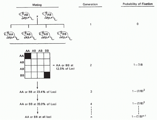
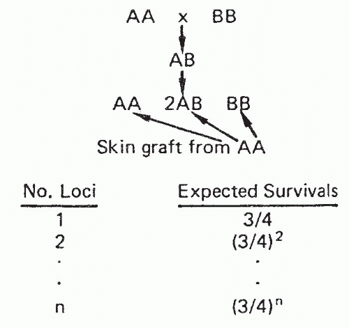
Experimental models for acute rejection include nonprimarily vascularized skin grafts, heart graft fragments, artificial “sponge” allografts, or islet transplants in rodents, which may not accurately reflect the processes of rejection for primarily vascularized organs. While there are models of heart, kidney, liver, and other types of primarily vascularized organ transplants in rodents, these types of transplants are more tolerogenic and hence more easily accepted than similar transplants in large animals and humans. Studies of primarily vascularized organ transplants in large animals, such as monkeys or pigs, have obvious clinical relevance, but are expensive and require many special resources.

Acute GVHD is the counterpart of cellular rejection that involves graft-versus-host alloreactivity, usually in the context of HCT, but also sometimes with organ transplants that carry significant amounts of donor lymphoid tissue (eg, liver). Like acute rejection, acute GVHD is T cell-dependent. T-cell depletion of the donor hematopoietic cell graft prevents GVHD but is associated with increased rates of graft rejection and relapse of malignant diseases.

The concepts of “direct” and “indirect” allorecognition introduced previously must be considered at both the sensitization and effector phases of an immune response. The definition of “indirect” recognition used in this chapter is based on which set of APCs (donor versus recipient) is presenting donor antigen. “Cross-priming” is a term specifically denoting sensitization of CD8 T cells through the indirect pathway. As shown in Figure 46.9, there are three major T-cell pathways to consider in relation to graft rejection. These include 1) direct recognition of donor alloantigens by CD4+ T cells, which generate effector CD4 cells and provide help for the generation of effector CD8 cells; 2) direct activation of CD8 T cells by donor APCs; and 3) CD4+ T-cell activation by recipient APCs presenting re-processed donor antigens (the indirect pathway of sensitization). This pathway is important in providing help for immunoglobulin production by B cells. A role in rejection for cross-primed CD8+ T cells is not included in Figure 46.9, because this pathway has not been shown to play a role in allograft rejection except under certain circumstances, as described in the following. The multiplicity of T-cell sensitization and effector pathways involved in graft rejection and GVHD is demonstrated by the frequent observation of rejection or GVHD when only CD4+ or CD8+ T cells are depleted.166,167,168 As a result of the high precursor frequency of T cells that respond to allogeneic MHC antigens directly, populations of T cells that ordinarily have minimal significance become functionally important.


Sensitization and Cell Trafficking during Rejection and Graft versus Host Disease

Transplanted tissue contains passenger leukocytes of donor origin that have the characteristics of immature DCs.169 In response to the inflammatory signals that are triggered by retrieval and transplantation, both within the tissue itself as well as in the recipient,170 the donor-derived passenger leukocytes rapidly leave the graft and migrate to the secondary lymphoid tissues of the recipient.171 Secondary lymphoid tissues comprise the spleen, lymph nodes, and gut- or mucosal-associated lymphoid tissue, and depending on the location of the graft, the passenger leukocytes will migrate to the tissue that drains the graft site where they encounter naïve T cells. After transplantation, both in situ within the tissue and during migration, the passenger leukocytes
acquire the phenotypic and functional characteristics of mature DCs, expressing high levels of MHC class I and II molecules as well as other cell surface costimulatory molecules necessary to fully activate naïve CD4+ and CD8+ T cells.172 Once in the secondary lymphoid tissues they act as professional APCs, presenting antigens expressed in the transplanted tissue to recipient T cells via the direct pathway of allorecognition.173


FIG. 46.9. Model of T-Cell-Mediated Rejection. A: Interactions between cluster of differentiation (CD)4+ Th, donor antigen-presenting complex, and CD8+ cytotoxic T lymphocyte. B: Additional pathways of T-cell sensitization that can lead to rejection. MHC, major histocompatibility complex.

Naïve T cells recirculate around the body and are constantly moving through the secondary lymphoid tissues sampling the APC, both host- and (after transplantation) donor-derived, for antigen.174 If a naïve T cell with a TCR that can recognize a donor MHC molecule encounters the donor-derived passenger leukocyte in the draining lymphoid tissue as it recirculates, it will stop, interact, and differentiate into an antigen-experienced effector T cell. In support of the secondary lymphoid tissue being the primary site for sensitization of naive T cells and initiation of rejection after solid organ transplantation, Lakkis and colleagues175 showed that cardiac allografts were not rejected in splenectomised aly/aly mice that lack secondary lymphoid tissue as a result of a mutation in the gene encoding NF-kB-inducing kinase176 and suggested that in this situation permanent graft acceptance was due to immunologic ignorance. Other studies supporting the concept that secondary lymphoid tissues draining the graft are the key site for initiation of the immune response have followed the fate of T cells of a known specificity for donor antigen as they respond.177

Similarly, after bone marrow transplantation (BMT) the initiation of GVHD also takes place in the secondary lymphoid tissue, with evidence for the initial proliferation of donor CD4+ T cells followed by CD8+ T cells in secondary lymphoid organs with subsequent homing to the intestines, liver, and skin.178,179,180 Visualization of T cells responding during the initiation of GVHD showed that while Peyer patches are involved, other secondary lymphoid tissues contribute to the activation of T cells that can home to the gut; mesenteric lymph nodes and spleen are also sites where gut homing T cells were activated.179

In solid organ transplantation, exclusive initiation of rejection in the secondary lymphoid tissues conflicts with the earlier hypothesis that rejection was initiated within the graft itself by donor endothelial cells lining the vessels that could activate T cells directly as they passed through the graft.181,182 Since these early papers, there have been a number of studies that support this hypothesis. For example, human endothelial cells have been shown to activate naïve T cells in vitro.183 In the mouse, APCs that are not of hematopoietic origin have been shown to activate CD8+ T cells in vitro and in vivo,184 thus supporting the concept that T cells may be activated in the graft rather than in the secondary lymphoid tissue. Moreover, splenectomized lymphotoxin α and lymphotoxin β knockout mice that also lack secondary lymphoid tissues were found to reject cardiac allografts, albeit at a slower than normal tempo.185 Each of these models is subtly different immunologically, and therefore different components of the immune response to an allograft may be differentially affected by the presence or absence of secondary lymphoid tissues. Clearly, in the absence of secondary lymphoid tissue, the initiation of the rejection response by naïve T cells is less aggressive.

While antigen presentation via the direct pathway plays a dominant role in initiating the response to a transplant, a finite number of donor-derived passenger leukocytes is transferred within a transplanted organ. Thus the role of the direct pathway initiated by passenger leukocytes may diminish with time as eventually only “nonprofessional” APCs, including endothelial cells, remain to stimulate direct pathway T cells. Thus the role of endothelial cells within the graft may assume a greater significance with time after transplantation both for the initiation of the response and as a target for direct pathway effector cells. While activation of naïve T cells may occur predominantly in the secondary lymphoid tissues after transplantation, activation of memory T cells in presensitized recipients is quite different. Unlike naïve T cells, memory T cells can migrate to nonlymphoid tissues in the periphery186 and can trigger rejection through pathways that are independent of secondary lymphoid tissues.187
Thus, in humans, where there are likely to be both naïve and memory T cells that can recognize or cross-react with donor MHC molecules, rejection may be initiated both within the secondary lymphoid tissue as well as within the allograft by naïve and memory T cells, respectively.

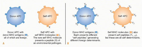
At the same time that donor-derived passenger leukocytes are leaving the graft, recipient leukocytes including APCs are attracted to the graft by the inflammatory mediators and chemokines released in the vicinity of the transplanted tissue. As these cells traffic through the graft, they phagocytose debris arising from tissue damage at the time of transplantation before migrating to the draining lymphoid tissue. The ingested antigens are processed and presented on recipient MHC molecules to T cells in the recipient lymphoid tissue.188 In addition, soluble antigens released from the graft will also be transported in the blood to the draining lymphoid tissue, where they will be taken up and presented by resident APCs. Common antigenic peptides presented by the indirect pathway are the hypervariable peptide binding regions of MHC molecules.189 Indirect pathway responses undoubtedly contribute to acute rejection, although the tempo of rejection may be slower due to the lower frequency of T cells that can respond. However, unlike direct pathway allorecognition, the indirect pathway is available for antigen presentation for as long as the graft remains in situ, and therefore becomes the dominant mode of allorecognition long term.

A third pathway of allorecognition has been described more recently, the so-called semidirect pathway that involves the capture of donor MHC-peptide complexes by host APCs. The exchange of fragments of cell membrane between cells that interact with one another is a well described phenomenon in cell biology. In the context of the immune response to an allograft, the transfer of membrane fragments from allogeneic cells expressing donor MHC molecules can result in the presentation of intact donor MHC molecules by recipient APCs to T cells. The significance of the semidirect pathway is still under investigation.190

Traffic of naïve lymphocytes is usually restricted to recirculation between the blood and lymphatic systems. However, once they have been primed in the secondary lymphoid tissues, activated lymphocytes as well as other activated leukocytes must be able to migrate into the graft in order to destroy the transplanted tissue, a process known as leukocyte recruitment.

The inflammatory processes at the site of transplantation generate chemotactic cytokines called chemokines, and upregulation of chemokine receptor expression by activated leukocytes enables them to migrate along the chemoattractant gradient to reach the graft.191

Inflammatory signals also affect blood vessels in the vicinity of the transplant, causing vasodilation and endothelial activation. Activated endothelial cells rapidly externalize preformed granules called Weibel-Palade bodies that contain the adhesion molecule P-selectin192 and rapidly upregulate expression of vascular cell adhesion molecule and CD62E (E-selectin). At the same time, chemokines released from the graft become tethered to the endothelium, and these alterations in endothelial surface markers advertise to passing leukocytes that an inflammatory process is occurring in the neighboring tissue.

Leukocytes are usually conveyed within the fast laminar flow at the center of blood vessels, but once activated leukocytes reach postcapillary venules in proximity to the graft, they are able to leave this rapid flow and move toward the edge of the vessel. This occurs in response to the local chemokine gradient and is assisted by the slower blood flow in the vasodilated blood vessels near the graft. Leukocyte extravasation is a multistep process. Initially, low-affinity interactions develop between endothelial P-selectin and sialyl-LewisX moieties that are present on the surface of activated leukocytes. These interactions continually form and break down, and the leukocyte “rolls” along the endothelial surface. If chemokines are present on the endothelial surface, conformational changes in leukocyte integrin molecules occur that allow them to bind other endothelial adhesion molecules such as ICAM-1. These higher-affinity interactions cause arrest of the leukocyte on the endothelial surface, allowing it to commence extravasation. Having entered the tissues, the activated leukocytes continue to migrate along chemokine gradients in order to invade the graft.


Antigen Recognition and T-Cell Help in Graft Rejection and Graft-versus-Host Disease

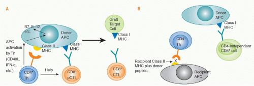
Role of Direct Cluster of Differentiation 4 Allorecognition. Priming of naïve, directly alloreactive T cells requires professional APCs that leave the graft and enter the recipient’s lymphoid tissues. Direct CD4 T-cell sensitization by donor class II MHC antigens may both generate CD4+ effector cells and provide help for the activation, differentiation, and proliferation of cytotoxic CD8+ cells that directly recognize donor class I MHC antigens and destroy the graft (see Fig. 46.9). Depletion of donor APCs can markedly prolong graft survival,193,194,195,196,197,198 illustrating the importance of direct allorecognition in inducing rejection. The CD4 help for CD8 cells consists of both cytokine (eg, IL-2) production and “conditioning” of the APC, for example by interactions of CD40 on the APC with CD40L on the activated CD4 cell. These interactions upregulate APC expression of CD80 and CD86 costimulatory molecules and cytokines such as IL-12 and MHC, making the cell a more effective APC. Studies of antiviral immunity indicate that CD4 help is needed for development of full effector function,199 and for CD8 memory cell survival200 and function.201

Studies involving very limited (not clinically relevant) antigenic disparities between donors and recipients suggested that a “three-cell cluster” model involving interactions between helper T cells, effector T cells, and APCs was essential for rejection.202,203,204,205 However, studies involving more extensive, clinically relevant histoincompatibilities206,207 suggest that CD4 helper cells sensitized by antigen presented on recipient APCs can provide help for directly alloreactive CD8+ effector cells. It remains possible that a “three-cell cluster” is still essential for CD4 cells to provide help to CD8 cells mediating rejection, and that donor class I MHC/peptide complexes are transferred and picked up by recipient APCs. Recipient APCs with directly alloreactive CD8 T cells would thereby encounter their ligands on the same recipient APC that an indirectly alloreactive CD4 cell recognizes. Such transfer of class I/peptide antigens, resulting in this type of
“semidirect” antigen presentation, requires consideration in transplant models.208,209,210,211

CD4+ T cells alone can cause graft rejection (without CD8 cells) in the setting of class II or multiple minor histoincompatibilities,198,212,213,214,215 indicating that CD4 T cells can mediate rejection effector functions. In BMT recipients, they can induce GVHD in the absence of CD8 cells in the setting of class II, full MHC, or multiple minor histoincompatibilities,216,217,218,219 and can reject class II and minor antigen-mismatched bone marrow.220,221

Role of Indirect Cluster of Differentiation 4 Cell-Mediated Allorecognition. Indirectly alloreactive CD4 T cells have roles in skin and solid organ graft rejection,210,222,223,224,225,226,227,228,229,230,231,232 including the provision of help for class-switched alloantibody responses.233 This help requires cognate interactions between recipient class II-restricted indirectly alloreactive CD4 cells and host B cells that recognize donor MHC molecules through their immunoglobulin receptors, process them, and present donor MHC peptides with their class II molecules. CD4 cells also contribute to rejection of bone marrow grafts differing only at class I MHC loci, possibly implicating indirect allorecognition.220,221

Rejection by CD4 cells of skin grafts lacking class II MHC shows the strength of the indirect pathway of rejection.206,222 Rejection of islet xenografts in mice may depend on indirectly xenoreactive CD4+ T cells.234 Sensitization of indirect CD4 responses to donor MHC-derived peptides has been demonstrated in patients undergoing graft rejection, and these may be correlated with poor outcomes.235,236,237,238,239,240,241,242 A major role for indirect allorecognition has been suggested in the setting of chronic rejection,230,243,244,245,246 in part because of its role in inducing antibody responses, which are implicated in chronic rejection.247,248,249,250,251 Moreover, the eventual replacement of donor APCs by recipient APCs implicates the latter in long-term graft recipients.196,241,252,253 Consistently, direct alloresponses tend to subside over time in transplant patients.254,255,256

Nevertheless, donor APC depletion or the lack of donor class II MHC can prevent rejection in some situations.193,194,195,196,197,198 An essential role for indirect allorecognition has not been demonstrated for acute rejection.21,253,257,258 Indirectly alloreactive CD4+ cells alone fail to reject skin grafts with minimal class I or minor histoincompatibilities,202,259,260 or to induce GVHD against class I MHC or minor histocompatibility barriers alone.216,217 With rodent primarily vascularized allografts, donor APC depletion may, by preventing the strong direct alloresponse, allow the inherent tolerogenicity of the organ to prevail.

Role of Helper-Independent Cluster of Differentiation 8+ T Cells. CD8 T cells can readily reject skin and bone marrow allografts in the absence of CD4 cells,166,202,220,221,261,262 and alloreactive CD8 T-cell memory can be generated and maintained without CD4 cells.263 CD8 cells can also induce GVHD without CD4 T cells in the setting of full MHC, class I only, and minor antigen histoincompatibilities.264,265 Direct recognition of recipient MiHAs on recipient APCs is essential for the induction of CD8-dependent, CD4-independent GVHD in MHC-identical, lethally irradiated mice,266 but indirect267 or “semidirect”
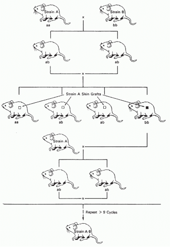
211 CD8 recognition of recipient antigens presented by donor APCs amplifies the process.

Together, these studies show that CD4 help is not critical for CD8 cell-mediated rejection or GVHD. However, the requirement for CD4 help may increase in the absence of inflammatory stimuli, as indicated by marked differences in the need for CD4 help for CD8-cell activation and GVL effects in the presence and absence of inflammation.268,269 Grafts expressing only class I antigen disparities are usually rejected quite slowly, and CD4-independent rejection is relatively easily suppressed by cyclosporine.270,271,272 Many primarily vascularized grafts that express only a class I antigen disparity require CD4+ cells to initiate rejection, and, when it occurs, CD4-independent rejection by CD8+ cells is dependent on the number of donor APCs in a graft.166,222 CD4-indepenent CD8+ cells do not reject grafts expressing only a small number of minor antigen disparities and generate only weak helper responses even in the presence of multiple MiHA disparities. CD8+ helper cells also differ from CD4+ helper cells in being unable to provide help for other cell populations.273 CD8+ cells alone cannot reject skin grafts with only limited class II antigen disparities.202,218,259,274

Cross-Primed Cluster of Differentiation 8 Cells. Peptides of exogenous antigens were originally thought to be presented by MHC class II antigens, whereas those of endogenous cellular antigens are presented by MHC class I molecules.275,276 However, it is now clear that class I presentation of exogenous peptides (cross-presentation) is essential for many immune responses, including those against microbial and tumor antigens.277,278,279,280,281 Several pathways have now been delineated for cross-presentation by class I molecules.282,283,284 CD8 cell crosspriming was originally demonstrated in a transplantation model by Bevan70 when minor antigen-disparate grafts with MHC antigens of type A were placed on MHC (A × B) F1 recipients and CD8+ cells became sensitized to the minor antigens presented by both A and B types of class I MHC molecules. Activation of cross-primed CD8 cells is strongly dependent on CD4 help and IL-2.285 Cross-primed CD8 cells recognizing donor MiHAs and MHC-derived peptides are most likely to participate in rejection when there is sharing of class I alleles between the donor and recipient. Without such sharing, the self-class I/allogeneic peptide epitope cannot be presented by the parenchymal or endothelial cells of the graft.286 However, even without class I sharing, indirect CD8+-cell sensitization can lead to skin allograft rejection, perhaps due to recognition of donor peptides presented by recipient endothelial cells on host-derived vessels that revascularize the graft.287,288 Cross-primed CD8 cells might also contribute to graft rejection via indirect effector mechanisms upon antigen recognition on host APCs in the graft or by producing inflammatory cytokines.289 Some of the rejection processes previously attributed to cross-primed CD8 cells may in fact be mediated by CD8 cells seeing intact donor MHC-peptide complexes on recipient APCs (“semidirect” presentation).


Effector Mechanisms of Rejection and Graft-versus-Host Disease

While cytotoxic T cells are important effectors of graft rejection and GVHD, additional mechanisms involve effector cells of the innate immune system and cytokines as final mediators of tissue destruction. The net result of this multiplicity of pathways is considerable redundancy of mechanisms of graft rejection and GVHD.

Cytotoxic Mechanisms of Graft Rejection and Graftversus-Host Disease. Rejecting organs contain proteins and messenger ribonucleic acid (RNA) encoding perforin, granzymes, and proteases associated with cell-mediated cytotoxicity.290,291,292,293,294,295,296,297 The presence in urine of RNA encoding perforin and granzyme B has been associated with renal allograft rejection in humans.298 Although the perforin/granzyme pathway is the major cytolytic pathway for CD8 T cells and CD4 cells tend to utilize the Fas/FasL pathway,299 both subsets are capable of both types of cytolytic activity,300,301 and the perforin pathway is available to both T-cell subsets mediating GVHD.302 All of these cytotoxic proteins play contributory roles, and no single protein has been found to be critical for solid organ graft rejection,303,304,305,306 GVHD,307,308,309,310 or bone marrow graft rejection311 in the presence of clinically relevant mismatches. Critical cytotoxic interactions have been identified in less relevant animal models involving Fas-dependent GVHD directed at isolated class II MHC disparities310 and perforin-dependent rejection of Kb mutant class I-only mismatched heart allografts.303 Fas ligand promotes lymphoid hypoplasia312 and skin and liver GVHD,312 and both Fas ligand and TRAIL are required for GVHD-related thymic destruction.313 While the perforin-granzyme pathway contributes to GVHD,310,312 the Fas pathway appears to be of greater overall importance. In contrast, the perforin/granzyme and TRAIL pathways predominate in antileukemic effects, especially of CD8 cells, and selective blockade of the Fas/FasL pathway may ameliorate CD8-mediated GVHD without eliminating GVL effects.309,314,315,316,317,318

Non-Cytotoxic T-Lymphocyte Effector Mechanisms in Graft Rejection and Graft-versus-Host Disease. T cells can effect rejection of grafts whose parenchymal cells do not express the TCR ligand, indicating the existence of “indirect” effector mechanisms. Entire skin grafts can be rejected when only the APCs are foreign,319 indicating that nonselective destruction of grafted tissue can occur. Several studies have implicated indirect CD4 cell-mediated rejection of skin320,321 and cardiac322 allografts. Replacement of graft endothelium by the host was shown to be needed for rejection through this indirect effector mechanism.322 GVHD of the liver and intestine can be induced by donor T cells in MHC-deficient hosts receiving wild-type host DCs, suggesting that indirect effector mechanisms may also mediate tissue injury of GVHD.323,324 However, CD4-mediated GVHD against MiHAs is markedly attenuated when the target antigens are expressed only on hematopoietic cells.325 Thus, “indirect” effector mechanisms can destroy transplanted tissue or recipient tissue in the case of GVHD, but less efficiently than direct cytotoxic mechanisms.

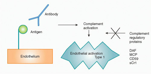
Another non-CTL graft rejection mechanism involves antibodies, which cause hyperacute rejection, acute humoral rejection, or chronic rejection through Fc receptor, complement-mediated, and other inflammatory pathways. B cells326,327 and antibodies327 contribute to cGVHD in animal models and are implicated in human cGVHD.328,329,330 B-cell depletion with rituximab has been reported to have efficacy against cGVHD.330,331,332

Cytokines as Mediators of Graft Rejection and Graftversus-Host Disease. Interactions between alloreactive CD4 helper cells producing cytokines of the “Th1” type and alloreactive cytotoxic CD8+ effector cells can mediate rejection and GVHD via direct cytotoxicity.333 The “indirect” mechanisms of graft rejection and GVHD are likely to include cytokines.298 “Th17” cells producing IL-17 and other proinflammatory cytokines promote rejection and GVHD.334,335,336,337,338,339,340,341,342,343,344,345,346,347,348 The generation of Th-17 cells is antagonized by Th-1 cells and promoted by IL-23, transforming growth factor (TGF)-β, and IL-6. A great redundancy of rejection pathways is suggested by studies detecting both Th1 (IL-2, IFNγ) and Th2 (IL-4, IL-5, IL-10) cytokines in rejecting allografts.296,349,350,351,352,353,354,355,356 Th2 can also mediate GVHD and rejection. There appears to be strain-dependent tissue specificity to the type of GVHD induced by the various Th subsets.337,357,358 Thus, while the concept that Th2 cytokines are anti-inflammatory attracted interest in the transplantation field,359,360,361,362,363,364,365,366,367 Th2 responses can clearly contribute to both graft rejection359,368,369,370 and GVHD.357,371,372,373 With a few special exceptions,374,375,376,377 studies using various cytokine knockout mice as recipients have failed to reveal any single molecule that is essential for rejection378,379,380,381,382,383,384 or GVHD.371,385,386

In GVHD, cytokines such as tumor necrosis factor (TNF)-α and IFNγ play a role. Macrophages are activated by lipopolysaccharides from the damaged gut epithelium and by IFNγ to release TNF-α, nitric oxide, and other mediators of tissue injury.387,388,389,390,391 In certain models, TNF-α had been shown to be critical for wasting disease and intestinal GVHD.392,393 While the relative contribution of cytokine-dependent mechanisms versus direct cell-mediated cytotoxicity to GVHD is still a matter of debate, GVHD is induced by T cells incapable of both perforin-mediated and Fas-mediated cytotoxicity, even in recipients lacking TNF receptor 1-mediated signaling,310,394,395 demonstrating the redundancy of GVHD effector mechanisms.

Graft-Infiltrating Cells. Many types of cells infiltrate rejecting grafts, including CD4+ and CD8+ T cells, NK cells, and macrophages.396,397,398,399,400,401,402,403,404 While B cells may be less prominent,405 their presence has been associated with both acute and chronic rejection, and they are attracting increasing interest for their role not only as producers of antibody effectors of rejection, but also as APCs.406 B cells may be located in tertiary lymphoid organs found in chronically rejecting allografts.407

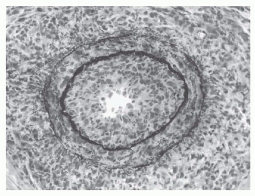
The number of invading T cells in a graft is not necessarily correlated with the speed of rejection.405 This finding has
suggested that certain critical elements of the graft, such as its blood vessels, are the actual site of graft destruction and, indeed, endothelialitis is an important hallmark of clinically significant rejection activity.408

Repertoire analysis of graft-infiltrating T cells in acutely rejecting grafts reveals marked polyconality,22,409,410,411,412 but only the donor-reactive CTLs show evidence of having been activated in vivo.413 Oligoclonal dominance has been suggested in studies of tolerated rodent allografts412 and in long-term rejected human kidneys.414 T cells infiltrating xenografts included a broad TCR repertoire.415,416,417 T cells mediating GVHD in the setting of multiple minor histoincompatibilities demonstrated a markedly skewed repertoire involving several different Vβ families.418,419 Clinical studies suggest that the anti-MiHA TCR repertoire is most often polyclonal.420

Role of Natural Killer Cells. Although the role of NK cells in marrow rejection is well established in mice, the amount of resistance mediated by NK cells to allogeneic hematopoietic stem cells is limited and can be readily overcome by increasing the dose of donor stem cells administered.421,422 Furthermore, a role for NK cells in resisting human allogeneic marrow engraftment has not been clearly demonstrated, although they might be expected to be important in recipients of reduced toxicity conditioning regimens. Indeed, patients with severe combined (T- and B-cell) immunodeficiency who have functional NK cells require cytotoxic conditioning to permit engraftment of haploidentical marrow, whereas those lacking NK cells do not.423

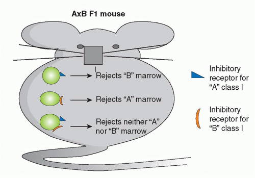
The ability of NK cells to be triggered by “missing self” may have utility in HCT.424 Donor-derived NK cells with graft-versus-host reactivity due to the lack of donor class I MHC inhibitory ligands in the recipient can kill residual host leukemia cells and alloreactive cells that resist the marrow graft without causing GVHD. The alloreactive donor NK cells may also reduce susceptibility to GVHD by killing recipient APCs needed to activate donor T cells.424,425 While striking antileukemic effects of KIR mismatching were detected in heavily conditioned patients receiving high doses of haploidentical CD34+ stem cells,424 the effect of KIR incompatibility has been more variable in other clinical studies,426,427,428,429,430,431 and the antitumor benefit is most evident for acute myelogenous leukemia.432

The possible role of NK cells in rejecting solid organ grafts is somewhat controversial. NK cells are prominent among cells infiltrating rejecting organ allografts and may be the earliest producers of inflammatory cytokines and chemokines and inducers of DC maturation.433,434,435,436 If NK cells make an important contribution to solid organ allograft rejection under normal circumstances, they must be dependent on T cells, as mice lacking T cells are unable to reject nonhematopoietic allografts. Furthermore, whereas bone marrow allografts from class I deficient donors (β2m-/-) are subject to potent NK-mediated rejection (because these cells cannot trigger inhibitory receptors on host NK cells437), β2m-/- skin grafts are not rejected by β2m+ recipients.438 NK cells have recently been reported to play a critical role in cardiac allograft rejection in CD28 knockout mice,439,440 and NK cells can mediate a particular form of chronic allograft vasculopathy in a murine cardiac allograft model.435 This lesion may be triggered by viral infection.441

Inhibitory receptors on NK cells are quite broad in their class I specificity,442 and fully allogeneic class I MHC marrow is less susceptible to NK-mediated marrow destruction compared to class I-deficient marrow.437,443 Because of the increased disparity of xenogeneic compared to allogeneic MHC molecules, NK cells may receive fewer inhibitory signals from xenogeneic than allogeneic cells. Indeed, transduction of HLA molecules into porcine endothelial cells reduces NK cell-mediated xenogeneic cell adhesion and cytotoxicity.444,445,446 However, some inhibitory receptors, such as killer cell lectin-like receptor G1, do recognize xenogeneic ligands such as e-cadherin.447 NK cells may also be activated by interactions of activating receptors with ligands on xenogeneic cells,448,449 of which several examples have been identified.450,451 On balance, activating xenogeneic NK cell-target interactions are more effective than inhibitory interactions. Indeed, NK cells resist xenogeneic marrow engraftment to a greater extent than allogeneic marrow.421,452,453,454 NK cells have also been implicated in the acute vascular rejection455 that can destroy solid organ xenografts that have escaped hyperacute rejection (see following discussion) and in xenogeneic skin graft rejection.456 As one mechanism by which NK cells mediate cytolysis is antibody-dependent cell-mediated cytotoxicity, it is possible that immunoglobulin G natural antibodies play a significant role in initiating NK cell-mediated rejection. NK cells also release cytokines, such as IFNγ, and TNF-α, which activate macrophages and endothelial cells, and induce inflammation.455

Role of Natural Killer T Cells. While NKT cells have apparent inhibitory effects on graft rejection457,458,459,460 and GVHD,461,462 they have also been reported to participate in rejection of tissues and bone marrow in mice.463,464,465 The latter is due to the ability of NKT cells to activate NK cells.465 NKT cells promote skin graft rejection by cross-primed CD8 cells via their ability to produce IFNγ.466

Role of Monocytes/Macrophages and Eosinophils as Effectors of Rejection. Classical delayed-type hypersensitivity (DTH) responses are thought to depend on the activation of macrophages by helper T cells through production of IFNγ. It is likely that proinflammatory cytokines and chemokines produced by activated monocytes and macrophages play a role in endothelial cell activation and lymphocyte recruitment. Additionally, activated macrophages may damage tissue through the production of toxic molecules such as nitric oxide.467

Macrophages play an especially important role in the rejection of cellular xenografts such as islets468,469 in a T cell-dependent manner.470 Macrophages cause almost immediate rejection of xenogeneic bone marrow, even in the absence of adaptive immunity.471,472,473 Human macrophages can phagocytose porcine cells in an antibody- and complementindependent manner.474 Additional studies have implicated macrophages in solid organ and skin xenograft rejection.475,476,477,478,479,480 This prominent role for xenogeneic macrophages may reflect the combined ability of certain xenogeneic
receptors to activate macrophages,481 whereas important inhibitory interactions, such as that between CD47 and its macrophage ligand SIRPα, are not effective.482 Surprisingly, a system for monocyte-mediated recognition of allogeneic non-MHC nonself has been described.483

Eosinophils recruited to allografts by Th2 T-cell responses have been reported to be effectors of graft rejection in some experimental models, and eosinophils are often found clinically in rejecting allografts.333 Th2-derived IL-4 and IL-5 recruit and activate eosinophils, which release highly cytotoxic substances from granules into the tissue.


Chronic Rejection and Chronic Graft-versus-Host Disease

Most experimental studies of rejection are performed without immunosuppression and, therefore, graft destruction usually occurs within the first several days or weeks by one of the mechanisms described previously. In clinical practice, however, the use of immunosuppression usually allows graft survival for much longer periods of time. Nonetheless, clinical survival statistics reveal that even when 1-year graft survival has been achieved, the loss of transplanted organs continues to occur at a rate of about 3% to 5% per year, and a significant proportion of this delayed or late graft failure appears to be due to immunologic mechanisms.

The term “chronic rejection” is commonly used to describe this later process of delayed graft destruction, although in kidney transplantation the Banff classification schema has proposed to replace this term with interstitial fibrosis and tubular atrophy.484 As immunosuppressive reagents have become more effective at controlling acute rejection, chronic rejection has emerged as one of the most important problems in clinical practice. Indeed, while there has been ongoing improvement over the past 30 years in the 1-year graft survival rates for kidney transplants, the halflife for organs that have survived for 1 year has not changed significantly over that entire period of time; as a result of this ongoing loss, only about 50% of transplants are still functioning 10 years later.

Although almost every type of organ transplant suffers from deterioration in function over time, the pathologic manifestations are different in each case. Kidney biopsies tend to show interstitial fibrosis along with arterial narrowing from hyalinization of the vessels—hence the terminology interstitial fibrosis and tubular atrophy. In the heart, the process is manifested principally as a diffuse myointimal hyperplasia, proceeding to fibrosis of the coronary arteries that has often been referred to as “accelerated atherosclerosis” or “transplant arteriosclerosis.” Chronic rejection in lung transplants primarily affects the bronchioles with progressive narrowing of these structures and is referred to as “bronchiolitis obliterans.” The liver may be the one type of organ transplant that is relatively resistant to chronic rejection, but the progressive destruction of bile ducts referred to as the “vanishing bile duct syndrome” may be another manifestation of this process.

Some of the causes of chronic graft destruction may not be immunologic in origin.485,486 Analysis of sequential kidney transplant biopsies suggests that chronic rejection represents cumulative and incremental damage to the graft from time-dependent nonimmunologic and immunologic causes.487,488 Potential nonimmunologic factors that have been considered to contribute to the development of chronic rejection include the initial ischemic insult, the reduced mass of transplanted tissue (especially in the case of kidney transplants leading to hyperfiltration injury), the denervation of the transplanted organ, the hyperlipidemia and hypertension associated with immunosuppressive drugs, the immunosuppressive drugs themselves, and chronic viral injury, amongst others. Nonetheless, while these factors undoubtedly contribute to the process, there is a marked difference in survival between syngeneic and allogeneic transplants in experimental models. Thus, there is almost certainly an important immunologic component in most cases of chronic rejection.

Several important observations regarding the pathogenesis of chronic rejection have emerged from clinical practice, particularly the analysis of biopsy samples. In kidney transplants, two distinctive phases of injury of chronic allograft nephropathy have been described.487 Previous studies have suggested that there is a high correlation between the onset of chronic rejection and a history of early acute and subclincial rejection episodes.489,490 Analysis of protocol biopsies has revealed that the onset of mild chronic rejection by 1 year after kidney transplantation is associated with an initial phase of early tubulointerstitial damage from ischemic injury that occurs before severe rejection is detected. Beyond 1 year, a later phase of chronic allograft nephropathy was characterized by microvascular and glomerular injury.487 Importantly for long-term outcomes, the clinical data show that the process of chronic rejection is usually refractory to increases in immunosuppressive therapy, in contrast to acute rejection episodes that almost always respond to treatment. The development of chronic rejection has also frequently been associated with the presence of antidonor antibodies,491,492 and the deposition of complement component C4d in the allograft.

Taken together, these clinical observations have suggested to some that chronic rejection is the result of donorspecific alloantibody production.493 Moreover, there are also now data emerging to suggest that components of the innate immune system such as NK cells can also contribute,494 in some cases in conjunction with alloantibody.495 All of these suggestions may be correct, but neither the logic nor the evidence fully supports the exclusive involvement of one mechanism. The relationship between alloantibody, C4d deposition, and neointimal fibrosis is complex.496 In the first place, alloantibody production often reflects activation of indirect pathway T cells (see previous discussion), and hence it might equally well be a marker for other rejection mechanisms as opposed to a cause of chronic rejection. Moreover, the presence of alloantibody and C4d may be transient, and there are clear examples of chronic changes in the graft in their absence.496 In addition, early rejection episodes probably reflect primarily the degree of antidonor immunoreactivity, and there is no proven direct link with later deterioration of graft function. Therefore, even if sufficient
immunosuppression were given to prevent acute rejection, chronic rejection might still occur when the levels of immunosuppression are reduced over the long term, even if acute rejection had never occurred. Finally, experimental studies have suggested that the mechanisms of chronic rejection are not absolutely dependent on either antibody formation or on the occurrence of acute rejection episodes.

The uncertainties that arise from the interpretation of the clinical data make it important to develop experimental models for studying the mechanisms of chronic rejection. It is difficult in the laboratory, however, to mimic a process that may take 5 or 10 years to develop in patients treated with immunosuppressive drugs. Thus, the effort to study chronic rejection experimentally has depended on surrogate short-term pathologic markers that are thought to predict the long-term changes of chronic rejection. In particular, these studies have concentrated on the development of the myointimal proliferation that is thought to be the precursor of the chronic vascular changes typically observed in patients. In rodents, pigs, and primates, this has often been done with grafts after an initial period of immunosuppression that prevents acute rejection.497,498,499 All of these experimental studies are subject to the caveat that the surrogate pathologic lesion occurs much earlier than the typical changes of chronic rejection in patients. Thus, the process being studied experimentally may not be the same as the clinical process.


Pathologic Manifestations of Experimental Chronic Rejection

The typical pathologic features of the experimental lesion associated with chronic rejection are shown in Figure 46.10.500 The marked narrowing of the vascular lumen is caused by the substantial proliferation of endothelial and then smooth muscle cells that can be host-derived.501 Associated with this proliferation is progressive destruction of the media. In time, the cellular proliferation becomes less pronounced and is replaced by concentric fibrosis that narrows the vascular lumen. Immunohistologic staining indicates that there is increased expression of several adhesion molecules,500 intracellular proteins such as vimentin,502,503 and easily detectable levels of several molecules, including nitric oxide synthase,504 acidic fibroblast growth factor, insulin-like growth factor, IFNγ,505 and endothelin,506 each of which may play a role. The abnormal expression of self-molecules in allografts undergoing chronic damage can lead to autoantibody formation.507 Ultimately, the ischemia resulting from vascular occlusion results in fibrosis in the parenchyma of the organ and consequent organ dysfunction.508 In the case of the lung or the liver, chronic injury may cause changes most prominently in the bronchioles or the bile ducts, but this is also associated with arterial lumen loss, which may be the primary lesion causing bronchiolitis obliterans or bile duct fibrosis, respectively.508


FIG. 46.10. Histology of Chronic Rejection.


Immunologic Mechanisms of Chronic Rejection

Rejection requires a dialogue between the innate and adaptive immune systems.509 Innate immunity is most likely involved at the outset of the process that leads to the development of chronic rejection, and there is evidence to suggest that NK cells494,510 and complement activation can be involved.511 At the level of the adaptive response, because it is assumed that stimulation of direct pathway T cells is likely to diminish over time as donor APCs are replaced by recipient APCs (see previous discussion), it is commonly assumed that the predominant immune response that causes chronic rejection occurs through the indirect pathway.512

Studies in pigs have suggested that the vascular changes of chronic rejection are more apt to develop when there are class I antigenic disparities than when there are only class II disparities and that the development of the lesion depends especially on CD8+ T cells.513 In mouse models, in contrast, there is evidence that either CD4+ or CD8+ T cells can produce the lesion and that either class I or class II antigenic disparities are sufficient to stimulate chronic rejection.514 The finding that class II antigenic disparities are themselves sufficient to induce this pathology is consistent with the observation of class II MHC expression on the vascular endothelium and medial smooth muscle cells of mouse cardiac allografts with these vascular lesions.515 Because class II MHC is not constitutively expressed by mouse vascular endothelial cells, indirect recognition of donor class II transferred from passenger leukocytes may be responsible for inducing an inflammatory response that leads to subsequent upregulation of class II on the donor vascular endothelium. In keeping with the prediction of many clinical studies, adoptive transfer experiments into mice with severe combined immunodeficiency have shown that alloantibodies in the absence of T cells can induce the typical pathologic vascular changes, and lesions can develop in T-cell-deficient mice.516 However, T cells without B cells have also been shown to cause the lesion, although there may be somewhat less tendency to progress to end-stage fibrosis.517

Several studies have indicated that the induction of donor-specific tolerance can prevent the development of the vascular changes of chronic rejection, although not all of the short-term manipulations that have been effective in preventing acute rejection have necessarily prevented the later onset of chronic rejection. Remarkably, mice rendered
tolerant by neonatal injection of donor splenocytes, or by the induction of high levels of lasting, multilineage mixed chimerism with demonstrated central deletion of donorreactive T cells and permanent acceptance of donor-specific skin grafts, demonstrate graft vasculopathy in donor cardiac allografts.510 Thus in the complete absence of antidonor T-cell reactivity, other cell types such as NK cells may induce these types of lesions in cardiac allografts. In addition, T-cell recognition of cardiac-specific antigens presented by donor MHC and not shared by donor hematopoietic cells could play a possible role in the development of these lesions in immunocompetent, tolerant mice.494

From these data, it seems likely that multiple immunologic mechanisms may be capable of creating the graft arteriosclerotic lesions that are characteristic of chronic rejection, and that T-cell alloreactivity is not essential for their induction. Whether there is a critical final common mediator involved in all of these pathways is not currently known. However, IFNγ has been shown to play an important role in the development of lesions in several models,518,519 and signal transducer and activator of transcription (STAT)4-deficient mice, which do not respond to IL-12 and therefore cannot generate Th1 responses, show markedly reduced severity of graft vasculopathy compared to wild-type mice.520 TGF-β has been shown to attenuate the lesions, but has also been detected within the lesions and implicated in the development of fibrosis.521


Chronic Graft-versus-Host Disease

cGVHD is the most common and severe complication among patients surviving for more than 100 days after allogeneic BMT. Clinically, acute and cGVHD can be distinguished on the basis of the time of onset, clinical manifestations, and distinct pathobiologic mechanisms. Acute GVHD usually occurs within 2 to 6 weeks following allogeneic BMT and primarily affects the skin, liver, and the gastrointestinal tract with T-cell infiltration of the epithelia of the skin, mucous membranes, bile ducts, and gut. However, acute GVHD has been noted to occur later in recent protocols involving nonmyeloablative conditioning for HCT. In contrast, cGVHD involves a wider range of organs and clinical manifestations include scleroderma, liver failure, immune complex disease, glomerulonephritis, and autoantibody formation.

The pathogenesis of cGVHD, like chronic rejection, is poorly understood. The disease involves T-cell responses to alloantigens or autoantigens.522 Because most BMT is performed between HLA-identical or closely HLA-matched pairs, alloreactivity may be directed against miHAs presented by shared MHC molecules, or against MHC alloantigens when present. T cells developing de novo in a recipient thymus that is damaged due to GVHD may result in the emergence of autoreactive T cells into the peripheral repertoire. The injury to target organs is poorly understood, but may involve inflammatory cytokines and fibrosis, as well as B-cell activation and production of autoantibodies.

The main risk factors for the onset of cGVHD are HLA disparity, donor and patient age and sex, source of progenitor cells, graft composition and previous acute GVHD. cGVHD can be treated providing it is identified sufficiently early after initiation. Even with treatment, extensive skin involvement, thrombocytopenia, and progression are poor prognostic factors.


PHYSIOLOGIC INTERACTIONS THAT MODULATE GRAFT REJECTION AND GRAFT-VERSUS-HOST DISEASE

Although graft rejection and GVHD often involve exceptionally strong immune responses, these responses are still accompanied by downregulatory components that can be manipulated to promote graft survival. While many of these manipulations are described in the section on tolerance, some of the regulatory components of the rejection response are briefly described here.


Downregulating Signals Following T-Cell Activation

Interactions between Fas and Fas ligand (FasL), which is upregulated during rejection responses, can mitigate GVHD and graft rejection by killing activated T cells and APCs.306 FasL-deficient recipients are more susceptible than normal mice to the development of GVHD,523 and FasL can promote resistance to rejection of tissues transplanted to some “privileged sites,” such as the testis or the anterior chamber of the eye.524 However, forced overexpression of FasL causes a nonspecific inflammatory syndrome associated with prominent neutrophil infiltration525 and can promote graft destruction.526,527 Overexpression of FasL has, however, been reported to promote survival of heart allografts in recipients of FasL-expressing donor-specific transfusions (DSTs),528 and of bone marrow529 and islet530 allografts.

CTL antigen (CTLA)4 helps maintain self-tolerance, as evidenced by the T-cell lymphoproliferative autoimmune syndrome that develops in CTLA4 knockout mice.531,532 CTLA4 has been shown to play a role in T-cell tolerance in many systems.384,533,534,535,536,537,538,539,540,541,542,543,544,545,546 Blockade of CTLA4 accelerates cardiac allograft rejection547 and increases GVHD.548 PD1, an additional downregulatory molecule expressed by activated T cells that recognizes the B7 family members PDL-1 and PDL-2, also mitigates graft rejection549,550,551,552,553,554 and GVHD.555,556,557 Interaction of one of the PD1 ligands, PD-L1, with its alternative receptor B7.1, also mitigates graft rejection.558 While PD1 plays an important modulatory role in the presence of extensive antigenic disparities between murine heart graft donors and recipients, the B- and T-lymphocyte attenuator (BTLA)-herpesvirus entry mediator (HVEM) inhibitory pathway predominates in the presence of more restricted antigenic disparities.559 BTLA-HVEM interactions may also control GVHD,560 but the opposing effects of BTLA and HVEM expressed by T cells on T-cell activation complicate interpretation of experiments, which have produced conflicting results with respect to GVHD560,561 and islet allograft survival.562,563


Immunomodulatory Effects of Cytokines

There is considerable evidence of roles for IL-10 and TGF-β in downmodulating graft rejection564,565,566 and GVHD,372,567,568,569,570 and these cytokines may have therapeutic utility.571,572,573,574,575 These
activities may reflect the important role of these cytokines in regulatory T (Treg)-cell and B-cell generation and function. On the other hand, IL-10 can enhance cytolytic mechanisms of islet graft rejection,576 and high doses can accelerate GVHD.372 Moreover, TGF-β is an important mediator of fibrotic pathologies in both chronic rejection and GVHD.577,578 TGF-β, in concert with proinflammatory cytokines such as IL-1 and IL-6, also promotes the development of proinflammatory Th-17 cells.579

The inflammatory condition that develops in IL-2 knockout mice is clear evidence of the important immunomodulatory role of this cytokine, and studies in several graft rejection580 and GVHD581 models have confirmed such a role in transplantation. Much of this effect is due to the important role of IL-2 in survival and expansion of Treg cells,582,583 as is discussed elsewhere in this chapter. IL-2 can also promote activation-induced cell death of alloreactive CD8 cells.582,584

While IFNγ can also promote graft rejection, rejection is rapid or even accelerated in IFNγ knockout mice.383,564,585,586,587 IFNγ has also been shown to play a downregulatory role in GVHD.371,385,588 IFNγ has antiproliferative effects on T cells,384,587 increases activation-induced cell death via the Fas/FasL pathway,589,590,591 upregulates nitric oxide production,592,593,594,595 and is necessary for Treg-cell function in certain conditions.596,597,598 Consistently, the major cytokines that induce IFNγ, IL-12 and IL-18, can inhibit graft rejection382,384,599 and GVHD.385,600,601 IL-12 and IL-18 act in an IFNγ and Fas-FasL-dependent manner,385,601,602 which preserves or enhances GVL effects.588,603,604,605,606 IFNγ promotes the GVL effect of CD8 T cells,385,607,608 apparently by promoting lymphohematopoietic grat-versus-host responses while inhibiting tissue GVHD,609 perhaps due in part to increased PDL1 expression by APCs,610 promotion of donor Treg-cell expansion,610 and reduced Th17 differentiation.337

Despite interest in the notion that Th2 cytokines are antiinflammatory and may suppress rejection and GVHD,359,360,361,362,363,364,365,366,367 IL-4 deficiency does not accelerate graft rejection564,611 and can actually downmodulate GVHD.371 However, there clearly are situations in which an immunomodulatory effect is achieved by Th2 cytokines. For example, the use of total lymphoid irradiation can alter the balance between NKT cells and conventional T cells in BMT recipients, and IL-4 production by enriched recipient NKT cells downmodulates GVHD.612 This approach, which also apparently involves Treg cell enrichment when combined with antithymocyte serum,613 has recently been extended to clinical trials of HLA-identical HCT for treatment of hematologic malignancies and renal allograft tolerance induction.614,615,616,617


The Presence of the Transplanted Organ

Vascularized organ allografts may be accepted spontaneously618,619,620,621 or with a short course of immunosuppression in rodents or pigs.621,622,623,624,625,626,627,628,629 The long survival of these transplanted organs can prevent rejection of other allografts from the same donor.618,630,631 In clinical transplantation, long survival of a transplanted organ may diminish the rejection response, as much less immunosuppression is required late after transplantation than in the early period. Studies in transiently chimeric monkeys and patients achieving tolerance with HLA-mismatched combined kidney and BMT strongly implicate a role for the kidney itself in promoting tolerance.632,633,634,635,636 Treg cells have been implicated in many of these models (see following discussion). The capacity of transplanted organs to regulate their own survival is often confused with the capacity of a treatment to induce tolerance. For example, many transient immunosuppressive regimens achieve vascularized allograft survival in rodents, but the role of the immunosuppression is to allow a strong Treg cell response induced by the graft to predominate under the controlled experimental conditions. These conditions do not usually apply to human transplantation, thus explaining the failure to translate the many tolerance regimens that succeed in rodents into clinical practice.


Role of Graft and Tissue Injury

Graft injury, such as that associated with ischemia-reperfusion and host tissue injury induced by conditioning therapy in the case of HCT, play an important role in promoting graft rejection and GVHD, respectively. Local inflammatory processes activate innate and consequently adaptive immunity and T-cell activation. Inflammation plays an important role in promoting APC migration from tissues to lymph nodes637,638,639,640,641 and also promotes trafficking of activated T cells into tissues, as is illustrated dramatically in HCT models. Administration of large numbers of nontolerant donor lymphocytes to established mixed bone marrow chimeras (ie, animals not recently treated with conditioning therapy) leads to a graft-versus-host response that attacks only lymphohematopoietic tissues and does not cause GVHD, a disease of epithelial tissues such as skin, intestines, and liver.642,643 In contrast, similar numbers of T cells cause rapidly lethal, severe GVHD in freshly irradiated hosts.642,644 Conditioning rapidly induces production of chemokines in the GVHD target tissues, promoting immigration of T cells that then elicit a further cascade of chemokines that amplifies the response.645 Upregulated adhesion molecules also promote leukocyte infiltration through the microvasculature of these tissues. Lethal total body irradiation (TBI) and cyclophosphamide, for example, upregulate the proinflammatory cytokines IL-1, IL-6 and TNF-α,646,647,648 which can upregulate endothelial cell E-selectin, P-selectin, ICAM-1, and vascular cell adhesion molecule-1.649,650 In the absence of such host target tissue inflammation, mature, activated graft-versus-host-reactive effector T cells are unable to traffic into skin and induce injury.644 Provision of a local tolllike receptor (TLR) stimulus promotes the entry of such cells into the skin and induces localized GVHD,644 indicating that tissue inflammation provides a critical checkpoint for T-cell recruitment to GVHD target tissues. A systemic TLR stimulus in this setting promotes severe, multiorgan GVHD.644 GVHD can also occur when very large numbers of nontolerant parental T cells are administered to genetically tolerant F1 hosts,651 indicating that, in the presence of sufficiently powerful graft-versus-host responses, the need for tissue inflammation to induce GVHD can be bypassed, possibly due to inflammation induced by high systemic cytokine levels.


All forms of organ and tissue transplantation involve ischemic and traumatic injury to the donor tissue, which may be one of the reasons that rejection episodes occur most frequently early after transplantation. The surgical trauma associated with transplantation is associated with very early production of chemokines in the graft,433,652 promoting infiltration of NK cells433 and neutrophils,653 which in turn perpetuate inflammation that promotes subsequent T-cell infiltration.654 At least partly due to the influence of these cells of the innate immune system,433 chemokines are produced before T-cell infiltration is seen. IFNγ, whose early production may require CD8 T-cell activation,655 also activates macrophages to become effective APCs and release chemokines.655,656,657 These phenomena, along with microbial exposures that drive innate immunity, contribute to “danger” signals that promote graft rejection.658 Nevertheless, skin and cardiac allografts that are allowed to heal before being exposed to alloreactive T cells are rapidly rejected if there is sufficient antigenic disparity between donor and host.659,660 Similarly, patients with long-standing allografts are rarely able to terminate immunosuppressive therapy without rejection. Therefore, “danger” signals are not a critical requirement for graft rejection, and it is better to picture the antigenic disparity and the recipient’s immune responsiveness as the dominant features controlling graft rejection, while danger signals may influence the timing, intensity, or character of the rejection response.


Role of the Innate Immune System

The innate immune system comprises a group of cells and molecules (Table 46.5) that provide a first line of defense against pathogens and which also play an important role in allograft rejection. Primary adaptive immune system responses that rely on the activation and expansion of antigenspecific T and B cells take several days to reach maturity. In contrast, the innate immune system can be considered as a “preformed” defense mechanism that is immediately available to defend the host until either the dangerous stimulus is cleared or the adaptive immune system is able to mount an antigen-specific response.661 Clearly, this is a somewhat simplistic view; while many components of the innate immune system can be recruited very quickly after transplantation, their activity can be amplified after activation.


TABLE 46.5 Components of the Innate Immune System


Cell


Primary function


Macrophage/neutrophil


Phagocytosis, opsonisation, antigen presentation


Release of inflammatory mediators


Dendritic cell


Antigen uptake and presentation to lymphocytes


Natural killer cell


Release of cytokines


Cytotoxic to virally infected or mutated cells


Eosinophil


Release of inflammatory mediators


Killing of antibody-coated entities


Complement


Opsonisation, target cell lysis, and chemoattraction


The physical process of graft retrieval and implantation generates signals within the graft and the recipient that trigger rejection. The concept of “danger” triggering an immune response has evolved as an idea over many years.170,661 Pattern recognition receptors exist to detect the unwanted presence of bacterial or viral pathogen-associated molecular patterns, but after transplantation the TLRs that form part of the pattern recognition receptor family can also be used to detect the molecules produced as a result of implantation of the graft, so called damage-associated molecular patterns (DAMPS). These signals include heat shock proteins, reactive oxygen species, high mobility group protein B1, complement breakdown products, nucleic acids (deoxyribonucleic acid and RNA), mitochondrial components, and molecules associated with tissue fibrosis that activate cells of the innate immune system via TLR ligation.

The immune system monitors the health of cells and responds to ones that have been injured and killed. Cell death is an inevitable consequence of the ischemia and reperfusion injury that is caused by organ and tissue retrieval. Dying cells expose intracellular DAMPs that can be recognized by components of the innate immune system.662 The role of DAMPs may vary depending on the type of transplant performed and the degree of injury resulting from cell isolation or organ retrieval. Some DAMPs may be expressed in a tissue-restricted manner (eg, one class of DAMPs called alarmins include molecules such as β-defensins that are expressed primarily by leukocytes and therefore may be more relevant after hematopoietic stem cell transplantation or BMT). In addition, the location or type of injury may contribute to the outcome of dying cells triggering a response. Thus, the contribution of different DAMPs to triggering the innate immune system may vary with different types of transplant.

Macrophages and other phagocytic cells can ingest dying cells and necrotic tissue; when activated, they release cytokines such as TNF-α, IL-1, and IL-6, which all contribute to the local inflammatory environment. Production of active IL-1β requires proteolytic cleavage of the inactive form, pro-IL-1, a process linked to a multiprotein complex called an inflammasome that acts as a platform for caspase 1, the cysteine protease involved in the proteolytic maturation and secretion of IL-1β.663 A number of different inflammasomes exist, but the nucleotide oligomerization domain-like receptor protein 3 inflammasome is the best studied to date. IL-1β can be highly deleterious to the tissue in which is produced; therefore, inflammasome activity is tightly controlled. As a consequence of these events, the early infiltration of macrophages into a graft at the onset of rejection has been suggested to be a poor prognostic sign for transplant survival. Macrophage colony-stimulating factor, produced by tubular and mesangial cells, promotes macrophage infiltration and proliferation, and may play a pathogenic role in acute rejection.

Damaged tissue can also trigger complement activation in the absence as well as the presence of antibody; complement has been demonstrated to contribute to ischemia reperfusion
injury.664 Activated complement components constitute a proteolytic cascade that generates a range of effector molecules.170 The anaphylatoxins C5a and C3a are chemoattractant molecules that assist leukocytes to home to the graft while other soluble mediators are able to opsonise cells, targeting them for destruction by phagocytes.665 Recognition of C3b, C4b, or their fragments covalently bound to target cells by complement receptors on the surface of leucocytes facilitates antigen presentation and T-and B-cell activation.666,667 Generation of the terminal components of the complement cascade (C5b-9) results in formation of the membrane attack complex within the target cell membrane and initiation of target cell lysis. This has been demonstrated to play an important role in ischemia reperfusion injury.664 In addition to the potential of the damaged tissue itself to activate complement, there is also evidence that natural immunoglobulin M antibody can trigger complement activation via both the classical and mannose-binding lectin pathway.668 Studies in muscle reperfusion models initially identified natural immunoglobulin M as a major initiator of pathology through the activation of the complement system and recruitment of inflammatory cells. When the repertoire of natural immunoglobulin M antibodies was altered, significant protection of the myocardial tissue was observed with only limited apoptosis of cardiomyocytes and decreased neutrophil infiltration compared to when natural antibody was present.669 As mentioned previously, there is also increasing evidence that complement can influence graft outcomes, contributing to the development of acute and/or chronic rejection, either directly or through antibodydependent mechanisms.511,670

NK cells are innate immune mediators that express cell surface receptors, including activating receptors that bind to widely expressed carbohydrate residues on self-cells and inhibitory receptors that bind self-MHC class I molecules. Activating NK cell receptors including NKG2D recognize natural stress signal ligands, whereas the inhibitory receptors include the CD94-NKG2A complex, KIR family in humans, and Ly49 family in mice. The possible role of NK cells in graft rejection is discussed previously. Absence of an appropriate MHC class I ligand on an allogeneic cell informs the NK cell that the allogeneic cell should be killed. Some malignant or virally infected cells downregulate MHC class I expression or express altered class I molecules as a strategy to evade CD8+ T-cell cytotoxicity. As a result, they are unable to stimulate inhibitory receptors and are vulnerable to NK cell killing. Thus, NK cells could contribute to tissue damage following cell and solid organ transplantation. While the role of NK cells in rejecting bone marrow (at least in mice) is clearer than for solid organ transplantation,671 NK cells can have a marked and lasting impact in this setting,672 particularly with a form of chronic cardiac graft vasculopathy in mice.494 NK cells likely contribute to acute rejection in certain donor-recipient combinations where, even if they are not the major drivers of the responses, they have a significant impact by secreting IFNγ. Nevertheless the precise role of NK cells requires further elucidation as NK cells have also been shown to promote tolerance induction (see the following discussion) by killing donor APCs.673

Only gold members can continue reading. Log In or Register to continue

Stay updated, free articles. Join our Telegram channel

May 30, 2016 | Posted by in IMMUNOLOGY | Comments Off on Transplantation Immunology

Full access? Get Clinical Tree

Get Clinical Tree app for offline access