TISSUE RENIN-ANGIOTENSIN SYSTEM
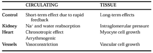
Local renin-angiotensin systems in multiple tissues can produce A-II independently of the circulating renin-angiotensin system (Table 79-1). Their function of these systems is probably to permit regional modulation of blood flow and also to assist in the action of growth factors. Most contractile cells (e.g., vascular smooth muscle, heart cells, mesangial cells, and sperm tails) have a well-defined local renin-angiotensin system. A-II is
formed in these cells either by renin or through other proteases such as chymase, cathepsin, or tonin.41,42 and 43
formed in these cells either by renin or through other proteases such as chymase, cathepsin, or tonin.41,42 and 43
Kidney Renin-Angiotensin System.
In addition to being the major site for release of systemic renin, the kidney has a local renin-angiotensin system that is involved in renal growth.44 ACE-I administration during pregnancy causes severe fetal renal abnormalities. The tubular epithelial cells contain ACE, and local A-II is found in proximal tubules and mesangial cells.45 The intrarenal A-II constricts afferent and efferent arterioles and directly increases Na+ reabsorption. Locally, A-II is also involved in renal pathologic conditions (e.g., the development of nephrosclerosis).
Cardiac Renin-Angiotensin System.
Evidence exists for a role for an intracardiac renin-angiotensin system in both normal and failing hearts.41,42 There is a direct expression of AGT and the A-II receptor (AT1) in cardiomyocytes. The exact source of the renin in cardiac tissue is controversial, because levels of mRNA are low.43 The heart can also express A-I, which may be converted by intracardiac ACE to A-II.43 Probably, cardiac renin-angiotensin system activity is regulated by the amount of AGT, ACE, or renin-binding protein in the cardiomyocytes. Consequently, local A-II can regulate vascular tone, contractility, growth, and cardiac hypertrophy.
Stay updated, free articles. Join our Telegram channel

Full access? Get Clinical Tree


