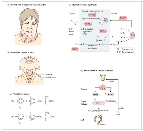
Nodular thyroid disease is common, affecting approximately 5% of the female population over the age of 50. Women are affected more commonly than men and the incidence increases with age. A 59-year-old lady, Mrs RB, presented with a long history of a swelling in the anterior neck. This had gradually increased in size over the years and had now become quite obviously visible. She had no dysphagia or dyspnoea, the thyroid gland was not painful, she did not have a hoarse voice and there was no history of previous radiotherapy treatment to the neck. She had no symptoms to suggest abnormal thyroid hormone production and on examination she was clinically euthyroid. The thyroid gland was asymmetrically enlarged with a 3 × 4 cm nodule palpable in the right lobe together with three other palpable nodules. The gland moved freely on swallowing and there was no associated lymphadenopathy. Her thyroid function tests were normal as follows: fT4 18.3pmol/L; TSH 0.85 mU/L; thyroid antibodies negative. Thyroid ultrasound scanning revealed a multinodular goitre. Fine-needle aspiration of the dominant nodule was performed and cytology of the aspirate showed no evidence of malignant cells. She decided to undergo conservative management with regular clinical follow-up.
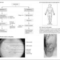
Clinical management of patients with nodular thyroid disease depends upon excluding the presence of malignant disease and then treating the goitre according to its size, patient preference and the likelihood of compression of other structures in the neck and mediastinum (Fig. 13a). In patients with compressive symptoms, CT scanning will reveal the extent of pressure on adjoining structures in the neck.
Thyroid gland: anatomy and structure
In humans, the thyroid gland is situated anteriorly in the neck (Fig. 13b), and its function is the synthesis and secretion of the thyroid hormones thyroxine (T4) and tri-iodothyronine (T3
Stay updated, free articles. Join our Telegram channel

Full access? Get Clinical Tree


