Fig. 51.1
(a–c) Note the sensitivity to insulin in lean subjects with PCOS is similar to that of normal obese control. It is apparent that the extreme lowest insulin sensitivity is seen in obese women with PCOS, while the highest is present in normal lean subjects
Case
A 22-year-old Caucasian female is referred to your office for evaluation of secondary amenorrhea. She reports menarche at age 14 and irregular menses for about 1 year. Her menses became regular and occurred monthly until age 17 menstruating only once every 2–3 months. For the past 6 months she has had no menstrual periods at all, and after taking several home pregnancy tests that were normal, she presented to her internist. He confirmed that she was not pregnant with a urine β-Hcg and referred her to your office for evaluation. She currently takes no medications. She notes that at age 16 she weighed 140 lbs, but gained about 40 lbs over the next 2- to 3-year period. Her weight has been stable at 180 lbs for the past few years. She is not exercising over concern for worsening her excessive axillary perspiration. She admits to some bothersome facial and abdominal hair growth and significant facial acne. She regularly waxes her facial hair but denies any other form of hair removal. The patient tends to snore and has some degree of daytime somnolence. She has been married for 1 year and is not using any form of contraception at this time. She and her husband are very interested in having children in the future.
On physical exam, she weighs 182 lbs and is 66 in. tall, with a BMI of 29.4. Her waist circumference is 38 in., waist:hip ratio is 0.84. Her blood pressure is 125/70 mmHg and pulse is 72 beats/min. She has notable acanthosis nigricans on the back of her neck and in her axillae bilaterally; skin tags are present in the nape of the neck. There is a mild degree of excess terminal (thick, pigmented) body hair on the upper lip and chin and significant acne vulgaris on the face. In addition, moderate periareolar and linea alba thick, terminal hair growth is present. There is no thyromegaly. Heart and lung exams are unremarkable. On abdominal exam, there is central adiposity without striae and there is no peripheral edema or cliteromegaly. Her liver edge was minimally tender.
Laboratory evaluation reveals a mild microcytic anemia, normal chemistries, an elevated ALT (SGPT) of 60 U/L (normal 10–45) with otherwise normal liver function tests, fasting blood glucose of 85 mg/dL and free testosterone of 6.2. Serum testosterone is 80 pmol/L, prolactin is 17 mIU/L; DHEAS, Androstenedione, 17-hydroxyprogesterone, and 24 h urinary-free cortisol are all within normal limits. The fasting plasma insulin level is 19 μU/mL (normal is less than 12 μU/mL), and sex hormone-binding globulin (SHBG) is 12 nmol/L (normal 40–120). Lipid panel reveals a total cholesterol of 246 mg/dL, triglycerides of 190 mg/dL, HDL of 42 mg/dL, and LDL of 166 mg/dL. A standard 2 h oral glucose tolerance test is performed and her fasting glucose of 88 mg/dL rises to 180 mg/dL and her insulin to 110 μU/mL) (after 2 h). An abdominal ultrasound reveals moderate fatty infiltration suggesting nonalcoholic fatty liver disease (NAFLD), and the adrenal glands were of normal size with no nodules.
The patient is started on metformin 500 mg daily, which is titrated up over a few weeks to 1,000 mg BID with meals. She is also advised to use an effective form of birth control until she desires pregnancy. In addition, she receives extensive counseling on diet and exercise for weight loss and diabetes prevention.
Review of Diagnosis, Treatment, and Complications
Diagnosis
The diagnostic criteria for PCOS are the subject of continuing controversy. In 1990 after an international consensus conference, the NIH published the following criteria for the diagnosis of PCOS (all three criteria are required):
1.
Menstrual irregularity due to chronic anovulation
2.
Evidence of clinical and/or biochemical hyperandrogenism (i.e., hirsutism, acne, or male-pattern alopecia)
3.
Exclusion of other causes of hyperandrogenism and menstrual irregularities (i.e., hyperprolactinemia, Cushing’s syndrome, androgen-producing tumors of the ovary or adrenal gland, adult-onset congenital adrenal hyperplasia)
4.
These criteria were based mainly on “expert opinion,” with limited scientific evidence. Notably, there is no mention of the presence of polycystic ovaries on ultrasonography.
In the years after the NIH criteria were released, it became apparent that there were women with normal menstrual cycles who had evidence of hyperandrogenism and polycystic ovaries on ultrasound and likely had PCOS. However, based on the NIH criteria, they would not be classified as having PCOS due to ovulatory menstrual cycles. Therefore, at a consensus conference in Rotterdam in 2003, the diagnostic criteria for PCOS were modified [6]. The new criteria require the presence of two of the following three:
1.
Oligo-ovulation (fewer than nine times per year) and/or anovulation
2.
Clinical and/or biochemical signs of hyperandrogenism
3.
Polycystic ovaries (meeting specific criteria on transvaginal ultrasound)
The criteria specify that other etiologies of hyperandrogenism and amenorrhea must be ruled out, as PCOS is a diagnosis of exclusion. When applying the Rotterdam criteria, compared with the NIH criteria, the prevalence of PCOS in the population increases to over 20 %. A 2007 Position Paper by the Androgen Excess Society contends hyperandrogenism (clinical or biochemical), is the major criterion distinguishing PCOS from other etiologies manifesting oligo-amenorrhea, with the inclusion of polycystic ovaries on ultrasonography as a criterion [7]. Similarly, they require exclusion of other etiologies that may mimic PCOS in establishing the PCOS phenotype (Table 51.1).
Table 51.1
Diagnostic criteria for PCOSa
Criteria | NIH 1990 “classic” | Rotterdam 2003 | AE-PCOS |
|---|---|---|---|
Oligomenorrhea | + | +/− | +/− |
Clinical or biochemical hyperandrogenism | + | +/− | + |
Polycystic ovaries on ultrasound | +/− | +/− | +/− |
Previous ultrasound criteria for polycystic ovaries included the presence of 8–10 follicles (measuring 2–8 mm in diameter), peripherally oriented with increased volume of stroma compared with the number of follicles. The Rotterdam ultrasound criteria include the presence of 12 or more follicles in each ovary (2–9 mm in diameter) and/or increased ovarian volume.
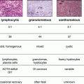
Nonetheless, sonographically apparent polycystic ovaries are not sufficient to diagnose PCOS as they can be demonstrated in many women who have other causes of hyperandrogenism or amenorrhea, as well as in up to 23 % of “normal” women. Some investigators believe the presence of a hyperandrogenic state may cause ovarian cyst formation, leading to the polycystic ovarian appearance on ultrasonography. Because of this, there continues to be controversy surrounding the inclusion of the polycystic ovarian morphology into the diagnostic criteria.
The menstrual dysfunction in PCOS typically presents around puberty with a normal or slightly delayed menarche and subsequent irregular menstrual cycles. Another pattern seen in PCOS women is initially regular cycles with subsequent menstrual irregularity associated with weight gain; often improved with weight loss. As ovulation is often infrequent, and first trimester pregnancies frequently abort, difficulty conceiving is often present.
The pathogenesis of PCOS is not completely understood. Current evidence suggests PCOS is a complex genetic trait influenced by genetic variants and environmental factors [2]. Numerous genes associated with PCOS or traits of PCOS have been identified. For example, PCOS susceptibility loci have been identified in Chinese women with PCOS and recently two of these PCOS susceptibility loci were also identified in European populations (DENND1A and THADA) [3]. Additionally, twin studies have established an inherited basis of PCOS with an increased inheritance in first-degree female relatives of women with PCOS.
In the normal ovary, the theca cells produce the androgens androstenedione and testosterone. These are converted to estrone and estradiol by aromatase activity in the granulosa cells. Androgen production by the theca cells is under the control of pituitary LH and the conversion to estrogens is under the control of FSH. LH and FSH production are in turn under the control of hypothalamic GnRH. It is the pulsatility of GnRH production that controls the relative amount of LH and FSH secreted by the pituitary. A rapid frequency of GnRH pulses favors the production of LH and slower pulse frequency favors the production of FSH. Abnormal gonadotropin secretory dynamics including altered LH action may be a component of the pathogenesis of PCOS. Women with PCOS have higher mean serum LH concentrations, possibly due to an increase in GnRH pulsatility. The serum FSH concentration may be normal or low in PCOS, causing an elevated LH/FSH production compared with young normal women in the early follicular phase with normal cycles. The likelihood of detecting an elevated serum LH depends on various factors such as last menstrual period, body mass index, and oral contraceptive pill use and as such, the diagnostic criteria for PCOS do not include an elevated serum LH and/or increased LH/FSH ratio.
The causative defect may also lie within the ovaries themselves. There may be higher LH action at the ovary in PCOS, resulting in hyperstimulation of thecal cells and premature expression of LH receptors in granulosa cells causing premature luteinization. There is evidence from several studies that the theca cells in women with PCOS may be more efficient at producing androgens than those of normal women. Therefore, these ovaries would produce a greater amount of androgen in response to a given level of LH, leading to the hyperandrogenic state of PCOS and secondary changes in GnRH pulsatility would be secondary. Of note, an elevated LH level is not mandatory in PCOS as increased ovarian androgen secretion and/or polycystic ovarian morphology can be seen without an elevated LH level. An elevated LH/FSH ratio is, however, more likely in nonobese women with PCOS. A slight elevation in prolactin may be seen, of unclear significance; however, a significant prolactin level elevation (>40 mg/dL) warrants an additional evaluation.
PCOS is also associated with the metabolic syndrome, insulin resistance, and compensatory hyperinsulinemia. The National Health and Nutrition Examination Survey (NHANES III) reported between a 40 and 50 % incidence of the metabolic syndrome (using the National Cholesterol Education/ATP III criteria) in women with PCOS. These women also have a higher incidence of insulin resistance than the general population, independent of their weight. Obesity is a frequent concomitant with PCOS and exacerbates insulin resistance, menstrual abnormalities, and higher likelihood of adverse pregnancy outcomes. Insulin resistance alone is not sufficient to develop PCOS, as hyperandrogenism is central to the disorder (via stimulation of androgen biosynthesis in the ovarian theca cell). Physical exam findings from hyperinsulinemia include the occurrence of acanthosis nigricans (darkening of skin most often on the back of the neck, in the axillae and in the groin) as was noted in our patient.
Insulin causes stimulation of theca cell secretion of androgens and suppresses hepatic SHBG production, leading to an increase in free androgens and clinically apparent as the hyperandrogenic phenotype typical of women with PCOS. Due to this phenomenon, treatment with weight loss and insulin sensitizers (such as metformin, thiazolidinediones) are often used in women with PCOS to decrease insulin resistance, decrease androgen levels, and improve ovarian function.
The metabolic syndrome has been linked to an increased risk of vascular disease (including myocardial infarction and stroke) and diabetes [4]. Therefore the link between PCOS and the metabolic syndrome has significant health implications. Thus many professional organizations recommend screening women with PCOS for the metabolic syndrome including impaired glucose tolerance with a baseline 2-h 75-g OGTT, prior to treatment and if a diagnosis is made every year or 2 thereafter.
The patient in our clinical vignette has impaired glucose tolerance as reflected by a 2 h OGTT glucose between 140 and 200 mg/dL (greater than 200 mg/dL is a sign of diabetes mellitus). The Rotterdam consensus meeting recommended against specific testing for insulin resistance given that tests for insulin resistance are not necessary to diagnose PCOS nor identify treatment. They did recommend screening all obese women with PCOS for components of the metabolic syndrome [assess waist circumference, measure blood pressure, fasting lipids and perform a 2-h oral glucose tolerance test (OGTT)]. The 2 h 75-g OGTT is recommended as normal fasting glucose levels do not identify women with impaired glucose tolerance who are at highest risk for future development of diabetes.
Related posts:

Stay updated, free articles. Join our Telegram channel

Full access? Get Clinical Tree


