T-Lymphocyte Developmental Biology


T-Lymphocyte Developmental Biology


Ellen V. Rothenberg

Ameya Champhekar


OVERVIEW OF T-CELL DEVELOPMENT


Introduction

T-cell development is a distinctive branch of hematopoiesis as well as a crucial input to immune system function. T-cell precursors, like other blood cells, are continuously generated from hematopoietic stem cells through a process that involves loss of access to alternative hematopoietic fates as well as gain of lineage-specific characteristics. In addition, like B cells, T cells must undergo programmed rearrangement of gene segments in order to assemble the genes that will encode their receptors for antigen, the &agr;, &bgr;, &ggr;, and &dgr; T-cell receptor (TCR) chains. T-cell development is more distinctive in other ways. Unlike almost all other hematopoietic cell types, T-cell precursors must leave the bone marrow in order to adopt their fate. Most of them must migrate to the thymus for their differentiation: a specialized epithelial organ that provides a framework for intensive signaling that guides the cells to become T cells at the expense of other developmental fates. The T-cell program itself is also remarkable because of the variety of different types of T cells that it produces. Despite indelibly establishing common T-cell features, the cells that pass through T-cell development retain great functional plasticity and eventually fan out into a spectrum of different effector subtypes. Some of their diversification begins in the thymus, even while their universal T-cell properties are being acquired. Remarkably, as described in the following, some of the same transcription factors simultaneously play roles both in universal T-cell gene expression and in competitive oppositional mechanisms that determine particular effector subtype choices.

Thus, the T-cell program is more than a simple progression toward maturity: it is a mosaic of developmental processes that on the one hand forge a durable T-lineage commitment and on the other hand open up many different routes to useful T-cell functionality. T-cell function depends on recognition of antigen, and so a crucial part of the arming of the T-cell precursor for function is assembly of the TCR complex. To generate cohorts of T cells with various different recognition specificities, this process depends on individual cells’ random choice of TCR gene segments for rearrangement, and on additional chance incorporation of somatic mutations in the TCR genes, potentially different for every cell. The process is capable of generating autoreactive receptors or defective receptors as well as useful ones. Thus, the T-cell development program also incorporates several discrete filtering steps to eliminate newly made cells if they have dangerous or useless TCR specificities. These “repertoire selection” checkpoints are major landmarks in the T-cell development process, and they often provide choice points in developmental fate as well.

Both TCR recognition specificity and effector function subdivide T cells, and one of the most notable features of T-cell development is that particular functional subtypes become preferentially associated with particular specificities of antigen recognition. For one major divergence, the cluster of differentiation (CD) 4+ “helper” versus CD8+ “killer” decision, a gene network explanation has emerged, but explanations for differentiation of TCR&ggr;&dgr; versus TCR&agr;&bgr; and “innate-type” versus “conventional” T cells of both receptor types remain a challenge.

This chapter covers T-cell differentiation both as a hematopoietic developmental program and as an immunological process that trains new cohorts of cells for appropriate discrimination between recognition of those targets that should elicit attack and those that should elicit protection. It begins with an introduction to the molecules that define T-cell identity and distinguish stages in the T-cell development process; a global synopsis of intrathymic T-cell development is then presented as a framework for the rest of the chapter. Subsequent sections zero in on particular steps in the process, developmental branch points, and checkpoints, in which crucial aspects of mechanism have been illuminated. The chapter then closes with a survey of some open challenges that are faced in the next few years.


Key Molecules for T-Cell Identity: Receptors, Stage Markers, and Effector Molecules

One goal of T-cell development is to arm mature T cells with a full set of the molecular criteria for T-cell identity. The mature T cell requires not only recognition apparatus, centered around the TCR complex, but also signal transduction apparatus to convey quantitative recognition information to the nucleus, and a poised transcriptional response system that can induce an appropriate combination of effector genes in response.


T-Cell Receptor Complex Components

In all jawed vertebrates, there are two forms of TCR complex: one with antigen recognition mediated by an &agr;&bgr; heterodimer and one using a &ggr;&dgr; heterodimer. Individual T cells use either &agr;&bgr; or &ggr;&dgr; receptors, but not both. All four chains, &agr;, &bgr;, &ggr;, and &dgr;, are multidomain immunoglobulin superfamily proteins, type I transmembrane proteins (single-pass proteins
with N termini on the exterior of the cell). The highly variable N-terminal domain sequences of TCR chains mediate recognition of antigen, usually a peptide bound to a major histocompatibility complex (MHC) molecule on the surface of an antigen-presenting cell (APC). The variable domains are encoded by combinations of gene segments that assemble variably during a specific developmental phase when somatic mutation/recombination occurs. TCR&agr; and TCR&ggr; coding genes are assembled by joining segments of two types (V to J), while TCR&bgr; and TCR&dgr; genes are assembled by joining V, D, and J segments. Once the rearrangements have finished, the receptor sequences and recognition specificities of the T cells are fixed forever afterwards. As a rule, each T cell expresses only a single TCR&agr; (or &ggr;) and only a single TCR&bgr; (or &dgr;) chain. TCR heterodimer presentation at the cell surface then depends on assembly with a complex of signaling chains that do not themselves mediate antigen recognition, called CD3&ggr;, &dgr;, and &egr; and TCR&zgr; (or CD3&zgr;). Like the TCR genes themselves, expression of the CD3 and &zgr; chains is T-cell specific and a result of gene activation induced by the T-cell developmental program.


T-Cell Receptor Coreceptors and Signal Modulators

TCR engagement with MHC-peptide ligands on APCs is greatly enhanced by coreceptors called CD4 and CD8 that interact with the most conserved parts of the MHC molecules. CD4, a linear, monomeric immunoglobulin superfamily molecule, interacts with class II MHC, while CD8, a dimeric molecule (either CD8&agr;&bgr; or CD8&agr;&agr;), interacts with class I MHC. The cytoplasmic domains of both molecules provide docking sites for a Src-family protein tyrosine kinase, Lck, that plays a key role in T-cell signaling. Thus, CD4 and CD8 not only stabilize TCR-MHC complex interactions but also ferry a major signaling mediator to the site of TCR-MHC interaction.

Lck activation upon TCR engagement triggers a complex cascade of signaling pathways. While some of the pathway components are universal mediators of calcium, PI3-Kinase, Ras/MAP kinase, and NF-&kgr;B activation, T-cell responses also use specialized components such as the adaptor LAT, protein kinase C&thgr; and the T-cell-specific kinases Zap70 and Itk. In addition, other cell surface receptors also feed into these pathways to modulate the signals. One signal-amplifying receptor is CD28, an immunoglobulin superfamily disulfide-linked dimer. Another transmembrane signaling molecule, CD5, is induced by TCR signaling and appears to provide feedback signal-damping functions. These molecules are not strictly T-cell specific but are tightly developmentally regulated within the T-cell pathway.


Activation Markers/Developmental Stage Markers

When T cells are activated, three additional cell-surface molecules are usually upregulated with different kinetics. CD69 is usually first, expressed transiently, followed by CD25 and then by CD44, which is the most persistently expressed. CD25 can be used as a subunit of the receptor for the growth factor IL-2 (as IL2R&agr;) but can also be expressed alone. In mature T cells, persistent CD44 expression marks memory and antigen-experienced effector cells, whereas CD25 is most often a marker for Foxp3+ Treg cells. However, all three markers have different roles in T-cell development, where their expression patterns usefully distinguish various stages.

Thy-1, a glycolipid-linked membrane receptor, is highly expressed in all murine T-lineage cells from an early stage on, though not in human. CD24 or “heat-stable antigen,” another cell surface molecule expressed in many hematopoietic precursors, is highly expressed in all TCR-negative stages, gradually downregulated in a stepwise fashion throughout T-cell development, and finally turned off when intrathymic maturation is complete.


Growth Factor Receptors

Despite the importance of interleukin (IL)-2 and IL-4 in mature T cells, complete IL-2 and IL-4 receptors are not required for most aspects of T-cell development. However, receptors for IL-7 are crucial for the initial expansion of T-cell precursors in early stages of T-cell development, indispensable for TCR&ggr;&dgr; cell development, and probably also important for divergence of CD4+ (helper) and CD8+ (killer) branches of TCR&agr;&bgr; cell development. At the earliest stages of T-cell development, IL-7R function is anticipated and supplemented by Kit, which is the receptor for “stem cell factor” (steel factor, Kit ligand), that is also essential for early T-cell development. Kit and IL-7R are dynamically regulated with distinct patterns of expression across early stages of T-cell development.


Effector Response Molecules

Mature T cells are armed for particular stereotyped responses to antigen recognition. Most CD4 helper cells make their first antigen response by turning on expression of the growth factor IL-2. Killer T cells respond to activation by expressing the membrane-perforating molecule perforin, granzymes A and B, and relatively toxic cytokines like interferon (IFN)&ggr;. Th1 cells do so by expressing IFN&ggr; and TNF&agr;, but much less perforin. Th2 cells respond by expressing IL-4 and in some cases IL-5 and/or IL-13. Natural killer T (NKT) cells rapidly activate IL-4 expression together with IFN&ggr;. Th17 cells, often found defending epithelia, express inflammatory cytokines such as IL-17A, IL-17F, and IL-22. Additional subsets, follicular helper T cells and Th9 cells, also have particular canonical effector programs that they use to respond to TCR signals. Whereas all these subtypes promote immune activation upon stimulation, regulatory T cells (Treg cells) respond to stimulation by blocking the effector responses of neighboring T cells, at least in part through expression of IL-10, TGF&bgr;, and specific cell-surface interaction molecules.

In mature T cells, the poised state of distinct sets of cytokine genes that distinguishes each subset is maintained, in between rounds of stimulation, by transcription factor expression patterns and site-specific chromatin modification. Particular transcription factors provide distinctive, crucial regulatory inputs in particular cell types. Thus, Runx3, T-bet, and eomesodermin are important to maintain killer function, T-bet to maintain Th1 function, GATA-3 to promote Th2 function, PLZF to promote NKT-cell function,
ROR&ggr;t to promote Th17 function, Bcl6 for follicular helper T-cell function, and Foxp3 to promote Treg function. Of particular interest is the fact that most of these factors also play pivotal earlier roles in the generation of T cells in general.

The differentiation of particular effector subsets is usually considered to start with antigen-inexperienced (naïve) mature T cells, after they have finished with their intrathymic development. The specialization itself is part of the mature T cell’s portfolio of possible responses to antigen, especially among CD4 T cells. Conventionally, one may consider these antigen-driven events to be a completely different type of developmental process from the intrathymic events that mold hematopoietic precursors into T cells. However, we will see in the following that effector programming can also originate in the thymus for subsets like NKT cells, certain classes of TCR&ggr;&dgr; cells, and some Tregs: the distinction is not absolute.


Narrative Summary: Major Stages of Intrathymic T-Cell Development

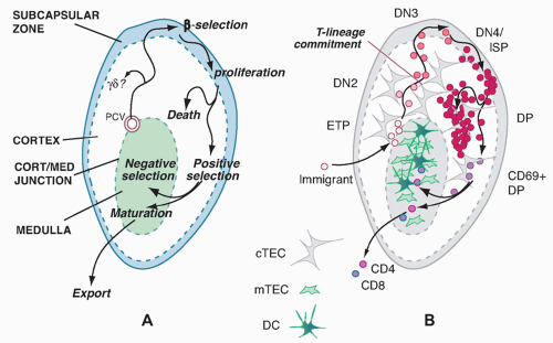
The major known site for T-cell development in all jawed vertebrates is the thymus.
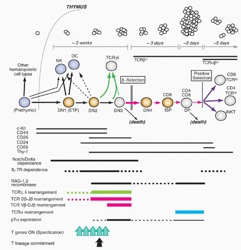
1 The thymus does not harbor its own long-term source of precursors but is continuously populated throughout life by new precursors that are replaced as their descendants complete maturation and selection. These precursors migrate to the thymus from bone marrow or fetal liver when they are still multipotent. The thymus not only enforces their commitment to a T-cell fate and T-lineage differentiation but also provides a proliferation-inducing environment. It is estimated that the numbers entering the thymus per day are quite modest, on the order of < 100 per day for a typical young adult mouse.2,3 The descendants of that small cohort of input cells expand over a 2-week period to a cohort of cells that have undergone T-lineage commitment, TCR gene rearrangement, and TCR expression, eventually yielding ˜50 × 106 new thymocytes a day before their proliferation stops.


Major Subdivisions Based on CD4, CD8, and T-Cell Receptor-&agr;&bgr; Status

The steps of T-cell differentiation in the mouse thymus are summarized in Figure 13.1.3,4,5,6 Important landmarks for the process are provided by the expression patterns of the TCR coreceptors, CD4 and CD8, and the timing of rearrangement of the TCR-coding genes themselves.

Cells in the early stages of development in the thymus are CD4- CD8- (“double negative” [DN]) and cannot yet express TCR. While the DN stages can be subdivided (described subsequently), the cells in all the DN stages are mostly determining their commitment to a T-cell fate, proliferating, and preparing for their first expression of TCR complexes. With successful expression of a TCR&bgr; chain, they turn on CD4, CD8&agr;, and CD8&bgr; genes together. The resulting CD4+ CD8&agr;&bgr;+ “double positive” (DP) cells are highly distinctive to the thymus, not found among mature T cells in peripheral lymphoid organs, and are diagnostic of a pivotal stage in T-cell development and survival. The DP cells in steady state constitute by far the major fraction of thymocytes (˜80%), and many of them come to express complete TCR&agr;&bgr; receptors as well. However, the overwhelming majority of these cells are fated never to go further in their differentiation. Only a small fraction are allowed to progress further through positive selection, to become either CD4+CD8- TCR&agr;&bgr;+ cells (“CD4 cells”) or CD4- CD8&agr;&bgr;+TCR&agr;&bgr;+ cells (“CD8 cells”). It is estimated that the cells have only about 3 days within the DP stage in which to achieve this success before their window of opportunity closes. Left in the cortex, the other DP cells of their cohort die “of neglect,” to be replaced by progeny of the next cohort of precursors, at turnover rates up to 50 × 106 per day.


T-Cell Receptor-&agr;&bgr; Rearrangement and Selection Checkpoints

The regulated process of TCR rearrangement and selection helps to explain why so many DP cells must be produced. TCR coding genes are assembled through a highly regulated but highly imprecise recombination process mediated by the recombinase complex RAG1/RAG2, the same recombinase that rearranges immunoglobulin VDJ gene segments in B cells. The imprecision is useful to add to the diversity of the eventual T-cell recognition pool, but it also results in many gene rearrangements that are out of reading frame or otherwise defective. To winnow out the useful cells, TCR gene rearrangement occurs in two sequential bursts, each followed by a quality control checkpoint at which all the cells with defective receptors can be eliminated.

The timing of different phases of recombination competence is determined by the specific activation and deactivation of the RAG1 and RAG2 gene products; the genetic loci on which their effects can be focused at any given time are determined by developmentally regulated unmasking via localized chromatin opening. For TCR&agr;&bgr; cells, the most numerous T cells in mice and humans, the TCR&bgr; gene must rearrange first, while the cells are still in the DN stage. The cells carrying out this rearrangement need to pause proliferation in order to allow the RAG complex to work, and most often they are never allowed to divide again unless they succeed in generating a good TCR&bgr; coding sequence through rearrangement at one allele. This strict condition is the “&bgr;-selection checkpoint.” Only cells passing this checkpoint are allowed to become DP cells, receiving a bonus of multiple rounds of cell division to expand the winners and dilute the stalled, dying losers. Through the process of &bgr;-selection, then, the cells also shift the gene loci that are accessible for rearrangement, losing the ability to rearrange their TCR&bgr; genes (and TCR&ggr; and TCR&dgr; genes) any further and acquiring the ability to rearrange their TCR&agr; genes.

Once they become DP cells, TCR&agr; rearrangement gets underway. As a successful in-frame TCR&agr; chain rearrangement occurs, the heterodimer TCR complexes of the new TCR&agr; chains with the previously generated TCR&bgr; chains become the next subject of testing at a second checkpoint. This second checkpoint is more draconian than the first, as the criterion for success here is not simply successful expression of a TCR&agr;&bgr; heterodimeric protein, but also the quantitative details of the recognition specificity of the new
receptor, as measured by its interaction with ligands in the thymic environment. The newly expressed TCR could fail to recognize anything, making it useless, or it could confer on the cell a dangerous autoreactivity that could lead to autoimmune disease. All of these failures must be eliminated before they appear in the mature T-cell population. The only successful cells are the ones that happen to have heterodimeric receptors with the right combination of functionality and low affinity for self-ligands. These are the ones that are allowed to become CD4 or CD8 cells through a process termed “positive selection,” with the CD4 cells generally becoming cytokine-producing “helpers” and the CD8 cells generally becoming “killers.” The positively selected cells permanently silence their RAG gene expression and undergo further maturation steps but little if any additional cell division before they are sent out to the periphery.


FIG. 13.1. Schematic Summary of T-Cell Developmental Stages. Top: Cartoons depict the approximate transit times and population expansion through the different stages. Middle: Recognized major stages of mouse T-cell development are shown, indicating CD4, CD8, and T-cell receptor (TCR) phenotype, and phases that are TCR&bgr;-dependent or TCR&agr;&bgr;-dependent. Development through positive selection of CD4, CD8, and natural killer T cells is shown; not shown are pathways to regulatory T cells, CD8&agr;&agr; innate-type cells, alternative &ggr;&dgr; pathways, or late-stage negative selection. White circles: nondividing or slowly dividing stages. Yellow circles: stages of extensive proliferation. Blue circles: prethymic cells and non-T developmental alternatives. Green arrows: enabled by TCR&ggr;&dgr; expression. Magenta arrows: enabled by TCR&bgr; expression. Purple arrows: enabled by TCR&agr;&bgr; expression and selection. Lower: timing of expression of additional markers, phases of TCR gene rearrangement, and specification and commitment events (see text for details).


The effects of the ordered TCR gene rearrangements and quality control checkpoints are seen clearly in the effects of different mutations on the progression of T-cell development. Figure 13.2A,B depicts the CD4/CD8 profiles and DN subsets in a normal young mouse thymus of about 2 to 3 × 10⁸ cells. Figure 13.2C,D shows the effects on thymus populations in mutant mice in which TCR gene rearrangement is impossible (eg, via mutation of Rag1 or Rag2 or Prkdc, the gene that encodes a deoxyribonucleic acid [DNA]-dependent protein kinase needed for rejoining of the gene segments during rearrangement [a very instructive mutant, Prkdcscid, causes severe combined immune deficiency]). Because TCR&agr;&bgr;-lineage cells are so predominant in the mouse thymus, the effect of mutations of Tcrb alone appears very similar (Fig. 13.2E). In contrast, mutations of the Tcra locus alone allow production of a full complement of DP cells, although CD4 and CD8 SP cells are completely missing (Fig. 13.2F).


FIG. 13.2. T-Cell Developmental Progression and Thymus Cellularity Controlled by T-Cell Receptor (TCR) Assembly. A,B: Normal wild-type mouse thymocytes: cluster of differentiation (CD)4/CD8 phenotype of all thymocytes (A), CD44/CD25 phenotype of double negative (DN) thymocytes only (B). C,D: Recombination-deficient mouse thymocytes: CD4/CD8 phenotype of all thymocytes (C), CD44/CD25 phenotype of the cells, which are all DN (D). Data for Rag2 knockout cells are shown, but Rag1 knockout phenotype is the same and similar phenotypes are seen for Prkdcscid or Tcrb-/- Tcrd-/- thymocytes. E: Cartoon depicting CD4/CD8 phenotype of TCR&bgr; knockout (&ggr;&dgr; still available). A few pseudo-double positive cells are generated through transient use of a &ggr;&dgr; TCR, but the great majority of TCR+ cells in these populations are TCR&ggr;&dgr;+ DN. F: Cartoon depicting CD4/CD8 phenotype of TCR&agr; knockout: development through &bgr;-selection is unimpaired, but no single positive populations are formed. Note the effects of each genotype on the cell numbers per thymus.


T-Cell Receptor-based Lineage Diversification for T-Cell Receptor-&agr;&bgr; Cells

The first functional distinctions between TCR&agr;&bgr; cell subsets apparently arise only during emergence from the DP stage. Positive selection itself probably triggers these divergences through distinctive modes of positive selection signaling, as we discuss in detail in a later section. The three main outputs of TCR&agr;&bgr; cell positive selection are CD4 cells, CD8 cells, and NKT cells, so called because they share some functional qualities and surface receptors with natural killer (NK) cells. DP cells first experiencing positive selection signals immediately turn on the activation marker CD69, start increasing the surface density of their newly validated TCR&agr;&bgr; complexes, upregulate CD5, and then begin the process that results in shutting off either CD8&agr;&bgr; or CD4 to generate TCR&agr;&bgr;-high “CD4 single positive” and “CD8 single positive” cells, respectively. As the cells arrive at one of these “single positive” (SP) phenotypes, they still retain expression of another marker of immaturity, the CD24 “heat-stable antigen” that is expressed from the early DN stages onward. However, they turn off both CD69 and CD24 as they reach completion of their functional maturity, in a process that takes 3 to 7 more days.7 NKT cells undergo a distinctive maturation sequence that is discussed separately in the following, but they too lose CD69 and CD24 as they mature.

The selection of these different subsets is based on TCR and coreceptor interactions with different ligands in the thymic environment. CD4 cells are positively selected by TCR and CD4 joint interaction with MHC class II molecules. CD8 cells are positively selected by TCR and CD8&agr;&bgr; joint interaction with MHC class I molecules. NKT cells are positively selected by nonclassical class I MHC molecules, using non-CD4, non-CD8 accessory molecules (signaling lymphocytic activation molecule [SLAM] family molecules) to assist in signal triggering. Although the primary trigger in each case is TCR&agr;&bgr;-ligand recognition, the effector programs of these different major lineages diverge significantly, through the consequences of using these different types of coreceptors to assist in “interpreting” the TCR signals. How this signal-dependent feature of T-cell maturation works is a major focus of later sections of this chapter.


Negative Selection of Autoreactive T-Cell Receptor-&agr;&bgr; Cells

Many cells with TCR that might be dangerously autoreactive can be eliminated at the DP stage, but another crucial period for this aspect of quality control is after positive
selection. As newly generated single-positive cells display increased surface density of their TCR&agr;&bgr;, raising their avidity for MHC-bound ligands, those that have strongly autoreactive receptors are generally killed off by interaction with specific subsets of thymic stromal cells in a process called “negative selection.” This continual purging of the newly made T-cell recognition repertoire is crucial to avoid autoimmune disease, as important mouse mutations and human disease models reveal. An alternative pathway to defeat autoreactivity, at least among CD4+ cells, is to alter the functional subtype of the autoreactive cells, to program them for function as tolerogenic regulatory T cells before they even have a chance to leave the thymus. Generation of such “natural Tregs” (nTregs) is but one case where TCR interaction properties direct the cells to a particular T-cell functional class.


Generation of T-Cell Precursors in the Double Negative Compartment

For years, the least well understood set of thymocytes was the DN cells. The DN-stage cells comprise only a small fraction of thymocytes in steady state, generally less than 5%, but in fact represent the generative compartment of the thymus in which the largest number of total cell cycles takes place. Profound developmental transformations also occur during transit through the DN stages.

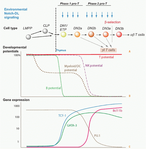
Understanding the basis of T-cell specification became possible only through the discovery of markers that could subdivide the DN compartment into successive stages (reviewed in refs. 2,3). The earliest stages of precursor differentiation within the thymus are represented by DN (CD4- CD8- TCR- or sometimes CD4low) cells that express high levels of the growth factor receptor Kit and the activation/adhesion molecule CD44 but low levels of CD25, termed Kit-high DN1 cells or early T-cell precursors (ETPs). Then, these cells turn on expression of CD25, signaling their definitive entry into the T-cell pathway, and the resulting Kit+ CD44+ CD25+ cells are called “DN2” cells. Both the DN2 and ETP stage cells proliferate, and IL7R is increasingly expressed in the DN2 stage. The DN2 cells then progress into the “DN3” stage (CD25high but now CD44low and Kitlow), and here proliferation slows or stalls, RAG protein levels rise, and efficient TCR&bgr; rearrangement can proceed. Development terminates at the DN3 stage unless the cells can pass &bgr;-selection or &ggr;&dgr;-selection.

Successful TCR&bgr; expression and passage of &bgr;-selection enable the cells to restart proliferation, shut off expression of CD25, and pass through a transitional “DN4” or “preDP” stage (CD25low, CD44low, Kitlow). They then proceed to acquire CD8 and CD4 on the cell surface, sometimes CD8 slightly before CD4 (“immature SP”), and finally culminate their differentiation into DP cells by finishing their proliferation (large DP to small DP transition). This extended &bgr;-selection-dependent transition from DN3 to small DP cells creates a unique regulatory state in the resulting DP cells that prepares them for selection. Notably, although some of these cells may be positively selected as described previously, only a minority may ever divide again until after they have left the thymus.


Distinctive Paths for T-Cell Receptor-&agr;&bgr; Cells

T cells that will use TCR&ggr;&dgr; instead of TCR&agr;&bgr; actually follow a special program or set of program options from the DN2 stage on. Whereas TCR&bgr; rearranges strictly before TCR&agr; and mostly in DN3 stage, the TCR&ggr; and TCR&dgr; genes can rearrange in parallel within the DN2 or DN3 stage. Thus, a RAG1/RAG2+ cell can in some cases acquire successful inframe rearrangements of both TCR&ggr; and TCR&dgr; genes before it is successful with TCR&bgr; alone. As a result, newly TCR&ggr;&dgr;+ cells arising within the DN2 or DN3 population leave the TCR&agr;&bgr; lineage mainstream, undergoing a &ggr;&dgr;-selection process that is different from &bgr;-selection. The &ggr;&dgr;-selected cells generally keep CD4 and CD8 silent, shut off RAG1/RAG2 permanently without promoting TCR&agr; rearrangement, and limit their proliferation to a much smaller number of cell cycles than &bgr;-selected cells.

It is not clear yet whether all TCR&ggr;&dgr; cells must go through two sequential checkpoints analogous to &bgr;-selection and positive selection. Like TCR&agr;&bgr; cells, TCR&ggr;&dgr; cells are now understood to emerge in multiple functional subtypes, and we will discuss in a separate section possible ways that these subtypes become differentially programmed.


CONTEXT OF T-CELL DEVELOPMENT


Anatomical Organization of T-Cell Development


Major Thymic Domains

The structure of the thymus plays a substantial role in the ordering and tempo of the steps in T-cell development.3 The epithelial framework of the thymus is a derivative of the fetal pharyngeal endoderm, emerging from the third branchial pouch in midgestation and only later descending from the neck region to its later position lying just over the heart. At its peak of function, the thymic epithelium looks very different from most endodermal epithelia, as it has become a highly porous, “lacy” three-dimensional lattice with few tight junctions, no obvious apical or basal polarity, and most of the spaces between epithelial cells crammed with developing lymphocytes. The structure is not uniform, as each lobe of the thymus is organized into cortical (outer) and medullary (inner) regions. The epithelial lattice appears quite open in the cortex but more compact in the medulla, where the lymphocyte:epithelial cell ratio is much lower. These different regions contain distinct epithelial cell types and different nonlymphoid hematopoietic cell types in addition to the developing T cells (Fig. 13.3).


Ordered and Checkpoint-Controlled Migration Pathways for Developing T Cells

Multipotent precursors follow a distinctive traffic pattern through the thymus8 (see Fig. 13.3). They enter through postcapillary venules at the cortical/medullary border, and they spend the bulk of the ETP stage proliferating quite close to this boundary. Then, as they enter the DN2 stage, they begin to migrate centrifugally, toward the outer cortex. They are thought to reach DN3 stage in the outer sector of the cortex and undergo the burst of proliferation triggered by &bgr;-selection at the extreme periphery of the cortex, just under the capsule and far away from the initial site of
precursor entry. Then, the DP cells generated during this proliferation start to fall back down through the cortex, covering the same ground as the DN cells that were their precursors, but now traveling in the opposite direction. The sheer number of these DP cells dominates the population in the cortex: in a healthy young adult mouse thymus, for the ˜2 × 10⁴ DN2 cells migrating toward the outer cortex, there are over 1.5 × 10⁸ DP cells falling back toward the medulla. Thus, the DN cells must work their way up through the vast excess of DP cells as they acquire T-cell character, and every other cell type in the cortex is embedded in a slow-moving sea of DP cells. As discussed in the next major section, this countercurrent migration may play an important role in helping the earliest precursors to receive inductive signals efficiently from the epithelium without competition.


FIG. 13.3. Anatomical Compartments of the Thymus and Migration Pathways of Developing T-Cell Precursors. A: Major compartments and migration pathways are depicted with key checkpoints in T-cell development indicated. Cort/med junction, cortical-medullary junction; PCV: postcapillary venule (immigration portal). B: Phenotypes of T-cell precursors (pink, red, violet) and stromal cell types in different compartments. cTEC, cortical thymic epithelial cells; DC, dendritic; mTEC, medullary thymic epithelial cells. For clarity, the “outbound” path from ETP to DN3 is separated from the “inbound” path from DN4 to CD4 or CD8 single positive maturation, but in vivo the “outbound” transit is surrounded with the “inbound” cells.

Despite their huge numbers, the DP cells are normally denied entry into the medulla. Positive selection signals are needed to induce new chemokine receptor expression (CCR7) on the fortunate minority of DP cells, and these are the only ones that can plunge into the medulla to continue their differentiation.2,9 After further maturation, once all signs of TCR signaling subside, the cells are finally allowed to upregulate emigration receptors that will allow them to leave the thymus entirely. It is also in the medulla, however, that even more stringent testing for TCR autoreactivity and purging of autoreactive cells takes place. Ultimately, less than 5% of the cells from each cohort of DP cells emerge from the thymus alive.


Thymic Anatomy Orders the Presentation of Inductive and Selective Signals

The pathway of T-cell precursors through the thymus helps to order their exposure to key differentiation-inducing stimuli.2,3,9 The cortical epithelium expresses high levels of integral membrane cell surface ligands for the Notch signaling receptor, mostly Delta-like 4 (DLL4, or DL4). This is important because Notch-Delta interactions play a central role in specification of T-cell progenitors from uncommitted precursor cells, as discussed in detail in the following section. In addition, the epithelium closest to the cortical-medullary junction makes the highest concentrations of the cytokine IL-7, which supports both early and late T-cell development. Epithelial cells expressing Kit ligand appear to be present in multiple domains of the thymus. As the precursors enter the thymus, therefore, they can rapidly engage in Notch-DLL4 signaling interactions in the presence of maximal concentrations of IL-7 and Kit ligand. These are ideal conditions for proliferation in the ETP and early DN2 stages. As the cells move outward, they encounter decreasing IL-7, which enables them to slow proliferation and begin to carry out efficient TCR gene rearrangement.

Thymic epithelium is one of the rare epithelia in the body that can express MHC class II molecules, which are normally limited to hematopoietic APCs, as well as the more broadly expressed MHC class I cell-surface molecules. Because TCR interactions with class II MHC are crucial for positive selection to CD4 fates and TCR interactions with class I MHC
usually direct selection to CD8 fates, this expression pattern makes it possible for the cortical epithelium to support positive selection to both CD4 and CD8 fates. At the cortical/medullary junction, there is a high concentration of thymic macrophages, which can rapidly dispose of the dead cells that are generated in large numbers by TCR-dependent selection every day.

Medullary thymic epithelium is distinctive in an additional way.9a Not only does it express class I and class II MHC, but also it is specialized for the detection and elimination of highly autoreactive cells. Medullary epithelial cells mature to an end stage in which they lift the repression of multiple silent loci in their genomes, genes that have no function in the thymus per se and would normally be expressed only in other organs in the body. These diverse tissue-specific self-antigens, presented on class I or class II MHC, are offered as “bait” to any newly positively selected cells whose receptors might recognize them. The presentation of these molecules is further enhanced by special populations of dendritic cells (DCs) in the thymic medulla, and these help to stimulate any autoreactive T cells to commit suicide. As we will see in the following sections, these medullary cell types can also suppress autoreactive T-cell precursors through an alternative mechanism, by directing their differentiation into nTregs.


Variations of T-Cell Development: Ontogeny and Species Differences

T-cell development in the thymus is common to all jawed vertebrates.10 A thymus, originating embryologically in the neck region and presenting Notch ligands to RAG1/RAG2+ lymphocyte precursors that turn on TCR gene expression, is found from cartilaginous fish to bony fish and amphibia through birds and all mammalian species. There is even an apparent equivalent structure newly discovered in the lamprey, a jawless vertebrate that probably shared its last common ancestor with mammals shortly after the Cambrian explosion (˜500 million years ago).11 Despite important commonalities, there are significant differences between T-cell development in mice and in other vertebrates, and even between mice and other mammals. In addition, even within the mouse, T-cell development undergoes significant changes between the first wave of production that occurs in the fetus and later waves of T-cell development in fetal and postnatal life, described subsequently. Thus aspects of T-cell development can be flexible even though they produce similar T-cell populations as outputs.


Ontogeny: Distinctive Features of the Mouse Embryonic Thymus

When the thymus rudiment is first formed in midgestation in the developing neck region of the embryo, it is not vascularized. Thus, the first wave of hematopoietic precursors that enter the thymus do so by migrating across the mesenchyme and penetrating the thymic rudiment from the outside.

There are two distinctive things about this first wave of thymic lymphopoiesis. First, the precursors of the first wave are intrinsically somewhat different from the cells that will populate the thymus in later waves. Descendants of early intraembryonic hematopoietic precursors, they have a unique capability to give rise to certain specialized types of TCR&ggr;&dgr; cells as well as more conventional kinds of TCR&ggr;&dgr; cells and TCR&agr;&bgr; cells.12,13 Second, the thymus epithelium itself is naïve in the sense that it has not yet been affected by signals from lymphocyte gene products, nor dilated into an open mesh by lymphoid proliferation. The medulla is not evident yet as a distinctive domain, and the spatial distribution of cortical epithelial products like IL-7 and Notch ligands is not necessarily ordered as it will later become.14,15

One result is that differentiation occurs much faster in the fetal mouse thymus than in the adult. Passage from DN1 to DN3 stage occurs within ≤ 2 days from embryonic gestation days E12.5 to E14.5 instead of 7 to 10 days in adults.16,17 Passage to DP occurs just 2 days later, requiring about 4 days instead of ˜2 weeks overall.3 The fetal-specific types of TCR&ggr;&dgr; cells are in fact produced as the first wave of mature T cells, by E15.5, even before DP cells emerge. Finally, the gene expression requirements for T-cell development in the fetal thymus are subtly different than in the adult (eg, a more stringent requirement for transcription factors Ikaros and E2A, and a reduced requirement for transcription factor TCF-1 and the IL-7R).

It is interesting that there is generally reduced TCR sequence diversity in the fetal T cells as compared to those made after birth. Normally, TCR sequences have two sources of variation: the combinatorial diversity based on the particular V, D, and J segments that are rearranged, and the sloppiness of cleavage and addition of untemplated nucleotides at the recombination breakpoints (ie, junctional diversity). One enzyme, terminal deoxynucleotidyl transferase (encoded by Dntt), is responsible for much of the junctional diversity. It is normally turned on in the earliest stages of prethymic precursor development and expressed throughout T-cell development until positive selection. However, the waves of lymphoid precursors developing in the fetus do not express it, and this situation only changes around the time of birth. As a result, some TCR types expressed in fetal thymocytes including the “first wave” TCR&ggr;&dgr; receptors are essentially invariant, with highly predictable reading frames, even though the genes that code for them are assembled by RAG-mediated recombination. It seems likely that these TCR specificities have some evolutionary selective advantage for the young mouse.

The production of TCR&ggr;&dgr; cells as the initial wave in the fetal thymus is remarkably shared between different classes of vertebrates: one of the initial reports of first-wave TCR&ggr;&dgr; cells (then called “TCR1 cells”) was in chickens.18 In the mouse, there is considerable evidence that the first-wave cells are distinctive in more ways than their use of a particular TCR&ggr;&dgr; receptor complex, as discussed in the section on &ggr;&dgr; cells.


Evolutionary Conservation of Diverse T-Cell Lineages but Flexibility of Roles

Focus on the mouse system may in fact handicap our ability to perceive the roles of TCR&ggr;&dgr; cells, because mice and humans are the two known mammalian species with the
most limited systemic use of TCR&ggr;&dgr; cells. In fact, an &agr;&bgr;/&ggr;&dgr; distinction between T-cell subclasses is an ancient feature of vertebrate lymphopoiesis. Even cartilaginous fish like skates and sharks have well-defined &ggr;&dgr; and &agr;&bgr; TCR complexes that are used by T cells in different anatomical domains.19 This suggests that two TCR-defined branches of T-cell development have been coselected for persistence over > 400 million years of vertebrate evolution. Major divisions within the TCR&agr;&bgr; lineages, between CD4+ helpers and CD8+ killers, are similarly conserved among all bony vertebrates. Thus although the genes for immunological receptors themselves undergo rapid evolutionary change and reorganization, the underlying programs for lymphocyte development appear to be ancient and conserved.


Evolutionary Flexibility at Close Range: Mouse versus Human

Most comparative information is only available for mouse and human T-cell development. The outlines of T-cell development in the thymus are strikingly similar in these closely related mammalian species.20,21,22 However, many details of T-cell development have proven to be different between man and mouse. The cell surface markers that define stages of human T-cell development are different from their mouse counterparts, and even some of the same molecules are expressed in different developmental patterns, as shown in Table 13.1. One surprising difference concerns the quantitative influences of different doses of Notch signaling on TCR&agr;&bgr; as compared to TCR&ggr;&dgr; cell production.23 While most of the major features of development are similar, this kind of variation is a caveat against overgeneralizing data from the mouse.


TABLE 13.1 Comparison of Markers in Human and Murine T-Cell Development


Stage


Mouse Phenotype


Human Phenotype


ETP-like:


CD44+ c-Kit+


CD34+ CD38lo CD7-


Intrathymic T/DC/NK precursors


CD25-


CD1a-


Specification stage


CD44+ c-Kit+


CD34+ CD38+ CD7+


(DN2-like)


CD25+


CD1a-


Committed


CD44+/low c-Kitint


CD34+ CD38+ CD7+


T precursors


CD25+


CD1a+


TCR&dgr;, &ggr;, &bgr;


CD44+/- c-Kit+/-


CD34+ CD38+


rearrangement: &agr;&bgr;/&ggr;&dgr; precursors


CD25+ (DN2, DN3)


CD1a+ CD4-/+


&bgr;-selection


CD44- c-Kit-


CD34+ CD38+


CD25+ CD4-CD8-(DN3) to CD25- CD4- CD8-


CD1a+ CD4+to CD4+ CD8&agr;+ (CD4 ISP, early DP)


TCR&agr;


CD25- CD4+


CD4+ CD8&agr;+ CD8&bgr;+


rearrangement


CD8&agr;&bgr;+ (DP)


(early DP to DP)


Mature cells


TCR/CD3hi and CD4+ or CD8&agr;&bgr;+


CD1a-TCR/CD3hi and CD4+ or CD8&agr;&bgr;+


CD, cluster of differentiation; DC, dendritic cell; DN, double negative; DP, double positive; ETP, early T-cell precursor; NK, natural killer cell; T, T cell.


Data from reviews by Blom & Spits283 and Taghon & Rothenberg.22


Creating an In Vitro Context for T-Cell Development

The three-dimensional structure of the thymus was for many years a challenge to the full definition of T-cell developmental mechanisms. It was clear after many attempts that dissociated thymic epithelial cells could not reconstitute thymic function if plated in monolayer culture, and thus it seemed that the migration of precursors through organized three-dimensional domains would be a crucial aspect of T lymphopoiesis. Redox conditions also seemed crucial for thymic function. There were two early breakthroughs in experimental dissection of T-cell development. First was the generation of fetal thymic organ cultures, which were found to require a high oxygen concentration to work, either by plating a thymic lobe at an air-medium interface, or by pumping up to 70% oxygen into the incubator in which a submerged fetal thymic lobe was cultured.24,25 Second was the discovery that in these excellent culture conditions, even dissociated, purified thymic stromal elements could regenerate a competent thymic microenvironment if mixed together in a reaggregate fetal thymic organ culture. The development of reaggregate fetal thymic organ culture has allowed the separate roles of distinct epithelial and hematopoietically derived elements of the cortical and medullary environments in positive and negative selection to be rigorously demonstrated, as discussed in later sections.26 Both fetal thymic organ culture and reaggregate fetal thymic organ culture systems promote T-cell development from prethymic precursor stages all the way through positive and negative selection to generate functional mature T cells.

The fetal and reaggregate thymic organ cultures both make use of natural thymic stromal elements, which makes them close to physiological conditions, but also a logistical challenge. Furthermore, the organ cultures still re-form into closed structures, making the thymocytes developing within them hard to count, track phenotypically, or experimentally manipulate. Finally, these organ cultures remain very small, hampering use for molecular biology.

Against this background, the development of stromal monolayer coculture systems for T-cell differentiation has revolutionized the field. Two lines of research converged to make this possible. First, Nakano, Kodama, and Honjo developed OP9 bone marrow stromal lines from myeloid colony stimulating factor-deficient mice (Op, osteopetrotic mice),27 which proved to be ideal for supporting lymphoid development. However, only B and NK lymphocytes, not T cells, develop on these original OP9 stromal cell lines. Second, crucial experiments by the groups of Freddy Radtke and Warren Pear showed that environmental activation of Notch signaling is crucial to induce hematopoietic precursors to become T cells.28,29 Engineering OP9 cells to express a potent Notch ligand, Schmitt and Zuniga-Pflucker developed the OP9-DL1 stromal line to support T-cell development.30


This created robust, highly efficient, high-yield conditions for driving and supporting T-lineage development in vitro from a wide range of uncommitted hematopoietic progenitor types, while keeping the cells accessible, easily monitored, and easily transferable throughout.

The OP9-DL1 system and variations of it have made possible enormous advances in understanding of the early, Notch-dependent stages of T-cell development. This system exposes to experimental dissection all the stages from initiation of the T-cell developmental program through commitment and up to the DP stage (ie, all the stages that DN precursors would normally undergo during their “outward bound” migration through the thymic cortex). It has also opened up the ability to track and experimentally perturb early stages of human T-cell development, starting from cord blood precursors, in a direct parallel to early mouse T-cell development.23 However, the “inward bound” phases of T-cell development after &bgr;-selection or &ggr;&dgr;-selection appear to depend on special properties of thymic epithelial cells that remain to be duplicated by OP9-DL1 or any stromal coculture.


EARLY LINEAGE CHOICES AND COMMITMENT


Notch and the Specification of T-Cell Identity

Notch signaling is crucial for induction of the T-cell program, for progression from stage to stage of the specification process, and for maintenance of viability of the developing T cells throughout the DN stages, all the way to &bgr;-selection. DN thymocytes gain privileged access to Notch ligands in the environment despite the large excess of DP cells. This is because the DP cells “deny themselves” the chance to bind efficiently with Delta-class Notch ligands in the environment by downregulating key Notch modifier genes, Lfng and Mfng, during &bgr;-selection,31 which are needed to compete for stromal DLL4 binding. Thus, in a healthy normal adult thymus, the masses of DP cells actually insulate individual DN cells from competition with each other, allowing Notch triggering to be renewed repeatedly from ETP to DN3 stage.

Notch-DLL4 signaling activates the regulatory cascade that turns on T-lineage genes from ETP stage to DN2 stage to DN3 stage. T-lineage-specific signaling mediators and invariant TCR components are upregulated starting by the DN2 stage or in the DN2-DN3 transition. The IL-7R (Il7r gene) is fully turned on by DN2 stage to support proliferation of the precursors as they are acquiring their T-cell identity. Between DN2 and DN3 stage, RAG1 and RAG2 recombinases and multiple TCR signaling complex components and mediators of TCR signal transduction, including the kinases Zap70 and Itk, are all induced or upregulated. The specialized surrogate T-cell receptor &agr;-like chain, pre-TCR&agr; (pT&agr;, encoded by the Ptcra gene), is also turned on to provide a partner for complex formation with any successfully-encoded TCR&bgr; chain. Thus, the cells that reach the DN3 stage are already fully armed with substantial levels of the T-cell specific components that will be needed to transduce signals through the TCR, as soon as they have made a productive TCR gene rearrangement.

Notch signaling accomplishes this large-scale transcriptional mobilization by first activating the expression of several T-lineage transcription factors that can help the cells to open up previously silent genetic loci.32,33,34 The most important T-cell-specific members of this group are GATA-3 (encoded by the Gata3 gene) and TCF-1 (encoded by the Tcf7 gene). These T-cell factors collaborate with the Notchactivated transcription factor RBPJ&kgr; (also called CBF1 – suppressor of hairless – Lag-1 = CSL), and together they interact with a set of transcription factors that is already present and active in multilineage hematopoietic progenitors. These preexisting factors include “E protein” basic helix-loop-helix factors E2A (encoded by Tcf3 = Tcfe2a) and HEB (Tcf12), Ikaros family members like Ikaros (Ikzf1) and Helios (Ikzf2), Runx family factors like Runx1 and Runx3, Ets family factors including PU.1 (SPI1 in human, Sfpi1 in mouse) and Ets1, and other key hematopoietic factors like Myb and Gfi1.35 The knockout of any one of these factors or factor families is sufficient to abort early T-cell development.

This complexity is important to acknowledge because despite the essential role of Notch, no one transcription factor alone turns on the whole T-cell program in a single stroke. In fact, different factors collaborate with each other in a balanced way throughout the DN stages, to coordinate differentiation with the right degree of proliferation before the cells reach the DN3 stage. Furthermore, the E proteins E2A and HEB not only regulate T-cell identity genes, but also serve throughout T-cell development to enforce TCR signaling checkpoints, undergoing cycles of inhibition and reactivation in response to TCR stimulation. We will see others of these factors that also have different, dose-dependent roles later on, working with or against each other to cause one T-cell lineage to diverge from another.


Options, Precursors, and Stages

The cells that populate the thymus are not yet committed to a T-cell fate when they arrive. Elegant single-cell culture experiments using either adapted organ culture systems or stromal coculture systems show that the same individual cells that can give rise to T cells would still be able to generate NK cells, myeloid cells, and DCs under other conditions, when they arrive in the thymus.36,37,38,39 Several lines of research also imply that many of them are initially able to give rise to B cells.40,41 The commitment process is the process through which the developing T cells decide permanently against these other options.42

Given how similarly T and B cells rearrange their immunoreceptor genes and observe developmental checkpoints, it is not surprising that B-cell potential should be present in T-cell precursors. It is notable how early B-cell potential is lost—soon after the thymus-settling precursors enter the thymus; many experiments even imply that access to the B-cell fate can be lost even before thymic entry.43,44,45 However, after losing the B-cell option, DN thymocytes maintain a clearly demonstrable ability to shift to a myeloid fate, either macrophage or granulocyte, and this persists into the DN2 stage.37,38,39 As long as the cells remain in an intact thymus,
these possibilities remain latent and development progresses overwhelmingly toward a T-cell fate, but all that is needed to reveal these options is to remove the cells from the thymus and offer them cytokines that support myeloid cell survival. Even into the DN2 stage, some T-cell precursors also appear to be able to develop into mast cells, yet another nonlymphoid cell type.46 The cells that start the T-cell program thus begin with many alternative possibilities.

A great deal of work has been carried out to link the developmental repertoire of newly arrived thymic immigrants with uncommitted cells in the bone marrow that are likely to be their precursors. This lively field of research has shown that thymic immigrants can be derived not only from cells that were mostly committed to some kind of lymphoid fate prethymically (ie, common lymphoid precursors), but also from cells that were broader-spectrum lymphoid-myeloid multilineage precursors (lymphoid-primed multipotent precursors).43,47,48,49,50 Furthermore, even cells that have already begun to be restricted to a nonlymphoid, myeloid pathway (granulocyte-macrophage precursors) can also develop into T cells, if transferred from the bone marrow into a thymic environment or OP9-DL1 coculture.51 Many T-cell precursors in the thymus in fact show evidence of having earlier activated a myeloid lineage differentiation gene, lysozyme M.52 Thus, the dominant developmental program imposed by the thymic environment is a kind of boot camp that can force cells of different developmental origins to converge on a common T-cell fate, erasing signs of their previous distinct identities.


Mechanisms of Commitment

The thymus uses different mechanisms to eliminate various fate alternatives for the cells. One of these is the environmental signal through Notch itself. As long as thymic immigrants and even ETP and DN2 cells are being exposed to Notch ligands in the thymic microenvironment, they cannot exhibit any alternative potentials efficiently. As differentiation progresses, however, the cells also give up the ability to access these alternatives under any conditions (Fig. 13.4).

Early B-cell development in particular is intensely inhibited by Notch signaling. Not only are precursors prevented from initiating B-cell development in the presence of Notch- Delta signals, but also exposure to Notch signals rapidly strips them of the capability to enter the B-cell pathway even if Notch-Delta signaling is removed.40 Perhaps because early B and T cells share many other early developmental requirements —the cytokine IL-7, the role of stroma, prominent use of E proteins among their crucial lymphoid transcription factors—the Notch signal is uniquely important to make these two developmental programs mutually exclusive. The thymus is too good an environment for B-cell development in every other way; in fact it fills up with developing B cells if Notch-DLL4 signaling is defective.41,53,54

The choice against other alternatives takes longer and involves more conditionality. The myeloid and DC-enabling transcription factor PU.1 is normally expressed into the DN2 stage and probably explains the access to myeloid potential up to this point. Under normal conditions, however, the thymus is not a good source of myeloid-cell growth factors or differentiation factors, and so myeloid development is a low-efficiency path in the thymus regardless of the programming of the cells. In addition, active Notch signaling appears to add a further conditional obstacle to myeloid development through interference with the action of myeloid transcription factors.52,55 This mechanism still allows cells to keep myeloid potential as a latent option through several cell divisions, accessible if they are removed from the thymus. But eventually the T-cell developmental program reaches a stage when the myeloid-enabling transcription factors themselves are repressed. This occurs in the later DN2 stage, and it marks the point when the myeloid paths too are permanently cut off.

The most persistent fate alternative is the NK fate, which falls into a somewhat different category. The choice against becoming an NK cell appears to be made about the same time as the choice to forgo the DC fate option,36,56,57 but in fact one could argue that many CD8+ T cells never fully give up the NK possibility. There are many differences between NK cells and T cells: most obviously, the NK cells do not rearrange their TCR genes and utilize a substantially different antigen recognition system than T cells. Thus, any TCR+ cell would be classified as T rather than NK no matter how NK-like it was in other respects. In an apparent dichotomy, too, developing T cells require a class of transcription factors, the E proteins (E2A, HEB) that NK cells must inhibit: the E protein antagonist Id2 is an NK developmental requirement. However, NK cells mobilize effector programs that are almost completely congruent with those of effector CD8+ killer T cells. On persistent antigen stimulation, CD8 killer T cells turn on expression of NK-cell activation receptors as well as TCR and behave even more like NK cells in terms of promiscuity of killing. A recent gene knockout model suggests that only one transcription factor, Bcl11b, may stand between mature CD8 cells and NK-like behavior normally throughout their lifetimes as mature T cells.58 The loss, or reduction, of access to the NK program occurs at the specific stage of T-cell development, also in the DN2 stage, when Bcl11b is turned on for the first time.58,59,60

Bcl11b is turned on in a process that requires Notch signaling and TCF-1,61,62 possibly restrained until DN2 stage by other timing factors. DN2 cells that cannot turn on Bcl11b do not reach a normal DN2b or DN3 stage: instead, they keep prolonged access to myeloid fates and ongoing self-renewal programs as well as the NK program as long as cytokine levels permit.59,60 They are profoundly defective in generating TCR&agr;&bgr;-lineage thymocytes, DP, and later stages, as well.63,64 Thus, in addition to its special role in suppressing the NK-cell program, Bcl11b also appears to work in a gatekeeper capacity at the crux of the commitment process.

Commitment, therefore, involves at least three distinct mechanisms: the suppression of B-cell potential, the silencing of myeloid transcription factors, and the induction of Bcl11b. The last two of these happen roughly at the same point, in a transition that is now recognized to split the DN2 stage between uncommitted DN2a cells and committed
DN2b cells56,65 (see Fig. 13.4). DN2a cells have access to other fates and can survive when removed from Notch ligandrich environments; DN2b cells are committed and become acutely Notch signaling-dependent for viability. From commitment on, DN cells cannot survive deprivation of Notch ligands until they have successfully undergone &bgr;-selection or &ggr;&dgr;-selection. Transition to the DN2b stage also appears to prepare the way for shutting down precursor expansion and establishing the molecular conditions for enforcing the &bgr;-selection checkpoint.


FIG. 13.4. T-Lineage Commitment and Expression Patterns of Contributing Factors. A: Prethymic and intrathymic stages involved in T-lineage commitment. The thymus can be seeded by cells that retain myeloid potential (lymphoid-primed multipotent precursor) as well as by cells that have already reduced their access to this option (common lymphoid precursor). As shown in the figure, Notch-Delta signals are delivered to the cells repeatedly from their time of entry into the thymus until &bgr;-selection. B: Approximate times at which distinct cell-lineage options for T-cell precursors are excluded. Y axis represents remaining ability of T-cell precursor cells to enter alternative pathway at the indicated stages (relative units). C: Expression patterns of key regulatory genes noted in the text. Factors used in the T-cell program are depicted with solid lines, while PU.1, a factor used in multipotent progenitor, myeloid, and B-cell programs, is shown with a dotted line. Adapted from figures in Rothenberg42 and Rothenberg et al.279 Evidence for gene expression patterns reviewed in Rothenberg et al.32


PROTOTYPE OF A T-CELL RECEPTOR-DEPENDENT CHECKPOINT: &bgr;-SELECTION


&bgr;-Selection: Proliferation, Allelic Exclusion, and Creation of the Double Positive State

All TCR&agr;&bgr; cells, regardless of functional subset, are survivors and products of &bgr;-selection. The &bgr;-selection checkpoint is the first TCR-dependent control checkpoint that these cells encounter, and the &bgr;-selection process is the first TCR-dependent signaling response that they undergo. The outcome shapes the whole population upon which later selection and differentiation mechanisms must operate.

TCR&bgr; rearrangement and TCR-complex signaling are crucial for both the proliferation and differentiation events of &bgr;-selection. Thus recombinase-deficient (Rag1 or Rag2 knockout), DNA repair-deficient (eg, Prkdcscid, “SCID”), TCR&bgr; constant region knockout, or CD3&ggr; or &egr; knockout mice all leave T-cell development blocked at DN3 stage (see Fig. 13.2). In normal development, successful TCR V&bgr; joining to D&bgr;-J&bgr; joints in a productive reading frame is the limiting event, as CD3 chains begin to be synthesized and expressed on the surface at a low level irrespective of rearrangement. It is the ability to deliver a signal through these complexes that really drives &bgr;-selection, as both in vivo and in vitro a response that strongly mimics natural &bgr;-selection can be triggered even in RAG-deficient thymocytes if monoclonal antibodies are used to crosslink the CD3 chains on their surfaces to deliver an artificial signal.


Fulfilling the Requirements for &bgr;-Selection

By the DN3 stage, developing T cells are already under proliferative restraint, in part to make further development conditional and in part to enable the RAG1/RAG2 complex to work efficiently. Although many ETPs already have limited D&bgr;-J&bgr; TCR gene rearrangements due to low-level prethymic RAG expression,66 these partial rearrangements cannot encode functional receptors. By the DN3 stage, however, efficient recombination of V&bgr; segments to the joined D&bgr;-J&bgr; products begins, and some of these rearrangement products now encode functional TCR&bgr; chains.

The success of TCR&bgr; rearrangement is sensed by the cell when the newly made &bgr;-chain is assembled into a complex at the cell surface and transduces back a lowintensity signal through essentially the same kinase and adaptor mechanisms used in mature TCR signaling. Isolated TCR&bgr; chains cannot assemble with CD3 and TCR&zgr; chains on their own, and there is no TCR&agr; available yet, but the invariant pre-TCR&agr; chain provides a surrogate. The Ptcra gene does not require rearrangement and is turned on strongly as a direct Notch signal-induced gene by the DN3 stage. The pre-TCR&agr;/TCR&bgr; heterodimer thus forms a “pre-TCR” containing a full complement of CD3 and TCR&zgr;, which is readily transported to the cell surface to trigger signaling.

Unlike all later uses of the TCR&bgr;, this signaling does not depend on any component of ligand recognition from its heterodimer partner, but the pre-TCR&agr; chain can mediate spontaneous clustering that may help to trigger signaling.67,68,69 The rate-limiting criterion for successful &bgr;-selection is thus simply the structural integrity of the TCR&bgr; chain that allows it to participate in a well-formed signaling complex.


The &bgr;-Selection Response

Pre-TCR spontaneous signaling triggers a dramatic cascade of events. In hours, the cells not only begin to proliferate again but also set in motion a profound transformation. The cells become large blasts, almost 10 times the volume of most thymocytes. As they turn on CD4 and CD8&agr;&bgr; expression, and express the coreceptor CD28 for the first time, they shed their DN cell markers and everything that had sustained their viability as DN cells, losing CD25, IL-7R, and the last vestiges of Kit expression and also inactivating the Notch pathway as well.

The proliferation unleashed by &bgr;-selection is intense. In their 2 to 3 days of proliferation, the selected DN3 cells generate approximately 102-fold more DP cells. During this expansion, there are ample opportunities for clearing the cell surface of obsolete membrane proteins (eg, old CD25 molecules), clearing the nucleus of obsolete transcription factors (eg, the Notch-induced transcription factor Hes-1), and clearing the genome of obsolete states of chromatin modification.

During the proliferation triggered by &bgr;-selection, TCR gene rearrangement is temporarily suspended, as the cells downregulate Rag1 and Rag2 gene expression and inactivate RAG2 protein by cell cycle-dependent phosphorylation.70 RAG1 and RAG2 expression is restored by the time proliferation arrests in the DP stage, but by that time the cells have transformed the chromatin landscape in which the RAG1/RAG2 recombinase must find its targets. TCR&bgr; is now closed, establishing rigorous allelic exclusion for this TCR chain. TCR&agr; is now open for rearrangement, pioneered by some germline transcription as early as the late DN3 stage. At the same time, the potential for expressing TCR&ggr;&dgr; receptors is eliminated. TCR&ggr; genes become inaccessible, so that both germline transcripts from unrearranged loci and transcripts from any previously rearranged TCR&ggr; genes are silenced.71,72,73 TCR&dgr; genes, whether previously rearranged or not, are deleted as the TCR&agr; segments that straddle them rearrange. Thus, by the time proliferation stops, the DP cell is irreversibly confirmed in a TCR&agr;&bgr; lineage.


Creation of the Double Positive State: Setting the Stage for Positive and Negative Selection

With the first productive rearrangement of TCR&agr;, the developing DP cell will acquire the capacity to recognize unpredictable antigenic targets, including self-antigens. Until the new receptor specificity has passed quality control, a DP cell is therefore functionally disabled and “contained,” both within the thymic cortex and within a short default lifespan. DP cells not only lack emigration receptor expression but also acquire features that would cause them to be killed quickly if they did escape. As they pass through &bgr;-selection,
they downregulate their own MHC class I molecules and their surface glycoprotein sialylation74 pathways: they can even be purified on the basis of their resulting low MHC class I expression and preferential binding by the lectin peanut agglutinin. These changes make them into potential NK cell targets and ensure that they would be scavenged by macrophages via asialoglycoprotein receptors if they escaped. Also as &bgr;-selection begins, the antiapoptotic factor Bcl2 is repressed, replaced with the weaker or more conditional survival factors Bcl-xL and Bcl2A1.75,76 Even while they are still rapidly dividing, the emerging cells acquire an intense vulnerability to death signals, both glucocorticoid signaling and other strong signals.77

Cell surface expression of TCR&agr;&bgr; complexes are kept low on DP cells even when TCR&agr; chains are first expressed, partly due to uniquely high levels of Src-like adaptor protein (Sla gene product), which targets TCR&zgr; protein for rapid destruction.78 Nevertheless, DP thymocytes are, if anything, more sensitive to TCR-ligand interactions than mature cells with the same TCR&agr;&bgr; if an appropriate assay is used. Contributing factors may include their relative lack of sialic acid residues, thus reducing electrostatic repulsion, and their low expression of the negative feedback molecule CD5 that could otherwise damp their response to TCR&agr;&bgr; signals.79,80,81,82 Even though it is sensed, TCR signaling cannot turn on functional response genes like Il2 in DP cells. In part, this is because these cells have downregulated key activationtransducing transcription factors, c-Jun and c-Rel (for more information, see www.immgen.org/).83 &bgr;-selection also induces expression of the transcription factor, ROR&ggr;, from a distinctive promoter (ROR&ggr;t, also called RORC2), which actively blocks conventional effector responses even while maintaining Bcl-xL expression throughout the DP stage.84,85 ROR&ggr;t expression sets DP cells apart from all other major classes of thymocytes; however, it provocatively links them with Th17 cells in the periphery, with lymphoid tissue-inducing cells that organize lymph nodes in fetal life, and certain classes of newly recognized innate effector cells.86,87,88,89 The DP cells that emerge from the complex regulatory events of &bgr;-selection thus acquire a highly specialized physiology, and they are poised for selection.

The &bgr;-selection response is driven by the same major signaling pathways used in the activation of mature T cells. Lck, Ras pathway, PI3-kinase, and protein kinase C activation all play roles in the process, using virtually all the same adaptor molecules [LAT, Slp76 (Lcp2), GADS (Grap2)] that are used to coordinate signaling in positive selection later and in mature T cells.90,91,92 As in later stages of T-cell development, TCR signaling temporarily antagonizes effective activity of E proteins (E2A/HEB). A transient wave of immediate-early Egr factor activation results in transient upregulation of the E protein antagonist Id3, a response that will be echoed in positive selection later.93,94,95,96 As the cells reach the resting DP state, high-level E protein activity is restored.97 These factors cooperate with some of the same regulatory factors that are used in early T-cell development: initially Notch signaling, then the important T-cell transcription factor TCF-1.98,99,100,101 The fact that the cells emerging from this signaling response are idiosyncratic, functionally disarmed, short-lived DP thymocytes rather than robustly activated effector T cells must be explained as the result of the characteristic transcriptional and epigenetic regulatory state of the cells in this stage—the context within which the signaling pathways are sensed—rather than a unique biochemical pathway for the signals themselves.


A Clear and Present Danger: Creation and Enforcement of the &bgr;-Selection Checkpoint

The cell biology of the &bgr;-selection response is an obvious hazard for the organism. Intense polyclonal proliferation of this magnitude brings the cells close to a malignant state, and the program must include brakes to stop the proliferation as well as constraints to prevent it from being triggered inappropriately. In fact, most T-cell acute leukemias have precisely the phenotype of cells that cannot stop the &bgr;-selection response, continuously generating immature DP cells or DN-DP intermediates. Under normal conditions, a limiting factor for the process is the programmed downregulation of Notch responsiveness, as the extent of population expansion during &bgr;-selection is strongly influenced by Notch signaling.102,103 Indeed, by far the most dominant and consistent feature of T-cell acute lymphoblastic leukemias is the presence of gain of function mutations in the Notch1 locus, especially mutations that make Notch signaling ligand-independent.104 The result is particularly grave when the cells can join viability signals from pre-TCR complexes and Notch alike to proliferate without restraint.

Another dangerous combination is if the very rapid proliferation program induced by &bgr;-selection is superimposed on any continuing expression of hematopoietic progenitor self-renewal genes, inherited from earlier stages of T-cell specification, including SCL (Tal1), Lyl1, Lmo1, Lmo2, and Hhex.105,

Only gold members can continue reading. Log In or Register to continue

Stay updated, free articles. Join our Telegram channel

May 30, 2016 | Posted by in IMMUNOLOGY | Comments Off on T-Lymphocyte Developmental Biology

Full access? Get Clinical Tree

Get Clinical Tree app for offline access