T-Cell Antigen Receptors


T-Cell Antigen Receptors


Mark M. Davis

Yueh-Hsiu Chien


T-lymphocytes expressing &agr;&bgr; or &ggr;&dgr; T-cell antigen receptors (TCRs) are found together with B-lymphocytes in all but the most primitive vertebrate animals. These three cell types are the only ones that use random variable (V), diversity (D) in the case of TCR-&bgr; and -&dgr;, joining (J), gene rearrangement to generate diverse antigen receptors. During the last two decades, there has been a great deal of progress in identifying the molecules and genes of TCRs, and there is considerable information on their biochemistry and structure. While TCRs share structural and genetic similarities with B-cell antigen receptors (immunoglobulins [Igs]), they also possess a number of unique features pertinent to their specific functions. The first major difference was suggested by the experiments of Zinkernagel and Doherty, who found that cytotoxic cells specific for a viral antigen could only lyse infected cells that expressed a particular major histocompatibility complex (MHC) molecule.1,2 This phenomenon of “MHC-restricted recognition” is in marked contrast to the recognition of intact antigens by Igs.3,4 Later work demonstrated that what was being recognized by these T cells, which were of the &agr;&bgr; TCR type, were fragments of antigens or peptides bound to a characteristic groove in MHC molecules.5 These &agr;&bgr; TCRs are expressed on classical helper and cytotoxic T cells, which predominate in most lymphoid compartments (90% to 95%) of humans and mice.6 They are also expressed on natural killer (NK)T cells,7 regulatory T cells,8 and T cells in the mucosal sites such as the intestinal epithelial compartment (IEL).9 In most cases, the &agr;&bgr; TCR ligand is a peptide antigen bound to a class I or class II MHC molecule; but in the case of NKT cells, the antigen is a glycolipid bound to a nonclassical class I MHC molecule, cluster of differentiation (CD)ld.7

T cells bearing &ggr;&dgr; TCRs are less numerous than the &agr;&bgr; type in most cellular compartments of humans and mice (<5%). However, they make up a substantial fraction of T-lymphocytes in cows, sheep, and chickens.10 &ggr;&dgr; T cells coexist with &agr;&bgr; T cells but seem to be better represented in the mucosal compartments.10,11 Although &agr;&bgr; T cells perform most of the functions classically attributed to T cells, mice lacking &ggr;&dgr; T cells are clearly have a compromised immune defense indicating that &ggr;&dgr; and &agr;&bgr; T cells contribute to host immune defense differently.11,12 &ggr;&dgr; TCRs also recognize antigens directly, like antibodies, with no apparent need for antigen processing,13 at least in the most thoroughly studies cases. During the past few years, there have been considerable advances in our understanding of antigen recognition by &ggr;&dgr; T cells. This should lead to a better understanding of how &ggr;&dgr; T cells contribute to immune competence.


T-CELL ANTIGEN RECEPTOR POLYPEPTIDES

The search for the molecules responsible for T-cell recognition first focused on deriving antisera or monoclonal antibodies specific for molecules on T-cell surfaces. Ultimately, a number of groups identified “clonotypic” sera14 or monoclonal antibodies.15,16,17,18,19 Several of these antibodies were able to block antigen-specific responses by the T cells they were raised against or, when coated on a surface, could activate the T cells they are specific for. They were also able to immunoprecipitate 85,000 to 90,000 molecular weight (MW) disulfide-bonded heterodimers from different T-cell clones or hybridomas consisting of two 40,000 to 50,000 MW glycosylated subunits referred to as &agr; and &bgr;. Peptide mapping studies showed that there was a striking degree of polymorphism between heterodimers isolated from T cells of differing specificity, thus suggesting that these antigen recognition molecules might be akin to Igs.20,21

Work in parallel to these serologic studies exploited the small differences (approximately 2%) observed between B- and T-cell gene expression,22 and isolated both a mouse23,24 and a human25 T cell-specific gene that had antibodylike V, J, and C region sequences and could rearrange in T-lymphocytes.24 This molecule was identified as TCR-&bgr; by partial sequence analysis of immunoprecipitated materials.26
Subsequent subtractive cloning work rapidly identified two other candidate TCR complementary deoxyribonucleic acids (DNAs) identified as TCR-&agr;27,28 and TCR-&ggr;.29 It was quickly established that all antigen-specific helper or cytotoxic T cells expressed TCR-&agr;&bgr; heterodimers. Where TCR-&ggr; fit in remained a puzzle until work by Brenner et al.30 showed that it was expressed on a small (5% to 10%) subset of peripheral T cells together with another polypeptide, TCR-&dgr;. The nature of TCR-&dgr; remained unknown until it was discovered within the TCR-&agr; locus, between the V&agr; and J&agr; regions.31 Formal proof that the TCR-&agr; and -&bgr; subunits were sufficient to transfer antigen/MHC recognition from one T cell to another came from gene transfection experiments,32,33 and equivalent experiments have also been done with &ggr;&dgr; TCRs.34

As shown in Figure 11.1, all TCR polypeptides have a similar primary structure, with distinct V, D in the case of TCR-&bgr; and -&dgr;, J, and constant (C) regions exactly analogous to their Ig counterparts. They also share many of the amino acid residues thought to be important for the characteristic variable and constant domains of Igs.35 The C&bgr; region is particularly homologous, sharing 40% of its amino acid sequences with C&kgr; and C&lgr;. The TCR polypeptides all contain a single C region domain (versus up to four for Igs) followed by a connecting peptide. These usually contain the cysteine for the disulfide linkage that joins the two chains of the heterodimer (some human TCR-&ggr;&dgr; isoforms lack this cysteine and consequently are not disulfide-linked36). N-linked glycosylation sites vary from two to four for each polypeptide with no indications of O-linked sugar addition. C-terminal to the connecting peptide sequences are the hydrophobic transmembrane regions. These have no similarity to those of the IgH genes but instead have one (TCR-&bgr; and -&ggr;) or two (TCR-&agr; and -&dgr;) positively charged residues. As discussed later, these charged residues are critical for the association of the ligand-binding TCR polypeptides with the CD3 signaling polypeptides. This is important because the TCR polypeptides have very short cytoplasmic regions with no known role in signaling.


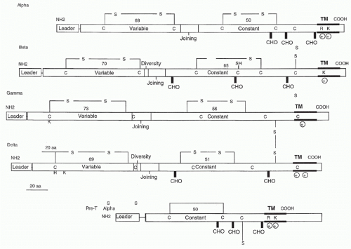
FIG. 11.1. Structural Features of T-Cell Receptors and Pre-T &agr; Polypeptides. Leader (L), variable (V), diversity (D), joining (J), and constant region (C) gene segments are indicated. Transmembrane and bold horizontal lines delineate the putative transmembrane regions; CHO indicates potential carbohydrate addition sites; C and S refer to cysteine residues that form interchain and intrachain disulfide bonds; R and K indicate the positively charged amino acids (arginine and lysine, respectively) that are found in the transmembrane regions.

A more recent member of the TCR polypeptide family is the pre-T&agr; chain, which serves as a chaperone for TCR-&bgr; in early thymocytes, similar to the role of &lgr;5 in pre-B cells.37 It was first identified and cloned by von Boehmer and colleagues.38 It has an interesting structure that consists of a single Ig constant region-like domain followed by a cysteinecontaining connecting peptide, a transmembrane region
containing two charged residues: an arginine and a lysine spaced identically to the TCR-&agr; transmembrane region. The cysteine in the connecting peptide is presumably what allows heterodimer formation with TCR-&bgr; and the similarity to TCR-&agr; in the transmembrane region is most likely to accommodate the CD3 polypeptides. Recently, Rossjohn and colleagues have presented a structure of the pre-T&agr; chain that is comprised of a single Ig-like domain that is distinct from the C domain of the TCR-&agr; chain; nevertheless, the mode of association between pre-T&agr; and TCR-&bgr; mirrored that mediated by the C&agr;-C&bgr; domains of the &agr;&bgr;TCR.39 The pre-TCR has a propensity to dimerize in solution, and the molecular envelope of the pre-TCR dimer correlated well with the observed head-to-tail pre-TCR dimer. This mode of pre-TCR dimerization enables the pre-T&agr; domain to interact with the V&bgr; domain through residues that are highly conserved across the V&bgr; and J&bgr; gene families, thus mimicking the interactions at the core of the &agr;&bgr; TCR’s V&agr;-V&bgr; interface. Disruption of this pre-T&agr;-V&bgr; dimer interface abrogates pre-TCR dimerization in solution and impaired pre-TCR expression on the cell surface. Their work suggests a mechanism of pre-TCR self-association that allows the pre-T&agr; chain to simultaneously “sample” the correct folding of both the V and C domains of any TCR &bgr;-chain, regardless of its ultimate specificity,39 which likely represents a critical checkpoint in T-cell development.

In both the mouse and humans, the cytoplasmic tail of pre-T&agr; is much longer than any of the TCR chains (37 and 120 amino acids, respectively), and the murine sequence contains two likely phosphorylation sites and sequences homologous to an SH3 domain binding region. These are not present in the human sequence, however, and so their functional significance is unclear.38 Thus, at least in principal, the murine pre-T&agr; molecule could function as signaling intermediate independent of the CD3 polypeptides, and it has recently been shown that at least one CD3 component (&dgr;&egr;, see the following) is not required for it to function normally in early thymocyte differentiation.40


CLUSTER OF DIFFERENTIATION 3 POLYPEPTIDES

Immunoprecipitation of the human TCR with anti-idiotypic antibodies after solubilization with the nonionic detergent, noniodet P-40 (NP-40), initially revealed only the &agr;- and &bgr;-chain heterodimer. However, the use of other detergents, such as digitonin or Triton-X100, revealed four other proteins.41,42,43,44 These are known as the CD3&ggr;, &dgr;, &egr;, and &zgr;. &ggr; and &dgr; form distinct heterodimers with &egr; within the TCR/CD3 complex (&ggr;&egr; and &dgr;&egr;), and &zgr; usually occurs as a disulfide-linked homodimer. In mouse T cells, NP-40 does not dissociate TCR heterodimers from CD3 molecules.44,45 In some cases, the &zgr;-chain can be part of a heterodimer in at least two forms. In mouse T cells, the &zgr;-chain can disulfide bond with a minor variant called the η (eta) chain.46,47 This latter chain is an alternate splicing variant of the &zgr;-chain gene.48 This alternatively spliced species of the &zgr;-chain is not found in significant quantities in human T cells.48 The second type of &zgr;-chain containing heterodimer contains the &ggr;-chain associated with the Fc&egr;RI (Fc&egr;RI &ggr;) and Fc&ggr;RIII (CD16) receptors.49,50 These CD3 subunits, in their various forms, are an integral part of TCR-mediated T-cell recognition because only they possess the immunoreceptor tyrosine-based activation motifs (ITAMs) that are necessary for cellular activation when the TCR engages ligand.


Characterization and Structural Features of the Cluster of Differentiation 3 Polypeptides

Figure 11.2 illustrates the principal structural features of the CD3 &ggr;, &dgr;, &egr;, and &zgr; polypeptides as derived from gene cloning and sequencing,42,51 and more recently by protein crystal structures of the extracellular domains of &ggr;, &dgr;, and &egr;.52,53,54,55 The extracellular domains of the &ggr;, &dgr;, and &egr; chains show a significant degree of similarity to one another. These domains retain the cysteines that have been shown to form intrachain disulfide bonds and each consists of a single Ig superfamily domain. The spacing of the cysteines in these domains produces a compact Ig-fold, similar to a constant region domain. The &ggr; and &dgr; subunits form distinct heterodimers with &egr; via highly conserved residues at the dimerizing interface.52,53,54,55 The connecting peptides of the CD3 &ggr;, &dgr;, and &egr; chains all contain highly conserved, closely spaced cysteines just before the membrane-spanning regions. These residues are likely candidates for the formation of interchain disulfide bonds and appear to play a role in the assembly of the CD3 and TCR polypeptides.52,56 The extracellular domain of the &zgr; chain consists of only nine amino acids and contains the only cysteine, which is responsible for the disulfide linkage of the &zgr;&zgr; homodimer or the &zgr; Fc&egr;RI&ggr; heterodimer. Each of the &ggr;, &dgr;, &egr;, and &zgr; polypeptides contain a conserved, negatively charged amino acids in their transmembrane region complementary to the positive charges seen in the TCR transmembrane regions.57,58,59,60

The cytoplasmic regions of the &ggr;, &dgr;, &egr;, and &zgr; chains are the intracellular signaling “domains” of the TCR heterodimer. Each of these molecules contains one or more amino acid sequence motifs that can mediate cellular activation.61 The intracellular sequences responsible for this activation are contained within an 18 amino acid conserved ITAM62 with the sequence X2YX2L/IX7YX2L/I. Both of the tyrosines in this motif are absolutely required to mediate signal transduction as mutation of either completely prevents the mobilization of free calcium or cytolytic activity.63 This sequence occurs three times in the &zgr; chain and once in each of the CD3 &ggr;, &dgr;, &egr;, and Fc&egr;RI &ggr; chains. There are also pairs of tyrosines present in the cytoplasmic domains of the &ggr;, &dgr;, &egr;, and &zgr; chains. This sequence motif is also present in the m&bgr;-1 and B29 chains associated with the Ig &bgr;-cell receptor and in the Fc&egr;RI &bgr;-chain but there are many more10 in TCR/CD3 than in any other receptors which use ITAMs. The tyrosines in these cytoplasmic sequences are substrates for the tyrosine phosphorylation that is one of earliest steps in T-cell signaling61 and is thought to occur aberrantly in nonproductive T-cell responses (eg, antagonism; see following). Serine phosphorylation of the CD3&ggr; also occurs upon
antigen or mitogenic stimulation of T cells64 and may play a role in T-cell activation as well.


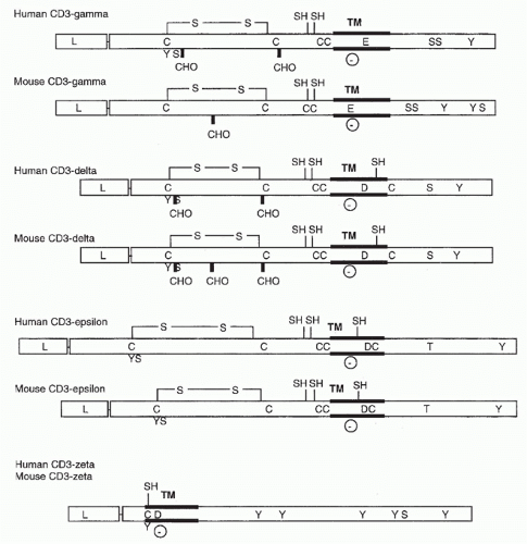
FIG. 11.2. Structural Features of the CD3 Molecules. As in Figure 11.1, transmembrane regions, carbohydrate addition sites, and cysteine residues are indicated. In addition, negatively charged transmembrane residues (D for aspartic acid and E for glutamic acid) and putative phosphorylation sites are shown.

Recently, live-cell imaging studies have shown a close interaction of the CD3epsilon cytoplasmic domain of the TCR with the plasma membrane, using fluorescence resonance energy transfer (FRET) between a C-terminal fluorescent protein and a membrane fluorophore.65 Electrostatic interactions between basic CD3epsilon residues and acidic phospholipids enriched in the inner leaflet of the plasma membrane were required for binding. Nuclear magnetic resonance studies of the lipid-bound state of this cytoplasmic domain revealed a deep insertion of the two key tyrosines of the ITAM into the hydrophobic core of the lipid bilayer, likely preventing their phosphorylation.65 Similar studies of CD3 zeta have confirmed that this is also the case for that component of the TCR/CD3 complex as well.66 This then likely defines the “off” state of the TCR/CD3 complex and is a novel explanation for how it can prevent tyrosine phosphorylation by lck. What is still mysterious is how ligand binding by the TCR disengages these signaling modules from the plasma membrane and triggers the kinase cascade needed to activate T cells.


Assembly and Organization of the T-Cell Receptor/Cluster of Differentiation 3 Complex

The assembly of newly formed TCR-&agr; and -&bgr; chains with the CD3 &ggr;, &dgr;, &egr;, and &zgr; chains and their intracellular fate have been studied in detail.39,40,41,61,63 Early studies have focused on mutant hybridoma lines, which fail to express TCR on their cell surface, and on transfection studies using complementary DNA for the different chains in the receptor; but recently, Wucherpfennig and colleagues have developed an elegant in vitro translation and assembly system that has clarified a number of important issues.54,55

Experiments in a nonlymphoid cell system67 have shown that TCR-&agr; can assemble with CD3 &dgr; and &egr; but not CD3 &ggr;
and &zgr;. In contrast, the TCR-&bgr; chain can assemble with any of the CD3 chains except the &zgr; chain. When the &zgr; chain was transfected with either &agr; or &bgr; chain genes, or any of the three CD3 chains, no pairwise interaction occurred.

Only when all six complementary DNAs were cotransfected was it shown that the &zgr; chain could be coprecipitated with the other chains.67 Based on these data, a model has been proposed that suggests that TCR-&agr; pairs with CD3 &dgr; and &egr; chains and that TCR-&bgr; pairs with the CD3 &ggr; and &egr; chains in the completed molecule. The &zgr; chain is thought to join the TCR and other CD3 polypeptides in that last stage of assembly.

Pulse-chase experiments have shown that all six chains are assembled in the endoplasmic reticulum (ER), transported to the Golgi apparatus, and then transferred to the plasma membrane. It also appears that the amount of &zgr; chain is rate limiting, as it is synthesized at only 10% the level of the other chains. This results in the vast majority of newly synthesized &agr;, &bgr;, or CD3 components being degraded within 4 hours of their synthesis. The remaining nondegraded chains are long lived due to the formation of complete TCR/CD3 complexes with the limiting &zgr; chain.68 TCR/CD3 lacking CD3 &zgr; chains migrate through the ER and Golgi intact but then are transported to and degraded in the lysosomes. The immunologic significance of this pre-Golgi degradation pathway is most evident in CD4+CD8+thymocytes where, despite high levels of synthesis of both messenger ribonucleic acid and protein for all the TCR, CD3, and &zgr; chains, surface expression is relatively low. The TCR chains in immature thymocytes seem to be selectively degraded.68 Thus posttranslation regulation appears to be an important means of controlling the cell surface expression of TCR heterodimers.

The TCR and CD3 &ggr;, &dgr;, and &egr; chains contain ER retention signals.68,69 If the &ggr; and &dgr; signals are removed, then the chains are transported through the Golgi and rapidly degraded in the lysosomes. In contrast, removal of the CD3 &egr; ER retention signal allows this chain, and any associated chains, to be transported to the cell surface. Thus, association of the TCR and other CD3 chains with &egr; renders their ER retention signals inoperative. However, the &egr; ER retention signal remains functional. This prevents the surface expression of partial complex intermediaries until CD3 &zgr; is incorporated into the complex, which then masks the &egr; ER retention signal and allows the transport of mature complexes to the cell surface.

The overall stoichiometry of the &agr;&bgr; TCR/CD3 complex is controversial. The work of Call and colleagues57 has shown the relationships between different CD3 dimers (&ggr;&egr;, &dgr;&egr;, and &zgr; &zgr;) and a single TCR &agr;&bgr; heterodimer (as shown in Fig. 11.3). Using mutagenesis, they found very specific interactions based on the positive charges in each TCR transmembrane domain with complementary negative charges in the transmembrane domains of the different CD3 components. The two positively charged residues of TCR-&agr; mediate interactions with the negatively charged residues of the of CD3&dgr;&egr; and CD3&zgr;&zgr; dimers, while the single positively charged residue of TCR-&bgr; mediates interactions with the negatively charged residues of CD3&ggr;&egr;. In addition, their data suggest a highly ordered assembly process as they found that the TCR/CD3&dgr;&egr; association facilitates the assembly of CD3&ggr;&egr; into the complex and that the association of the TCR and CD3 heterodimers was a prerequisite for incorporation of CD3&zgr;&zgr; into the complex. Importantly, the data show that there is only one TCR heterodimer per nascent TCR/CD3 complex in their in vitro expression system.

In contrast, a number of groups have found evidence that there can be two TCR heterodimers in a given TCR/CD3 cluster on T-cell surfaces. In particular, Terhorst and colleagues42 showed that in a T-T hybridoma, a monoclonal antibody against one TCR-&agr;&bgr; pair could comodulate a second &agr;&bgr; heterodimer. In addition, sucrose gradient centrifugation of TCR/CD3 showed a predicted molecular weight of 300 kDa, more than 100 kDa larger than expected from a minimal &dgr; subunit complex (&agr;, &bgr;, &ggr;, &dgr;, &egr;2, &zgr;2).70 Another study suggesting that there are least two TCRs in a given CD3 complex is the Scatchard analysis indicating that the number of CD3&egr; molecules on a T-cell surface equals the number of &agr;&bgr; TCRs.71,72,73 Finally, there is the work of Fernandez-Miguel et al.,74 who showed that in T cells which have two transgenic TCR-&bgr; chains, antibodies to one V&bgr; can immunoprecipitate the other. It was also found that they are often close enough to allow fluorescence energy transfer, meaning that the two TCR-&bgr;s in a cluster are within 50 Angstroms of each other.74 Interestingly, it appears that the TCR complexes with CD3 either have CD3 &ggr; or CD3 &dgr;, but not both, and these two
receptor types are expressed in different ratios in different cells. These data may not be irreconcilable, because while the initial TCR/CD3 assembly may involve only one TCR, these may dimerize or multimerize later on the cell surface.50,75


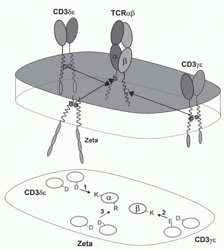
FIG. 11.3. A Model of the &agr;&bgr; T-Cell Receptor (TCR)-Cluster of Differentiation (CD)3 Complex. This shows the approximate positions of the &agr;&bgr; TCR chains and the CD3&agr;, &dgr;, &egr;, and &zgr; chains based on the membrane reconstitution and mutagenesis experiments of Call and Wucherpfennig.55 More recent experiments suggest that the CD3 polypeptides may be somewhat more clustered on the side of the TCR heterodimer.299

The composition of TCR/CD3 complexes on &ggr;&dgr; T cells is distinct from that of &agr;&bgr; T cells and changes with the activation state of the cell. Biochemical analysis showed that most murine &ggr;&dgr; TCRs contain only CD3 &ggr;&egr; dimers. Interestingly, a differentially glycosylated form of CD3 &ggr; was found to associated with &ggr;&dgr; TCRs dependent on the activation state of the cells.76 In addition, while C3&zgr;&zgr; is incorporated into the complexes of naïve cells, activation results in the expression and incorporation of Fc&egr;RI &ggr; into the &ggr;&dgr;TCR complex.76 Using quantitative immunofluorescence, Hayes and Love have derived data and proposed a model of murine &ggr;&dgr;TCR stoichiometry in which there are two CD3&ggr;&egr; dimers, as well as one CD3&zgr; dimer in each TCR complex.77 Taken together, these findings strongly suggest that signal transduction through the TCR will occur differently in &ggr;&dgr; versus &agr;&bgr; T cells.


T-CELL RECEPTOR GENES

As shown in Figure 11.4, TCR gene segments are organized similarly to those of Igs and the same recombination machinery is responsible for joining separate V and D segments to a particular J and C. This was initially indicated by the fact that the characteristic seven and nine nucleotide conserved sequences adjacent to the V, D, and J regions with the 12 or 23 nucleotide spacing between them, first described for Ig genes, are also present in TCRs.78 The most conclusive evidence of this common rearrangement mechanism is that both a naturally occurring recombination-deficient mouse strain (severe combined immune deficiency79) and mice engineered to lack recombinase activating genes 180 or 281 are unable to rearrange either TCR or Ig gene segments properly. Many of the other molecules involved in Ig gene rearrangement serve the same function in TCRs as well.82 As with Igs, if the V region and J region gene segments are in the same transcriptional orientation, the intervening DNA is deleted during recombination. DNA circles of such material can be observed in the thymus,83,84 the principal site of TCR recombination (see the following). In the case of TCR-&bgr; and TCR-&dgr;, there is a single V region 3′ to the C in the opposite transcriptional orientation to J and C. Thus, rearrangement to these gene segments occurs via an inversion. Variable points of joining are seen along the V, D, and J gene segments as well as random nucleotide addition (N regions) in postnatal TCRs. The addition of several nucleotides in an inverted repeat pattern, referred to a P element insertion, at the V-J junction of the TCR-&ggr; chains has also been observed.85


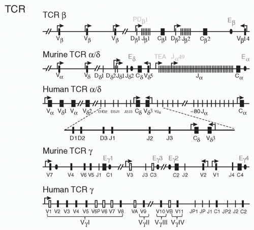
FIG. 11.4. T-Cell Receptor Gene Organization in Mice and Humans. Schematic of V, D, J, and C elements of the T-cell receptor genes. Transcriptional orientation is from left to right, except where noted. E designates enhancer elements, and S are silencer elements.


Organization of the T-Cell Receptor &agr;/&dgr; Locus

In humans and in mice, there is a single &agr;-chain C-region gene that is composed of four exons encoding: 1) the constant region domain, 2) 16 amino acids including the cysteine that forms the interchain disulfide bond, 3) the transmembrane and intracytoplasmic domains, and 4) the 3′ untranslated region (see Fig. 11.4). The entire &agr;/&dgr; locus spans about 1.1 MB in both mice and humans. There are 50 different J-region gene segments upstream of the C-region in the murine locus. At least eight of these J-regions are nonfunctional because of in-frame stop codons or rearrangement and splicing signals that are likely to be defective. A similar number of &agr;-chain J-regions are present in the human locus. This very large number of J-regions compared to the Ig loci may indicate that
the functional diversity contributed by the J segment of the TCR (which constitutes a major portion of the complementarity-determining region [CDR]3 loop) makes an important contribution to antigen recognition (see the following).

In both the murine and human loci, the C&dgr;, J&dgr;, and two D&dgr; gene segments are located between the V&agr; and J&agr; gene segments. In the murine system, there are two J&dgr; and two D&dgr; gene segments on the 5′ side of C&dgr; and the C&dgr; exons are approximately 75 kb upstream of the C&agr; gene and approximately 8 kb upstream of the most 5′ known J&agr; gene segments. The human organization is similar, with three D&agr; gene segments and three J&dgr;s. Surprisingly, in both species all of the D elements can be used in one rearranged gene rather than alternating as is the case with TCR-&bgr; or IgH. That is, in mice one frequently finds V&dgr; D1, D2, and J&dgr; rearrangements,86 and in humans V&dgr;, D1, D2, D3, and J&dgr;.87 This greatly increases the junctional or CDR3 diversity that is available, especially because of the potential for N-region addition in between each gene segment. This property makes TCR-&dgr; the most diverse of any of the antigens receptors known, with approximately 1012 to 1013 different amino acid sequences in a relatively small (10 to 15 amino acid) region.86 The implications for this and comparisons with other antigen receptor genes are discussed subsequently.

The location of D&dgr;, J&dgr;, and C&dgr; genes between V&agr; and J&agr; gene segments suggests that TCR-&dgr; and -&agr; could share the same pool of V gene segments. While there is some overlap in V gene usage, in the murine system, four of the commonly used V&dgr; genes (V&dgr;1, V&dgr;2, V&dgr;4, V&dgr;5) are very different than known V&agr; sequences and they have not been found to associate with C&agr;.88 The other four V&dgr; gene families overlap with or are identical to V&agr; subfamilies (V&dgr;3, V&dgr;6, V&dgr;7, and V&dgr;8, with V&agr;6, V&agr;7, V&agr;4, and V&agr;11, respectively).

The mechanisms that account for the preferential usage of certain gene segments to produce &dgr; versus &agr; chain are not known. While some V&dgr; genes are located closer to the D&dgr; and J&dgr; fragments than V&agr; genes (such as V&dgr;1), other V&dgr;s (such as V&dgr;6) are rarely deleted by V&agr; J&agr; rearrangements and thus seem likely to be located 5′ of many V&agr; gene segments.

One of the V&dgr; gene segments, V&dgr;5, is located approximately 2.5 kb to the 3′ of C&dgr; in the opposite transcriptional orientation and rearranges by inversion. Despite its close proximity to D&dgr; J&dgr; gene segments, V&dgr;5 is not often found in fetal &ggr;&dgr; T cells. Instead, the V&dgr;5 → DJ&dgr; rearrangement predominates in adult &ggr;&dgr; T cells.

An implicit characteristic of the &agr;/&dgr; gene locus is that a rearrangement of V&agr; to J&agr; deletes the entire D-J-C core of the &dgr;-chain locus. In many &agr;&bgr; T cells, the &agr;-chain locus is rearranged on both chromosomes and thus no TCR &dgr; could be made. In most cases, this is due to V&agr; → J&agr; rearrangement, but evidence suggesting an intermediate step in the deletion of TCR-&dgr; has been reported.89 This involves rearrangements of an element termed T early alpha (TEA) to a pseudo-J&agr; 3′ of C&dgr;. The rearrangement of TEA to this psuedo-J&agr; would eliminate the &dgr;-chain locus in &agr;&bgr; T cells. Gene targeting of the TEA element resulted in normal levels of &agr;&bgr; and &ggr;&dgr; T cells, but usage of the most J&agr;s was severely restricted,90 suggesting that its function is to govern the accessibility of the most proximal 5′ J&agr;s for recombination.


Organization of the T-Cell Receptor &bgr; Locus

The entire human 685 kb &bgr; chain gene locus was originally sequenced by Hood and coworkers91 (Fig. 11.5). One interesting feature is the tandem nature of J&bgr;C&bgr; in the TCR-&bgr; locus. This arrangement is preserved in all higher vertebrate species that have been characterized thus far (mouse, human, chicken, frog). The two C&bgr; coding sequences are identical in the mouse and nearly so in humans and other species. Thus it is unlikely that they represent two functionally distinct forms of C&bgr;. However, the J&bgr; clusters have relatively unique sequences, and thus this may be a mechanism for increasing the number of J&bgr; gene segments. Together with the large number of J&agr; gene segments, there is far more combinatorial diversity (J&agr; × J&bgr; = 50× 12=600) provided by J regions in &agr;&bgr; TCRs than in Igs.

Most of the V-regions are located upstream of the joining and constant regions and in the same transcriptional orientation as the D and J gene element, and rearrange to D&bgr;J&bgr; gene via deletion. Similar to the case of V&dgr;5, a single V&bgr; gene, V&bgr;14 is located 3′ to C-regions and in the opposite transcriptional orientation, thus rearrangements involving V&bgr;14 occur via inversion.

In the NZW strain of mouse, there is a deletion in the &bgr; chain locus that spans from C&bgr;1, up to and including the J&bgr; 2 cluster.92 In SJL, C57BR and C57L mice, there is a large deletion93 in the V-region locus from V&dgr;5-V&bgr;9. These mice
also express a V gene, V&bgr;17, that is not expressed in other strains of mice. Deletion of about half of the V genes (in SJL, C57BR and C57L mice) does not seem to have any particular effect on the ability of these mice to mount immune responses whereas mice that have deleted the J&bgr;2 cluster show impaired responses.94


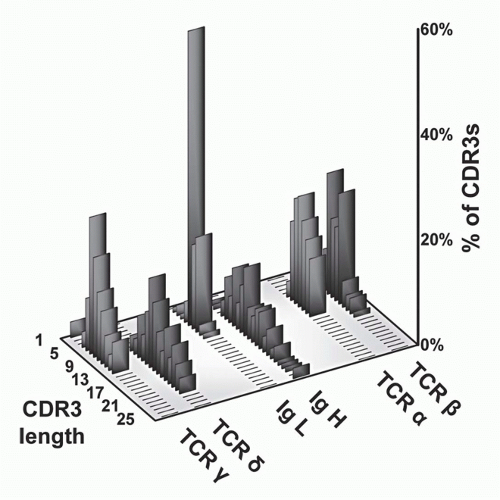
FIG. 11.5. Complementarity-Determining Region (CDR)3 Length Analysis of T-Cell Receptor (TCR) Polypeptides versus Immunoglobulin (Ig) Heavy and Light Chains. These data, modified from Rock et al.,129 show that whereas TCR-&agr; and -&bgr; CDR3 regions are relatively uniform with respect to each other, the other antigen receptor pairs show a marked asymmetry. Specifically, both Ig light chains (&kgr; and &lgr;) show very short CDR3s, as do TCR-&ggr; chains. In contrast, both IgH and TCR S TCRs are quite heterogeneous and tend to be longer. These data suggests that &ggr;&dgr; TCRs have a more antibody-like structure and binding properties. This has been borne out by subsequent analysis (see text).


Organization of the T-Cell Receptor &ggr; Locus

The organization of the mouse and human &ggr;-chain loci are shown in Figure 11.4. The human &ggr; genes span about 150 kb85 and are organized in a fashion similar to that of the &bgr; chain locus with two J&ggr;C&ggr; regions. There is more than one nomenclature commonly used to describe the &ggr; chain genes.85,96,97,98 Here, we use that of Lefranc and Rabbitts99 and Tonegawa and colleagues85 for the human and mouse &ggr; chains, respectively. The organization of the human &ggr; chain genes consists an array of V&ggr;s in which at least six of the V-regions are pseudogenes is located 5′ to these J&ggr;C&ggr; clusters, and each of the V genes are potentially capable of rearranging to any of the five J-regions. The sequences of the two human C&ggr; regions are very similar overall and only differ significantly in the second exon. In C&ggr;2, this exon is duplicated two or three times and the cysteine that forms in the interchain disulfide bond is absent. Thus, C&ggr;2-bearing human T cells have an extra large &ggr;-chain (55,000 MW) that is not disulfide bonded to its &dgr;-chain partner.

The organization of the murine &ggr; chain genes is very different than that of the human genes in that there are three separate rearranging loci that span about 205 kb.100,101 Of four murine C&ggr; genes, C&ggr;3 is apparently a pseudogene in BALB/c mice, and the J&ggr;3 C&ggr;3 region is deleted in several mouse strains including C57 Bl/10. C&ggr;l and C&ggr;2 are very similar in coding sequences. The major differences between these two genes is in the five amino acid deletion in the C&ggr;2 gene that is located in the C II exon at the amino acid terminal of the cysteine residue used for the disulfide formation with the &dgr; chain. The C&ggr;4 gene differs significantly in sequences from the other C&ggr; genes (in 66% overall amino acid identity). In addition, the C&ggr;4 sequences contains a 17 amino acid insertion (compared to C&ggr;1) in the C II exon located at similar position to the five amino acid deletions in the C&ggr;2 gene.101a Each C&ggr; gene is associated with a single J&ggr; gene segment. The sequences of J&ggr;1 and J&ggr;2 are identical at the amino acid level, whereas J&ggr;4 differs from J&ggr;1 and J&ggr;2 at 9 out of 19 amino acid residues.

The murine V&ggr; genes usually rearrange to the J&ggr; C&ggr; gene that is most proximal and in the same transcriptional orientation. Thus V&ggr;1 rearranges to J&ggr;4; V&ggr;2 to J&ggr;2; and V&ggr;4, V&ggr;5, V&ggr;6, and V&ggr;7 to J&ggr;1. Interestingly, some V&ggr; genes are rearranged and expressed preferentially during &ggr;&dgr; T-cell ontogeny and in different adult tissues as well.101 In particular, V&ggr;5+ and V&ggr;6+ T cells are generated in the fetal thymus with very limited/no junctional diversity. Instead, the adult thymus produces &ggr;&dgr; T cells expressing V&ggr;1, V&ggr;2, V&ggr;4, and V&ggr;7 gene segments with highly diverse junctional sequences. Moreover, &ggr;&dgr; T cells that localize to the secondary lymphoid organs tend to express V&ggr;4, V&ggr;1, and V&ggr;2, whereas those that localize to the intestinal epithelium express V&ggr;7.88,102,103,104,105,106 There are also reports suggesting that some intestinal epithelial &ggr;&dgr; T cells develop extrathymically.107 Regardless, it has been suggested that the V&ggr; gene rearrangement is a programmed process.108,109


Control of Transcription and Rearrangement

It has become increasingly apparent that transcriptional accessibility and rearrangement of TCR and Igloci are closely linked, following the early work of Alt and Yancopoulous.110 Factors governing accessibility and rearrangement include histone methylation,111,112,113,114 DNA methlyation, and the presence of enhancer and specific promoter elements.115 Even specific variations in the recombination signal sequences have been shown to elicit specific biases in V(D)J joining.86 With respect to enhancer elements in the TCR loci, these were first identified in the TCR-&bgr; locus, 3′ of C&bgr;2,116,117 and subsequently for the other TCR loci as well,115 as indicated in Figure 11.4. These TCR enhancers all share sequence similarities with each other. Some of the transcriptional factors that bind to the TCR genes are also found to regulate Ig gene expressions. It has been shown that TCR-&agr; enhancer (E&agr;) is not only important for normal rearrangement and expression for the &agr; chain locus but also is required for the normal expression level of mature TCR-&dgr; transcripts.118 Also interesting is the work of Lauzurica and Krangel,119,120 who have shown that a human TCR-&dgr; enhancer-containing minilocus in transgenic mice is able to rearrange equally well in &agr;&bgr; T cells as in &agr;&dgr; T cells but that an E&agr;-containing construct was only active in &agr;&bgr; lineage T cells. Similar to Ig genes, promoter sequences are located 5′ to the V gene segments. Although D → J&bgr; rearrangement and transcription occur fairly often in B cells and in B-cell tumors,121 V&bgr; rearrangement and/or transcription appears highly specific to T cells. In addition to enhancers, there also appear to be “silencer” sequences 3′ of C&agr;122,123 and in the C&ggr;1 locus.124 It has been suggested that these “repressor sites” could turn off the expression of either of these genes, influencing T-cell differentiation toward either the &agr;&bgr; or the &ggr;&dgr; T-cell lineage.

The murine TCR C&ggr;1 gene cluster comprises four closely linked V&ggr; gene segments, in the order V&ggr;7, 4, 6, and 5, which rearrange to a single common downstream J gene segment, J&ggr;1 (see Fig. 11.4). In early fetal thymocytes, rearrangements of V&ggr;5 and V&ggr;6 genes predominate, and the resulting V&ggr;3+ and Vg4+ cells migrate to the skin or reproductive tissue, respectively. Later in ontogeny, V&ggr;4 and V&ggr;7 rearrangements predominate, and cells expressing these V regions migrate from the adult thymus to the secondary lymphoid organs and the intestinal epithelium.100,101 At least two cis-acting, enhancer/locus control region (LCR) elements are present in the C&ggr;1 cluster. One is a T cell-specific transcriptional enhancer, 3&ggr;E&ggr;, located 3 kb downstream of the C&ggr;1 gene segment.125 A second element, “has,” was found between the V&ggr;7 and V&ggr;4 genes, based on DNAse I hypersensitivity.126 Similar enhancers have also been found to be associated with the C&ggr;2 and C&ggr;3 genes.95 Experiments suggest that simultaneous deletion of both enhancer elements in C&ggr;1 cluster severely diminishes TCR-&ggr; transcription, but only modestly reduces TCR-&ggr; gene rearrangement, while deletion of each element
separately has little effect.127 In contrast to these results in thymocytes, deletion of “has” alone reduces transcription of one V&ggr; gene specifically in peripheral &ggr;&dgr; T cells. Thus, the two elements not only exhibit functional redundancy in thymocytes but also have unique functions in other settings.


Allelic Exclusion

In Igs, normally only one allele of the heavy chain locus and one of the light chain alleles is productively rearranged and expressed, a phenomenon termed “allelic exclusion.” With respect to &agr;&bgr; TCR expression, while TCR-&bgr; exhibits allelic exclusion,128 TCR-&agr; seems much less constrained,129,130 and many mature T cells express two functional TCR-&agr; chains. As the chances of forming an in-frame joint with any antigen receptor is only one in three, the probability that a T cell would have two productively rearranged TCR&agr;s is only 1/3 × 1/3 = 1/9, or 11%. However, even when this happens, the two TCR-&agr; chains may not form heterodimers equally well with the single TCR-&bgr; that is expressed; thus, only one heterodimer may be expressed. But this simple calculation is complicated by the likelihood that only thymocytes that make at least one productive TCR rearrangement of each type will have a chance at maturation, which would eliminate almost half of the T cells (four-ninths, which is the product of a two-thirds chance of failure on one chromosome, followed by the same failure rate on the second). Secondly, it has been found that there is a mechanism called receptor editing, which means that a given rearrangement that is not productive, either because of an out-of-frame joint or for reasons of self reactivity, can induce a V region further 5′ or “upstream” of the initial VJ joint to rearrange to one of the remaining Jas.131 In any event, it has been reported that one-third of human T cells express two TCRs,132 which is slightly higher than what is expected from the probabilities discussed previously (approximately 20%), perhaps reflecting the effect of receptor editing.

There also appears to be an important role for the pre-TCR heterodimer (eg, pre-T&agr;:TCR-&bgr;) in blocking further TCR-&bgr; rearrangement and thus ensuring allelic exclusion at that locus.133,134 In particular, pre-T&agr;-deficient mice had a significant increase in the number of cells with two productive TCR=&bgr; rearrangements, compared with wildtype mice.133


T-Cell Receptor Diversity

Although the basic organization and V(D)J recombination machinery are shared between TCR loci and Igs, there are a number of striking differences. One of these is somatic hypermutation. In antibodies, this form of mutation typically raises the affinities of antigen specific Igs several order of magnitude, typically from the micromolar (10-6 M) to the nanomolar (10-9 M) range.135,136 We now know that most cellsurface receptors that bind ligands on other cell surfaces, including TCRs, typically have affinities in the micromolar range (see later section) but that they compensate for this relatively low affinity by engaging multiple receptors simultaneously (eg, increasing the valency) and by functioning in a confined, largely two-dimensional volume (eg, between two cells). Cells employing such receptors require weak, but highly specific, interactions so that they can disengage quickly.137,138 The rapid off-rates seen with TCRs (see later section) may even amplify the effects of small numbers of ligands.139,140

There has also been no enduring evidence for a naturally secreted form of either an &agr;&bgr; or &ggr;&dgr; TCR. Here again, it can be argued that such a molecule would have no obvious use as the affinities are too low to be very useful in solution. Thus, for most TCRs, the concentration of protein would have to be extremely high in order to achieve an effect similar to soluble antibodies (in the milligram/milliliter range).

A third mechanism seen in antibodies but not TCRs is CH switching, which allows different Ig isotypes to maintain a given V region specificity and associate it with different constant regions that have different properties in solution (such as complement fixation, basophil binding, etc.). As there is no secreted form of the TCR, this feature would also lack any obvious utility.

Where TCRs are equal—and in fact generally superior— to Igs is in the sheer number of possible receptors that can be generated through recombination alone. Table 11.1 summarizes the potential V region diversity that TCRs are capable of when the number of V region gene segments is multiplied by D, J, and N region diversity. It can be seen from this table that while the V region number is generally lower in murine TCRs, particularly TCR-&dgr; and TCR-&ggr;, this is more than compensated for by the degree of junctional diversity (where V and J or V, D, and J come together) and chain combinations, such that overall TCRs have orders of magnitude greater potential diversity than Igs. This junctional region corresponds to CDR3 as originally defined by Kabat and Wu for Igs.141 With respect to &agr;&bgr; TCRs, the concentration of diversity in this region (in both chains) can be explained by the key role that these sequences play in recognizing diverse peptides in MHC molecules (see later section), as supported by mutagenesis and structural studies. For &ggr;&dgr; TCRs and Igs, however, the diversity is almost all in just one chain (TCR&dgr; and IgH, respectively), and the implications of this are discussed subsequently. Recent work using high throughput sequencing techniques are in remarkable agreement with these crude early estimates of TCR-&bgr; diversity.142


The Complementarity-Determining Region 3 Length Distributions of &ggr;&dgr; T-Cell Receptors are More Similar to Those of Immunoglobulin than to Those of &agr;&bgr; T-Cell Receptors

Because CDR regions are loops between different &bgr; strands of an Ig or TCR V region (see later section), the configurations they adopt are generally very sensitive to their length, such that a difference of even one amino acid may produce a significant change in the overall structure.3,143 A comparison of CDR3 length distributions between the &agr;&bgr; TCRs, &ggr;&dgr; TCRs, and Igs (see Fig. 11.5)144,145 showed that those of TCR-&agr; and -&bgr; have a very constrained distribution of lengths and that these are nearly identical in size. These length constraints may reflect a requirement for both the &agr;
and &bgr; chains of TCRs to contact both the MHC molecules and bound peptides on the same plane, as borne out by structural studies (see later section). In contrast, the CDR3s of Ig heavy chains are long and variable, whereas those of Ig light chains are short and constrained. This may reflect the fact that Igs recognize both very small molecules (eg, haptens) as well as very large ones (eg, proteins). Surprisingly, &ggr;&dgr; TCR CDR3 length distributions are similar to those of Igs in that the CDR3 lengths of TCR-&dgr; chains are long and variable, whereas those of the TCR &ggr; chains are short and constrained. Thus, on the basis of this measure of ligand recognition, one might expect &ggr;&dgr; TCRs to be more similar to Igs than to &agr;&bgr; TCRs. This has been validated in subsequent biochemical and structural studies (see later sections).


TABLE 11.1 Sequence Diversity in T-Cell Receptor and Immunoglobin Genes


Immunoglobulin


TCR-&agr;/&bgr;


TCR-&ggr;/&dgr;


H


&kgr;


&agr;


&bgr;


&ggr;


&dgr;


Variable segments


250-1,000


250


100


25


7


10


Diversity segments


10


0


0


2


0


2


Ds read in all frames


Rarely


Often


Often


N-region addition


V-D, V-J


none


V-J


V-D, V-J


V-J


V-D1, D1-D2, D1-J


Joining segments


4


4


50


12


2


2


Variable region combinations


62,500-250,000


2,500


70


Junctional combinations


˜1011


˜1015


˜1018


Calculated potential amino acid sequence diversity in TCR and immunoglobulin genes without allowance for somatic mutation. The approximate number of V gene segments are listed for the four TCR polypeptides and contrasted with immunoglobulin heavy and light chains. CDR1 and CDR2 are encoded within the V gene segments. The pairing of random V regions generates the combinatorial diversity listed as “variable region combinations.” Because there are fewer TCR V gene segments than immunoglobulin V gene segments, the combinatorial diversity is lower in TCRs than in immunoglobulins. Estimates for the number of unique sequences possible within the junctional region are contrasted for TCRs and immunoglobulins. Amino acids within CDR3 are encoded almost entirely within the D and/or J region gene segments. (The last few amino acids encoded by a TCR V gene segment can contribute to diversity within the TCR CDR3-equivalent region, but the effects of these residues on junctional diversity are not included in these calculations.) The mechanisms for generation of diversity within the junctional region that are used for this calculation include usage of different D and J gene segments. N region addition up to six nucleotides at each junction, variability in the 3′ joining position in V and J gene segments, and translation of D region in different reading frames. Numbers are corrected for out-of-frame joining codon redundancy and N-region mimicry of germ-line sequences. Modified from Elliott et al.86


CDR, complementarity-determining region; TCR, T-cell receptor.


Chromosomal Translocations and Disease

The chromosomal locations of the different TCR loci have been delineated in both mouse and humans, and the results are summarized in Table 11.2. One significant factor in cancers of hematopoietic cells are chromosomal translocations that result in the activation of genes normally turned off or the inactivation of genes that are normally turned on. Thus, B- or T-lymphocyte neoplasia is frequently associated with inter- or intrachromosomal rearrangements of Ig or TCR loci or in some cases both.146,147

These translocations seem to mediated by the V(D)J recombinase machinery, indicating the inherent danger and need for tight regulation of this pathway. Such rearrangements are particularly common in the &agr;/&dgr; locus, perhaps because this locus spans the longest developmental window in terms of gene expression, with TCR-&dgr; being the first and TCR-&agr; the last gene to rearrange during T-cell ontogeny (as discussed in more detail in the following). In addition, the &agr;/&dgr; locus is in excess of 1 mb in size, and this provides a larger target for rearrangement than either TCR-&bgr; or TCR-&ggr;. Interestingly, in humans, TCR-&agr;/&dgr; is on the same chromosome as the IgH locus and VHJ&agr; rearrangements (by inversion) have been observed in some human tumor material.148,149 The functional significance of this is not known.

Particularly frequent is the chromosome 8-14 translocation [t(8;14) (q24;q11)] that joins the &agr;/&dgr; locus to the c-myc gene, analogous to the c-myc → IgH translocation in many mouse myeloma tumors and in Burkitt lymphomas in humans. In one cell line, a rearrangement occurred between the J&agr;-region coding sequences, and a region 3′ of c-myc.150 In both B- and T-cell malignancies, the translocation of c-myc into IgH or TCR-&agr;/&bgr; appears to increase the expression of c-myc and may be a major factor in the unregulated cell growth that characterizes cancerous cells. Other
putative proto-oncogenes that have been found translocated into the TCR-&agr;/&bgr; locus are the LIM domain-containing transcription factors Ttg-1151 and Ttg-2,152,153 which are involved in neural development; the helix-loop-helix proteins Lyl-1154 and Scl,155 which are involved in early hematopoietic development; and the homeobox gene Hox 11,156 which is normally active in the liver. How these particular translocations contribute to malignancy is unknown, but they presumably causes aberrations in gene expression that contribute to cell growth or escape from normal regulation. In patients with T-cell leukemia infected with the human T-cell lymphotrophic-I virus, there are large numbers of similar translocations; it is thought that human T-cell lymphotrophic-I itself is not directly leukemogenic but acts by causing aberrant rearrangements in the T cell that it infects, some of which become malignant.


TABLE 11.2 Chromosomal Locations of T-Cell Receptor, Immunoglobulin, and Related Loci in Mouse and Human


Mouse Chromosome


Human Chromosome


TCR-&agr;


14


14(q11-q12)


TCR-&dgr;


14


14(q11-q12)


IgH


12


14(qter)


TCR-&bgr;


6


7(q35)


CD4


6


12


CD8


6


2(p11)


Ig&kgr;


6


62(p12)


TCR-&ggr;


13


7(p14)


CD3-&ggr;


9


11(q23)


CD3-&dgr;


9


11(q23)


CD3-&egr;


9


11(q23)


CD3-&zgr;


1


1


Thy-1


9


11(q23)


Ig-&lgr;


16


22(q11.2)


MHC


17


6(p21)


Pre-T&agr;


17


6


CDR, complementarity-determining region; Ig, immunoglobulin; MHC, major histocompatibility complex; TCR, T-cell receptor.


Another disorder, which frequently associates with TCR and Ig locus translocation, is ataxia telangiectasia, an autosomal recessive disorder characterized by ataxia, vascular telangiectasis, immunodeficiency, increased incidence of neoplasia, and an increased sensitivity to ionizing radiation. Peripheral blood lymphocytes from patients with ataxia telangiectasia have an especially high frequency of translocations involving chromosomes 7 and 14.157 These sites correspond to the TCR-&ggr;, -&bgr;, and -&agr; loci, and the Ig heavy chain locus. Thus, it appears as though one of the characteristics of patients with ataxia telangiectasia is a relatively error-prone rearrangement process that indiscriminately recombines genes that have the TCR and Ig rearrangement signals.158


The Structure of &agr;&bgr; and &ggr; &dgr; T-Cell Receptors

As discussed previously, the sequences of TCR polypeptides show many similarities to Igs, and thus it has long been suggested that both &agr;&bgr; and &ggr;&dgr; heterodimers would be antibodylike in structure.24,25,159 The similarities between TCRs and Igs include the number and spacing of specific cysteine residues within domains, which in antibodies form intrachain disulfide bonds. Also conserved are many of the inter- and intradomain contact residues and, in addition, secondary structure predictions are largely consistent with an Ig-like “&bgr; barrel” structure. This consists of three to four antiparallel &bgr; strands on one side of the “barrel” facing a similar number on the other side, with a disulfide bridge (usually) connecting the two &bgr; “sheets” (sets of &bgr; strands in the same plane). All Ig variable and constant region domains have this structure, with slight variations in the number of &bgr; strands in variable region domains (by convention including V, D, and J sequences) compared with constant domains.


&agr; &bgr; T-Cell Receptor Structure

Efforts to derive x-ray crystal structures of TCR heterodimers and fragments of heterodimers presented many technical hurdles.160 One difficulty is that structure determination required engineering the molecules into a soluble form. A second problem is that many of the TCRs are heavily glycosylated, and it was necessary to eliminate most or all of the carbohydrates on each chain to achieve highquality crystals. An alternative is to express soluble TCRs in insect cells, where they have compact N-linked sugars, or in Escherichia coli, where they are unglycosylated. The first successes in TCR crystallization come from the laboratory of Mariuzza and collaborators who solved the structure of first a V&bgr; C&bgr; polypeptide161 and then a V&agr; fragment.162 In the following year, the first complete &agr;&bgr; TCR structures were solved.163,164 The structure of the 2C TCR, by Garcia and colleagues, is shown in Figure 11.6.163 In general, as predicted from sequence homologies, these domains are all Iglike, with the classical &bgr;-barrel structure in evidence in all three domains. At each end of the barrel in each V-region domain there are four loops between the &bgr; sheets, three of which form the CDRs of Igs, which are numbered in Figure 11.6. The fourth loop, between the D and E strands, has been implicated in superantigen binding. The six CDR loops from the two variable domains form the antigenbinding surface in both Igs and TCRs. The major anomaly in terms of similarity of TCRs to Igs is the structure of the C&agr;.165 C&agr; consists of one-half of the classical &bgr;-barrel, that is, one set (or “sheet”) of &bgr; strands while the rest of the partially truncated domain exhibits random coils. This type of structure is unprecedented in the Ig gene family. The functional significance of such a variant structure in unknown, but it has been suggested that this incompletely formed Iglike domain may be responsible for the observed lability of TCR-&agr;, and this may allow greater flexibility in the regulation of its expression. Another possible explanation is that this configuration is designed to accommodate one or more of the CD3 molecules.


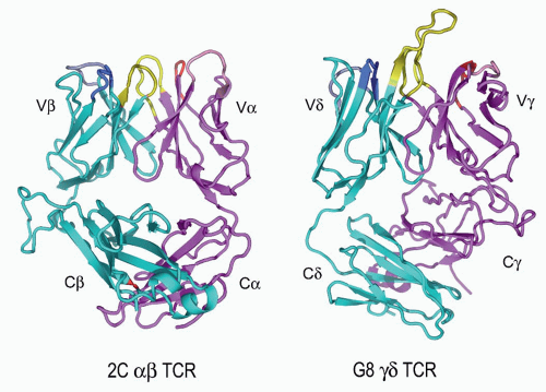
FIG. 11.6. Ribbon Diagrams of the T-Cell Receptor (TCR) Structures. This shows the structures of the 2C&agr;&bgr; TCR150 versus the G8&ggr;&dgr; TCR.153 The TCR-&bgr; and the -&ggr; chains are in cyan, and the TCR-&agr; and -&ggr; chains are in vermillion. The complementarity-determining regions (CDRs) of both are in yellow. The very long TCR-&dgr; CDR3 in G8, which binds the T10/T22 ligands, is very apparent here but is shorter in most other &ggr;&dgr; TCRs. Note the different C-region interactions with these TCRs and the deviations from the classic “&bgr; barrel” structure in both C&agr; and C&dgr;. The prominent C&bgr; loop to the left is also unusual and may mediate interactions with CD3 or other molecules on the T cell surface. (Figure courtesy of Dr. K.C. Garcia)


The now large number of solved &agr;&bgr; TCR structures can be compared to the three &ggr;&dgr; heterodimers (discussed in more detail in the following), and while these also resemble the Fab fragment of an antibody, there are several features that are unique to the &agr;&bgr; molecules, which may be significant. These include the following:


  • In one structure,165 four out of seven N-linked sugars diffracted to high resolution, indicating that they are not free to move very much and thus are likely to play a structural role, particularly in C&agr;:C&bgr; interactions. This correlates with mutagenesis data indicating that certain C&agr; sugars cannot be eliminated without abolishing protein expression166 and the disordered state of a C&agr; domain in the structure of a TCR lacking glycosylation.164


  • There is significantly more contact between V&bgr; and C&bgr; and between V&agr; and C&agr; than in the equivalent regions of antibodies.


  • The geometry of the interaction of V&agr; and V&bgr; more closely resembles that of the CH3 domains of antibodies than VHVL.


  • Between the CDR3 loops of V&agr; and V&bgr;, there is a pocket that can (and does in at least one case165) accommodate a large side chain from the peptide bound to an MHC.


&ggr;&dgr; T-Cell Receptor Structure

There are now three &ggr;&dgr; heterodimer structures: a &ggr;&dgr; TCR from a human T-cell clone G115,167 which can be activated by natural or synthetic pyrophosphomonoesters, a &ggr;&dgr; TCR from the murine T cell clone G8 together with its ligand, the nonclassical MHC class I molecule T22,168 and a human MHC class I chain-related-reactive &ggr;&dgr; TCR (&dgr;1A/B-3),169a which was determined as a single-chain Fv construct. The G8 structure is shown in Figure 11.6, alongside the 2C &agr;&bgr; TCR. The structure of a single human V&dgr; domain also has been determined.169 The V&dgr; domain of the G115 structure is similar to the isolated V&dgr; domain and the quaternary structure of G8 is similar to that of G115.168

The most distinctive feature of both the G115 and the G8 TCR, when compared with &agr;&bgr; TCRs and Igs, is that the C domains “swing out” from under the V domains. This unusual shape is highlighted by both a small elbow angle of 110 degrees, defined as the angle between the pseudo twofold symmetry axes that relate V to V and C to C, and a small V-C interdomain angle. This contrasts with an average of 149 degrees for &agr;&bgr; TCR structures. The small angle between the V&ggr; and C&ggr; domains shifts both C&dgr; and C&ggr; to one side. Moreover, the molecular surfaces of the constant domains are different than those of &agr;&bgr; TCRs with no clear similarities either in the shape or the nature of the C&agr;C&bgr; and C&ggr;C&dgr; surfaces; there are only a few solvent-exposed residues that are conserved in both C&bgr; and C&ggr; domains as well. Thus, it is unclear where or how the extracellular domains of the CD3 subunits interact with the extracellular portions of &ggr;&dgr; TCRs compared with &agr;&bgr; TCRs. This may explain why the CD3 components of &agr;&bgr; TCRs are so different from those of &ggr;&dgr; TCRs.

In terms of ligand binding surfaces, we note that the V&dgr; CDR3 of G8 protrudes significantly away from the other CDRs, as shown in Figure 11.6. This has significance in that this is the major region of contact with the T22 ligand (see later section). In the case of G115, both V&dgr; and V&ggr; CDR3 loops protrude from the rest of the putative binding surface and create a cleft between them. Portions of the CDR1&ggr; and &dgr; and CDR2&ggr; combine with the clefts between the CDR3 loops to form a pocket, which is surrounded by positively charged amino acid residues contributed by CDR2&ggr; and &dgr;, and CDR3&ggr;. The jagged surface of this TCR resembles the surface of an antibody that binds a small-molecule antigen. Although this would be consistent with the supposition that this TCR binds the negatively charged phosphate compounds,170 direct binding between the TCR and phosphoantigen including crystal-soaking and cocrystallization experiments have not been successful. Instead, a soluble G115 was found to bind a soluble form of adenotriphosphate (ATP) synthase F1 and apolipoprotein A-1.171

While the &dgr;1A/B-3 TCR maintains an overall fold similar to the other &ggr;&dgr; TCR structures, it was noted that unlike the G115 and G8 CDR3 regions, which are protruding out, the &dgr;1A/B-3 CDR loops together generate a nearly flat surface on the combining site. This difference is anticipated, as the CDR3 length distribution of the TCR&dgr; chains is quite variable as discussed previously, and like antibodies should have a broad range of binding site shapes.


&agr;&bgr; T-CELL RECEPTOR-LIGAND RECOGNITION


Binding Characteristics

Although it has long been established that this type of T cell generally recognizes a peptide bound to an MHC molecule, a formal biochemical demonstration that this was due to TCR binding to a peptide/MHC complex took many years to establish. Part of the difficulty in obtaining measurements of this type has been the intrinsically membrane-bound nature of MHC and TCR molecules. Another major problem is that the affinities are relatively low, in the micromolar range, which is too unstable to measure by conventional means.

To some extent, the problem of measuring the interactions of membrane-bound molecules can be circumvented by expressing soluble forms of TCR and MHC, which is also essential for structural studies (see previous discussion). For TCRs, many successful strategies have been described, including replacing the transmembrane regions with signal sequences for glycolipid linkage,172 expressing chains without transmembrane regions in either insect or mammalian cells,173 or a combination of cysteine mutagenesis and E. coli expression.164 Unfortunately, no one method seems to work for all TCR heterodimers, although the combination of insect cell expression and leucine zippers at the c-terminus to stabilize heterodimer expression has been successful in many cases.174 The production of soluble forms of MHC molecule has a much longer history, starting with the enzymatic cleavage of detergent solubilized native molecules175 as well as some of the same methods employed for TCR such as glycophosphatidylinositol (GPI) linkage,176 E. coli expression and refolding,177,178 and insect cell expression of truncated
(or leucine zippered) molecules.179 One interesting variant that seems necessary for the stable expression of some class II MHC molecules in insect cells has been the addition of a covalent peptide to the N-terminus of the &bgr; chain.180

The first measurements of TCR affinities binding to peptide/MHC complexes were performed by Matsui et al.181 and Weber et al.182 Matsui and colleagues used a high concentration of soluble peptide/MHC complexes to block the binding of a labeled anti-TCR Fab fragment to T cells specific for those complexes, obtaining an equilibrium binding affinity (Kd) value of approximately 50&mgr;M for several different T cells and two different cytochrome peptide/I-Ek

Only gold members can continue reading. Log In or Register to continue

Stay updated, free articles. Join our Telegram channel

May 30, 2016 | Posted by in IMMUNOLOGY | Comments Off on T-Cell Antigen Receptors

Full access? Get Clinical Tree

Get Clinical Tree app for offline access