Heritable thrombophilia
Possible heritable component
Acquired risk factors for venous thrombosis
Antithrombin deficiency
High factor VIII/VWF
Increasing age (over 35 in pregnancy)
Protein C deficiency
High factor IX
Pregnancy
Protein S deficiency
High factor XI
COC/HRT
F5G1691A (factor V Leiden)
High fibrinogen
Obesity
F2G20210A
(Sickle and thalassaemia disorders)
Factor XIII (qualitative)
High homocysteine
Hypofibrinolysis
Smoking
Immobility
Dehydration (hyperemesis)
Hospitalization
Antiphospholipid syndrome (APS)
Heart failure
Inflammatory disease
Chronic respiratory disease
Nephrotic syndrome
Cancer
Myeloproliferative disorders (PV, ET, PMF)
Paroxysmal nocturnal hemoglobinuria (PNH)
3.2 Heritable Thrombophilias Associated with an Increased Risk of Venous Thrombosis
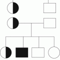
The heritable thrombophilias shown to be associated with at least a twofold increased risk of venous thrombosis are deficiencies of the natural anticoagulants antithrombin, protein C, and protein S, due to mutations in the corresponding genes SERPIN1, PROC, and PROS, and the two common mutations in genes encoding pro-coagulant factor: F5G1691A (FVR506Q, factor V Leiden) and F2G20210A (commonly referred to as the prothrombin gene mutation) (Table 3.1). The causal association between these heritable thrombophilias and venous thrombosis has been confirmed by comparing the prevalence of defects in patients with venous thrombosis and controls. The expression of heritable thrombophilia as a disease (venous thrombosis) is dependent on a strong gene-environment interaction and, in this respect, there is a strong interaction with pregnancy [1].
Numerous acquired medical conditions and environmental factors increase the risk of venous thrombosis (Table 3.1). Risk factors for venous thrombosis generally interact synergistically. This means that risk factors are not additive, rather that they multiply. For example, if two risk factors A and B each increase the risk of venous thrombosis threefold, then the combination of factors increases the risk nine times (3 × 3), not six times (3 + 3). Pregnancy is an independent risk factor for venous thrombosis, so the presence of additional heritable and acquired thrombophilias and environmental factors act synergistically to increase the risk of venous thrombosis in pregnancy. The baseline risk of venous thrombosis in women of reproductive age is low at approximately 1 per 10,000. Consequently, the relative increased risk of venous thrombosis associated with pregnancy translates into an absolute risk of only 1 per 1,000 live births overall. However, in an individual woman, a relative increase in risk due to multiple interacting factors may translate into a high absolute risk. This is the basis for assessing the risk of venous thrombosis and offering thromboprophylaxis in high-risk pregnancies (see Chap. 5).
3.2.1 Antithrombin Deficiency
Antithrombin is a protease inhibitor. Based on kinetic rates of inhibition, its primary targets are thrombin and factor Xa, and hence antithrombin both regulates generation of thrombin and inhibits thrombin that has been generated. Inhibition of target proteases is increased approximately 1,000-fold by glycosaminoglycan activation of antithrombin, which is the mechanism by which heparin acts as a pharmacological anticoagulant. The activation process involves an induced conformational change in the structure of antithrombin which enables formation of an irreversible covalent complex with the target protease [2]. The complex undergoes a further dramatic conformational change involving both the inhibitor and the inhibited protein which alters the properties of each, resulting in rapid clearance from the circulation.
Two laboratory (intermediate) phenotypes of heritable antithrombin deficiency are recognized. Type I is characterized by a quantitative reduction of antithrombin with a parallel reduction in function (measured as inhibitory activity against factor Xa or thrombin) and the level of protein in the plasma (measured immunologically as the antigenic level). Type 2 deficiency is due to the production of a qualitatively abnormal antithrombin protein characterized by disturbance of the complex inhibitory mechanism of protease inhibition as a result of a mutation in the SERPINC1 gene. The functional activity is discrepantly low compared to the antigenic level. Type 2 deficiency is subclassified according to the nature of the functional deficit:
Type 2 reactive site (RS) in which mutations alter the sequence of the mobile reactive center loop, thus reducing the ability to inhibit thrombin or factor Xa either in the presence or absence of heparin in a laboratory assay
Type 2 heparin binding site (HBS) in which mutations affect the ability of antithrombin to bind and be activated by glycosaminoglycans, resulting in reduced ability to inhibit thrombin or factor Xa only in the presence of heparin in a laboratory assay
Type 2 pleiotropic (PE) in which a single mutation produces multiple effects on the structure-function relationship of the molecule which is often associated with low plasma levels due to effects on either secretion or stability
Approximately 100 point mutations (missense, nonsense, or insertions or deletions causing frameshifts) and several whole or partial gene deletions have been identified as causes of type 1 deficiency. Numerous point mutations causing qualitative type 2 deficiency have been identified. Homozygous type 1 deficiency and type 2RS mutations are incompatible with life. Type 2 HBS and some PE mutations are associated with a lower risk of thrombosis; homozygosity, and compound heterozygosity, involving these mutations is compatible with life.
Functional activity assays typically use a chromogenic substrate and factor Xa as the target protease. The total amount of antithrombin protein can be measured immunologically with antibodies, for example, by enzyme-linked immunosorbent assay (ELISA). As antithrombin antigen levels may be normal or near normal in type 2 deficiency, immunological assays may fail to identify patients with these variants and so a functional assay should be used as the initial assay.
Although there is little reported variation in the plasma concentration of antithrombin both during healthy pregnancy and following delivery (as stated in Chap. 1), antithrombin levels may be slightly reduced in pregnancy and are reduced in women taking estrogen preparations, as well as in other situations. Consequently, the clinical significance of a low antithrombin level must be interpreted by an experienced clinician who is aware of all the relevant factors that may have influenced the test result in a specific patient.
3.2.2 Protein C Deficiency
Protein C is the zymogen precursor of activated protein C (APC). Protein C is activated to APC by thrombin bound to thrombomodulin on the endothelial surface. APC inactivates the activated cofactors (VIIIa and Va) and so inhibits thrombin generation. Factors VIII and V are activated by small amounts of thrombin during initiation of coagulation to nonenzymatic cofactors required for assembly of macromolecular complexes that are required for the full thrombin explosion. The enzymatic components of these complexes are factors IXa and Xa, and so inactivation of VIIIa and Va by APC leads to disassembly of the enzymatic complexes, thus attenuating thrombin generation.
Protein C deficiency is classified into type 1 and 2 defects on the basis of functional and antigenic assays. The relative risk of thrombosis in relation to type 1 and the various type 2 defects has not been characterized. Most heritable protein C deficiency is due to type 1 abnormalities. The majority of type 1 defects are due to point mutations. Multiple type 2 defects due to mutations in the PROC gene have been reported affecting the catalytic active site, the phospholipid-binding Gla domain, the propeptide cleavage activation site, and the sites of interaction with substrates or cofactors. In this case, there is discordance between the functional and antigenic levels.
The laboratory diagnosis of protein C deficiency is based on a functional assay. As protein C antigen levels may be normal or near normal in type 2 deficiency, immunological assays may fail to identify patients with these variants, so a functional assay should be used as the initial assay. Most commercially available functional assays use a snake venom to activate protein C and a chromogenic substrate to quantify APC activity. A chromogenic assay will detect type 1 and most type 2 defects. The diagnosis of type 1 protein C deficiency is problematic because of the wide overlap in protein C activity between heterozygous carriers and unaffected individuals. The diagnosis of type 2 defects is problematic because a chromogenic assay will only detect defects affecting the enzymatic site.
Protein C levels are not affected by pregnancy or estrogen exposure. Acquired low levels of protein C occur during anticoagulant therapy with oral vitamin K antagonists, vitamin K deficiency, disseminated intravascular coagulation (DIC), and liver disease. Consequently, the clinical significance of a low protein C level must be interpreted by an experienced clinician who is aware of all the relevant factors that may have influenced the test result in a specific patient.
3.2.3 Protein S Deficiency
Protein S is a vitamin K-dependent glycoprotein produced in the liver, endothelial cells, and megakaryocytes. Protein S is a nonenzymatic cofactor for APC-mediated inactivation of factors VIIIa and Va and additionally is involved with tissue factor pathway inhibitor-dependent natural anticoagulation. Approximately 60 % of protein S circulates bound to C4b-binding protein and is inactive. The remaining 40 %, designated free protein S, is uncomplexed and is the active form. Free protein S increases the affinity of activated protein C for negatively charged phospholipid surfaces on platelets or the endothelium and increases complex formation of APC with the activated forms of factors VIII and V (VIIIa & Va). However, the degree of C4b binding has not yet been shown to be a determinant of thrombosis risk. In addition to APC cofactor activity, protein S has an independent anticoagulant activity as a cofactor for TFPI (tissue factor pathway inhibitor).
Protein S is usually quantified immunologically rather than measured functionally. Nowadays, monoclonal antibodies that detect only free protein are used to quantify free protein S. Functional protein S assays are imprecise and are not used in the majority of coagulation laboratories.
Protein S levels are significantly lower in females, so much so that different normal reference ranges are required for males and females. There is a significant risk of a false-positive diagnosis of protein S deficiency in women. Protein S levels are reduced by estrogens and fall progressively during normal pregnancy. Acquired low levels of protein S occur during anticoagulant therapy with oral vitamin K antagonists, vitamin K deficiency, DIC, and liver disease.
Protein S defects are divided into three types:
In type I deficiency, both total and free protein S levels are low (and functional activity, if measured, is found to be low).
Type II defects are characterized by reduced activity in the presence of normal total and free levels of protein S. Type II deficiency is difficult to diagnose because functional protein S assays are imprecise.
In type III deficiency, the total protein S level is normal but the free protein S level is low. Some type III deficiency is thought to be a phenotypic variation of type 1 resulting from the same genetic mutations. However, it is now apparent that many patients with an apparent type III phenotype do not have heritable protein S deficiency. This may be related to an increase in C4b levels.
This complicated classification reflects the complexity of the biology of protein S but has no mechanistic reference to disturbance of natural anticoagulant activity. Given these limitations and the imprecision of laboratory methodology, the diagnosis of heritable protein S deficiency is less precise and the clinical implication of a low protein S level in an individual is more uncertain than it is for antithrombin or protein C.
3.2.4 F5G1691A (FVR506Q, Factor V Leiden)
Factor V is a cofactor required for thrombin generation. Factor V has no cofactor activity until cleaved by thrombin or factor Xa. Activated factor V (Va) is inactivated by APC (see Sect. 3.2.2 above). Resistance to activated protein C (APC resistance) is a laboratory phenomenon in which there is a suboptimal anticoagulant response to addition of APC to a patient’s plasma. In 95 % of cases of familial APC resistance, this is due to the same point mutation in the gene for FV, a guanine to adenine transition at nucleotide position 1691 in exon 10 (F5G1691A), resulting in a mutant protein FVR506Q. The mutation is known as the factor V Leiden mutation and the mutant factor Va has normal procoagulant activity, but substitution of glutamine for arginine at position 506 (which is an APC cleavage site) results in slower inactivation by APC. Nowadays, the mutation is frequently detected by direct DNA analysis (rather than by a clotting assay) to detect the presence of the mutant protein.
The mutation is present in around 4 % of the Caucasian population and around 15 % of unselected consecutive Caucasian patients with a first venous thrombosis. The prevalence is highest in Northern Europeans. The mutation is infrequent in other populations. The high prevalence and founder effect suggest positive selection, and this may relate to a favorable effect on embryo implantation and hence reproduction [3] rather than a lower risk of fatal hemorrhage in females during childbirth, as originally thought.
Acquired APC resistance is common, in part often due to increased FVIII levels, and is observed in pregnancy and in association with estrogen exposure.
3.2.5 F2G20210A
A single nucleotide change of guanine to adenine at position 20210 in the 3′ untranslated region of the prothrombin gene is a mild risk factor for venous thrombosis. The prevalence of the F2G20210A mutation is around 2 % in Caucasians with a higher prevalence in Southern compared to Northern Europeans. The mutation increases the plasma level of prothrombin by around 30 %, but the mechanism responsible for this has not been identified. No specific clotting test for the presence of the mutation has been described, and diagnosis depends on detection of the genetic mutation by DNA analysis.
3.2.6 Other Candidate Heritable Thrombophilias
A number of other anticoagulant proteins have been investigated as potential factors causing thrombophilia, but a relationship between venous thrombosis and low protein levels or associated gene mutations has not been established.
Increased levels of factors VIII, IX, and XI are associated with an increased risk of venous thrombosis, but a heritable basis for high levels associated with venous thrombosis is not established. There is equivocal evidence for a causal relationship between fibrinogen levels and venous thrombosis. Polymorphisms in the prothrombin gene have been described that may further increase the risk of venous thrombosis associated with the F2G20210A mutation, but the effect is mild. It was previously thought that deficiency of factor XII was a risk factor for venous thromboembolism, but subsequent investigation strongly indicates that this is unlikely. A protective effect against venous thrombosis has been reported for a polymorphism in the factor XIII gene (FXIIIV341L).
A causal relationship between levels of specific individual proteins involved in regulating fibrinolysis and venous thrombosis has not been established. However, in a case–control study using a global measure of fibrinolytic potential, there was an approximately doubled risk of venous thrombosis in patients with clot lysis times above the 90th percentile of controls [4]. Further analysis of a larger study confirmed this finding and demonstrated that hypofibrinolysis in combination with established acquired and genetic risk factors, such as F5G1691A, had a synergistic effect on venous thrombosis risk [5]. The genetic basis for hypofibrinolysis in these patients was not investigated.
Hyperhomocysteinemia may be caused by genetic abnormalities but only the severe inherited abnormalities of homocysteine metabolism (homozygous cystathionine beta-synthase deficiency and homozygous deficiency of methylenetetrahydrofolate reductase) result in congenital homocystinuria associated with an increased risk of both arterial and venous thrombosis, as well as premature atherosclerosis and mental retardation, epilepsy, and skeletal and eye problems. Fifty percent of patients present with venous or arterial thrombosis before the age of 30 years. The thermolabile variant of methylenetetrahydrofolate reductase (MTHFR), due to a common genetic polymorphism (C677T), is not a risk factor for venous thrombosis [6, 7].
3.2.7 Antiphospholipid Syndrome (APS)
The antiphospholipid syndrome (APS) is the most common acquired form of thrombophilia. APS is diagnosed when a patient with arterial or venous thrombosis (or pregnancy morbidity in women) is found to have antiphospholipid antibodies (anticardiolipin, aCL; and/or lupus anticoagulant, LA; and/or anti-beta-2-glycoprotein I, aß2-GPI). The updated international consensus (revised Sapporo) classification criteria for definite antiphospholipid syndrome [8] require the presence of a LA and/or IgG or IgM aCL present in medium or high titer (i.e., >40 GPL or MPL or > the 99th percentile) and/or aß2GPI (IgG and/or IgM) >99th percentile. These aPL should be persistent, defined as being present on two or more consecutive occasions at least 12 weeks apart. The international consensus criteria were originally designed for scientific clinical studies, and there remains a need for firm diagnostic criteria for routine clinical use which may differ from these. APS has conventionally been divided into primary and secondary forms, the latter being associated with systemic lupus erythematosus (SLE) or a related rheumatological condition. However, this distinction was abandoned in the revised Sapporo classification [8] on the basis that it is unknown whether APS and SLE are two diseases coinciding in an individual, underlying SLE offers a setting for the development of APS, or APS and SLE represent two elements of the same process.
Laboratory test results are subject to considerable pre-analytical variation. In addition, transiently abnormal results may be found in normal healthy individuals. For these reasons, for a patient to be considered to have antiphospholipid activity (aCL, LA, or aß2-GPI), test results must be positive on two separate occasions. The probability of misdiagnosing APS has been reduced by stricter criteria for antibody titers (>40 GPL or MPL for aCL or >90th percentile for aCL or aß2-GPI) and demonstration of persistence of antibodies (present on at least 2 consecutive occasions at least 12 weeks apart) [9]. Positivity in all 3 assays (aCL, LA, aß2-GPI) is associated most strongly with thrombosis and pregnancy complications. Recent evidence suggests that the antibodies most strongly associated with thrombosis and pregnancy morbidity are against domain I of β2-GPI; these antibodies are responsible for lupus anticoagulant activity specifically associated with clinical events and are responsible for positive aCL results. While the criteria for diagnosis of APS are unlikely to change again soon, it is possible that the laboratory identification of clinically relevant antibodies to domain I β2-GPI will eventually simplify the diagnosis and improve the clinical utility of laboratory tests.
3.3 Limitations of Laboratory Measurement and the Implications for Establishing a Causal Relationship and Developing Testing Strategies with Clinical Utility
The laboratory diagnosis of heritable thrombophilias is difficult as the tests are subject to considerable pre-analytical variables. Low levels of antithrombin, protein C, and protein S occur in a variety of circumstances and test results, and the clinical implications of both positive and negative results, are frequently misinterpreted. If testing is performed during pregnancy, results must be interpreted with reference to the effect of the pregnancy.
Functional assays should be used for which accuracy and imprecision are acceptable. However, no single method will detect all defects. Even in families with characterized defects, a phenotypic assay may fail to accurately discriminate affected and nonaffected individuals. True heritable deficiencies may not be detected and false positive diagnoses are common.
Low levels of antithrombin, protein C, or protein S may relate to age, sex, acquired illness, or drug therapy, so interpretation requires knowledge of the patient’s condition at the time of blood sampling. Low levels of antithrombin, protein C, or protein S suspected to be the result of heritable mutations should be confirmed on one or more separate samples. Demonstrating a low level in other family members supports a diagnosis of heritable deficiency, and characterization of the genetic mutation can be confirmatory.
As well as specific limitations relating to individual factors, there are a number of common generic issues which limit accuracy and precision of laboratory diagnosis and consequently contribute to limiting the clinical utility of thrombophilia testing. These can be summarized as follows:
The laboratory diagnosis of heritable thrombophilias is difficult as the tests are subject to numerous biological and pre-analytical variables.
The fact that venous thrombosis has a multiple genetic basis with incomplete penetrance and a strong gene-environment interaction makes counseling in relation to thrombophilia testing uncertain.
In families with known heritable thrombophilias, the risk of venous thrombosis can be increased in unaffected members as well as affected, so a negative thrombophilia result does not exclude an increased risk of venous thrombosis.
Even in families with characterized defects, a phenotypic assay may fail to accurately discriminate affected and nonaffected individuals.
True heritable deficiencies may not be detected and false positive diagnoses are common.
Low levels of antithrombin, protein C, and protein S occur in a variety of circumstances, and test results and the clinical implications of both positive and negative results are frequently misinterpreted.
Testing for heritable thrombophilias in selected patients, such as those with a strong family history of unprovoked recurrent thrombosis, may influence decisions regarding duration of anticoagulation. Unfortunately, in this regard, identifying patients for testing is not straightforward as criteria for defining thrombosis-prone families have not been validated and the association between family history of thrombosis and detection of inherited thrombophilia is weak.
In order to limit inaccuracy and imprecision, the British Society for Haematology has published clinical guidelines for testing for heritable thrombophilia [10] which include the following generic recommendations:
Testing at the time of acute venous thrombosis is not indicated as the utility and implications of testing need to be considered and the patient needs to be counseled before testing. As treatment of acute venous thrombosis is not influenced by test results, testing can be performed later.
The prothrombin time (PT) should be measured to detect the effect of oral vitamin K antagonists which will cause a reduction in protein C and S levels.
Functional assays should be used to determine antithrombin and protein C levels.
Chromogenic assays of protein C activity are less subject to interference than clotting assays and are therefore preferable.
Immunoreactive assays of free protein S antigen are preferable to functional assays. If a protein S activity assay is used in the initial screen, low results should be further investigated with an immunoreactive assay of free protein S.
Repeat testing for identification of deficiency of antithrombin, protein C, and protein S is indicated, and a low level should be confirmed on one or more separate samples. Deficiency should not be diagnosed on the basis of a single abnormal result.
Stay updated, free articles. Join our Telegram channel

Full access? Get Clinical Tree


