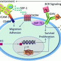
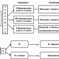
Fig. 1
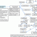
Our approach to MCL treatment
Table 1
Frontline regimens in patients ≤70 years
Frontline regimen | Age (years) | ORR (%) | PFS/FFS | OS |
|---|---|---|---|---|
R-Hyper-CVAD [16] | ≤81 | 97 | 3 years 64 % (≤65 3 years 73 %) | 3 years 82 % |
R-Hyper-CVAD [17] | <70 | 86 | 5 years 49 % | 5 years 63 % |
R-Hyper-CVAD [19] | ≤70 | 83 | 5 years 61 % | 5 years 73 % |
CALGB 59909 [21] | <70 | 88 | 5 years 56 % | 5 years 64 % |
CALGB 50403 [22] | <70 | 81 | 3 years 67 % | |
NLG MCL-2 [23] | ≤66 | 96 | 6 years 66 % | 6 years 70 % |
GELA [26] | ≤66 | 95 | 5 years 64 % | 5 years 75 % |
European MCL [27] | ≤65 | 95 | Median TTF 88 months | Median OS not reached |
Table 2
Frontline regimens in patients ≥70 years
Frontline regimen | Age (years) | ORR (%) | PFS/FFS/TTF/DOR | OS |
|---|---|---|---|---|
R-CHOP [34] | >60 | 78 | TTF 28 months, median DOR 36 months | 4 years 62 % |
R-FC [34] | >60 | 86 | TTF 26 months, median DOR 37 months | 4 years 47 % |
R-CHOP [35] | 70 (median) | 91 | 22.1 months (median) | Median not reached |
BR [35] | 70 (median) | 93 | 35.4 months (median) | Median not reached |
European older MCL [34] (R-CHOP + maintenance R) | >60 | 95 | Median DOR >6 years | 4 years 87 % |
References
1.
Swerdlow S, Campo E, Harris N et al (2008) WHO classification of tumors of hematopoietic and lymphoid tissues. World Health Organization, Lyon, pp 229–232
2.
3.
Zhou Y, Wang H et al (2008) Incidence trends of mantle cell lymphoma in the United States between 1992 and 2004. Cancerb 113(4):791–798CrossRef
4.
International Non-Hodgkin’s Lymphoma Prognostic Factors Project (1993) A predictive model for aggressive non-Hodgkin’s lymphoma. N Engl J Med 329:987–994CrossRef
5.
6.
Hernandez L, Fest T, Cazorla M, Teruya-Feldstein J, Bosch F, Peinado MA et al (1996) p53 gene mutations and protein overexpression are associated with aggressive variants of mantle cell lymphomas. Blood 87(8):3351–3359PubMed
7.
Greiner TC, Moynihan MJ, Chan WC, Lytle DM, Pedersen A, Anderson JR et al (1996) p53 mutations in mantle cell lymphoma are associated with variant cytology and predict a poor prognosis. Blood 87(10):4302–4310PubMed
8.
Martinez A, Bellosillo B, Bosch F, Ferrer A, Marce S, Villamor N et al (2004) Nuclear surviving expression in mantle cell lymphoma is associated with cell proliferation and survival. Am J Pathol 164(2):501–510PubMedCentralPubMedCrossRef
9.
10.
Cohen JB, Ruppert AS, Heerema NA et al (2012) Complex Karyotype (CK) is associated with a shortened progression-free survival (PFS) in patients with newly diagnosed mantle cell lymphoma. Blood A:2691
11.
Sarkozy C, Terré C, Jardin F et al (2014) Complex karyotype in mantle cell lymphoma is a strong prognostic factor for the time to treatment and overall survival, independent of the MCL international prognostic index. Genes Chromo Cancer 53(1):106–116. doi:10.1002/gcc.22123 CrossRef
12.
Nodit L, Bahler D, Jacobs S et al (2003) Indolent mantle cell lymphoma with nodal involvement and mutated immunoglobulin heavy chain genes. Hum Pathol 34:1030–1034PubMedCrossRef
Related posts:
 and Therapy of Chronic Lymphocytic Leukemia and Small Lymphocytic Lymphoma
 Lymphoproliferative Disorders
 of Non-Hodgkin Lymphomas: Diagnosis and Response-Adapted Strategies
 Expression Profiling in Non-Hodgkin Lymphomas
 Therapeutic Strategies and New Treatment Paradigms for Follicular Lymphoma
 of T-Cell Lymphomas: Diagnosis and Biomarker Discovery
Stay updated, free articles. Join our Telegram channel

Full access? Get Clinical Tree


