1
No uptake
2
Uptake ≤ mediastinum
3
Uptake > mediastinum but ≤ liver
4
Uptake moderately higher than liver
5
Uptake markedly higher than liver and/or new lesions
X
New areas of uptake unlikely to be related to lymphoma
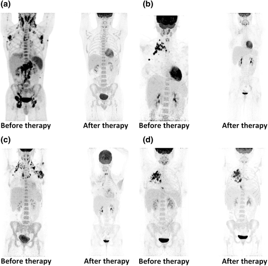
Fig. 1
Legend Examples of baseline and interim PET images with response assessed according to the Deauville five-point scale. a Deauville score 1. b Deauville score 1. c Deauville score 1. d Deauville score 1
3.1 Interim PET in Indolent NHL
There is a relative paucity of data on the value of interim FDG-PET in indolent lymphomas. Bishu et al. reported a retrospective series of 31 patients with advanced-stage FL, of whom 11 had FDG-PET scans midway through four cycles of induction chemotherapy. The number of patients was too small for any firm conclusions. Still, the results showed interesting differences: Four patients with some persistent FDG uptake had a mean PFS of 17 months and seven interim PET-negative patients had a mean PFS of 30 months [50]. There are only casuistic reports on the value of interim FDG-PET in localized FL or the more uncommon subtypes of indolent NHL, such as marginal zone lymphoma and small lymphocytic lymphoma.
Even if a clear correlation between interim FDG-PET and treatment outcome is shown in the future, the clinical implications are uncertain, since advanced-stage indolent lymphoma is very different from aggressive NHL. First of all, current first-line therapies for advanced-stage FL are not curative, and it is unclear whether early treatment intensification with autologous stem cell transplant (ASCT) or allogeneic bone marrow transplantation is of any benefit to poor-risk patients. Secondly, the long natural history of FL and the relative success of subsequent treatment lines in inducing lasting remissions mean that a longer time to first progression does not necessarily translate into longer survival.
3.2 Interim PET in Aggressive NHL
In aggressive NHL, PFS ranges from 10 to 50 % at 1 year for early PET-positive patients and from 79 to 100 % at 1 year for early PET-negative patients. The high risk of relapse seen among PET-positive patients is consistently shown in both early and advanced stages. Mikhaeel et al. studied a large cohort of 121 high-grade NHL patients, retrospectively and with a median follow-up of 28.5 months. They confirmed that response on FDG-PET after two or three cycles strongly predicts PFS and OS. Estimated 5-year PFS was 89 % for the PET-negative group, 59 % for patients with minimal residual uptake (MRU) on FDG-PET, and 16 % for the PET-positive group. Survival analyses showed strong associations between early FDG-PET results and PFS and OS [46]. Haioun et al. prospectively studied 90 patients with aggressive NHL, performing FDG-PET after two cycles of chemotherapy. They similarly found 2-year PFS rates of 82 and 43 % in early PET-negative and early PET-positive patients, respectively, and 2-year OS rates of 90 and 60 % [47]. Spaepen et al. compared interim FDG-PET after two cycles of chemotherapy with the International Prognostic Index (IPI). In multivariate analysis, FDG-PET at mid-treatment was a stronger prognostic factor for PFS and OS than was the IPI (P < 0.58 and P < 0.03, respectively) [42]. A number of other more recent studies have investigated the value of interim PET specifically in DLBCL cohorts and found negative predictive values similar to those found in the above-mentioned studies of mixed aggressive NHL populations. But the positive predictive value is variable and generally poor [51, 52]. A possible explanation for the low positive predictive value in aggressive NHL cohorts is the higher risk of inflammation and infections among patients who are treated with the more dose-dense and dose-intensive NHL regimens, as mentioned above. In order to overcome the low positive predictive value of interim PET, some investigators have proposed a semiquantitative approach (relative or absolute change in SUV from baseline to interim scan) as superior to visual PET assessment in patients with DLBCL [53, 54].
Aggressive NHL covers a number of lymphoma subtypes where first-line treatment of both localized and advanced disease stages is given with a curative intent. Aggressive NHL patients who respond poorly to first-line treatment or relapse soon afterward generally have a very poor prognosis even with high-dose salvage regimens. Such poor-risk patients could benefit from having their treatment failure recognized early during first-line therapy in order to switch a more intensive regimen as soon as possible. Much inspired by a similar and more widespread trend in Hodgkin lymphoma, a number of trials are currently investigating early PET response-adapted treatment strategies (Table 2). Most studies test whether early or mid-treatment PET-positive patients with aggressive NHL (mainly DLBCL) will benefit from early escalation to more intensive regimens or even high-dose therapy with autologous stem cell support (ASCT) [55–60]. One de-escalation study compares standard treatment with six cycles R-CHOP to good-risk DLBCL patients to abbreviated therapy with four cycles R-CHOP in patients with a negative PET after two cycles of therapy [61].
Table 2
PET response-adapted trials in aggressive NHL
Study title/description | Study group | Ref | Patients | Main PET-driven intervention | Study type |
|---|---|---|---|---|---|
Risk-adapted dose-dense immunochemotherapy determined by interim FDG-PET in advanced DLBCL | MSKCC | DLBCL | Salvage with HD + ASCT if PET positive after 4 × R-CHOP | Phase II | |
FDG-PET in predicting relapse in NHL patients undergoing chemotherapy with or without ASCT | Johns Hopkins | Aggressive NHL | Salvage with HD + ASCT if PET positive after 2–3 × (R-)CHOP | Phase II | |
Positron emission tomography-guided therapy of aggressive non-Hodgkin’s lymphomas (PETAL) | University Hospital, Essen | Aggressive NHL | (R-)CHOP versus Burkitt regimen if PET positive after 2 × (R-)CHOP | Phase III | |
Tailoring treatment for B-cell non-Hodgkin’s lymphoma based on PET scan results mid-treatment | British Columbia Cancer Agency | Advanced DLBCL | 4 cycles R-ICE if PET positive after 4 × R-CHOP | Phase II | |
FDG-PET-stratified R-DICEP and R-Beam/ASCT for diffuse large B-cell lymphoma (PET CHOP) | Alberta Cancer Board | DLBCL | Salvage with HD + ASCT if PET positive after 2 × R-CHOP | Phase II | |
A study of associations between rituximab and chemotherapy, with PET-driven strategy in lymphoma (LNH2007-3B) | LYSA | DLBCL | Salvage with HD + ASCT if PET positive after 2 × R-CHOP | Phase IIIa | |
Therapy for aaIPI intermediate or high-risk DLBCL | MSKCC | DLBCL | High dose if PET positive and biopsy positive after 3 cycles of immunochemotherapy | Phase II | |
Treatment adapted to the early PET compared to a standard treatment in low risk (aaIPI = 0) DLBCL | LYSA | DLBCL | 4 × R-CHOP versus 6 × R-CHOP for early PET-negative patients | Phase III |
4 PET/CT for Post-therapy Response Evaluation of Lymphomas
Response to treatment serves as an important surrogate for other measures of clinical benefit such as progression-free survival and overall survival. Response is also an important guide in decisions regarding continuation or change of therapy. Until recently, response evaluation in NHL was done according to the International Workshop Criteria [62], based mainly on morphological evaluation with a reduction in tumor size on CT being the most important factor. But after completion of therapy, CT scans will often reveal residual masses, and the presence of such masses is not highly predictive of outcome. By conventional methods, it is very difficult to assess whether this represents viable lymphoma or fibrotic scar tissue. An extensive number of studies have shown that FDG-PET performed post-treatment is highly predictive of PFS and OS with and without residual masses on CT. The vast number of studies in aggressive NHL has been summarized in a systematic review [63], while the data on indolent NHL are based on fewer studies [64–66].
Based on these findings, the International Harmonization Project developed new recommendations for response criteria for aggressive malignant lymphomas, incorporating FDG-PET into the definitions of end-of-treatment response in FDG-avid lymphomas [67, 68]. Subsequent retrospective analyses confirmed the superiority of the new PET/CT-based response criteria [69]. The recent revision of the international lymphoma staging and response criteria still uses PET response (according to the Deauville five-point scale) as the main determinant for post-treatment assessment, while measurements of the sum of the product of perpendicular diameters of measureable lesions are mainly important for the definition of progressive disease [3]. The negative predictive value of post-treatment PET/CT is high in most NHL subtypes, but due to a relatively low positive predictive value, treatment failure suspected due to a positive PET should be confirmed by biopsy before additional therapy is initiated. Alternatively, it is often preferable to wait and repeat the PET/CT after 1–2 months, provided the patient is not in clinical progression.
Unlike in Hodgkin lymphoma, the role of post-treatment PET/CT for selection of NHL patients for consolidation radiotherapy is unclear. Based on promising data showing a very high negative predictive value of PET/CT in primary mediastinal B-cell lymphoma [70], the International Extranodal Lymphoma Study Group conducts an experimental approach where consolidation radiotherapy is given only to patients with a PET-positive residual mass [71]. Also, the British Columbia Cancer Agency has since 2005 routinely used post-chemotherapy PET for all patients with residual masses larger than 2 cm. Only PET-positive patients receive consolidation radiotherapy. With 262 patients included in the series and a median follow-up of 45 months, only one of 179 PET-negative patients has received radiotherapy. The 4-year PFS and OS are 74 and 83 %, respectively. Since the data have still only been presented in abstract form, it is unfortunately unclear whether the relapses were predominantly located within or outside the sites of residual disease. If most relapses occur outside the sites of residual disease, there seems to be little value of consolidation radiotherapy in PET-negative patients, but this is not quite so clear whether a substantial number of relapses are seen within the sites of residual disease after first-line treatment [72].
5 Disease Surveillance After First Remission
5.1 Aggressive Non-Hodgkin Lymphomas
The value of routine surveillance imaging in aggressive NHL in first remission is controversial. Despite the lack of evidence, surveillance imaging is often performed with regular intervals during the first years in remission. The rationale behind this may be a presumed better outcome in patients with early and asymptomatic relapse as compared to those patients presenting with overt lymphoma symptoms. Early detection of relapse could in theory be advantageous for the following reasons: (1) lower tumor burden at relapse (and maybe reduced tumor heterogeneity and prevention of treatment resistant clones) and (2) less disease-related impairment of function and organs with better tolerability of treatment. Low disease stage and ECOG performance status 0–1 at relapse are associated with a better outcome in DLBCL [73]. With less than a quarter of the relapses being imaging detected, routine imaging did not contribute significantly to relapse detection in studies based on imaging modalities less sensitive than PET/CT [74–78]. Furthermore, the number of scans per relapse exceeded 100 per preclinical relapse for both DLBCL and PTCLs in first complete remission, and out of all patients in complete remission entering intensive follow-up protocols, only a very small minority (2–3 %) will experience imaging-detected relapse [78]. Few studies have investigated the value of PET/CT in the surveillance of aggressive NHL, mainly reporting on DLBCL. Generally, the rate of imaging-detected relapse seems to be higher when using regular PET/CT surveillance in first remission, and depending on the surveillance protocol, 31–100 % of relapses were detected by imaging [79–81]. However, with the number of PET/CT scans per relapse ranging between 35 and 120 [81, 82] and an estimated cost of up to US$85,550 per preclinical PET/CT-detected relapse, an indiscriminate use of PET/CT surveillance is not likely to be cost-effective. Restricting the use of PET/CT to high-risk patients (IPI > 2) decreases the number of PET/CT scans per relapse to 22 and may tip the balance toward better cost-effectiveness [81]. In patients with transformed indolent lymphomas undergoing routine imaging with PET/CT at least one time during follow-up, the PET/CT-detected relapses were all indolent histologies and less important from a clinical perspective. On the contrary, cases of relapsed DLBCL were symptomatic [83]. Since FDG uptake is unspecific and not restricted to lymphoma recurrence, the positive predictive value (PPV) of PET/CT surveillance in aggressive NHL has been reported low to moderate with values between 21 and 60 % [79, 81, 84]. The increasing use of PET/CT for lymphoma has probably led to safer recognition of uptake patterns characteristic for lymphoma and hence less false-positive reporting. Still, a confirmatory biopsy of lymphoma suspicious findings on PET/CT is mandatory whenever possible. As opposed to the problematic PPV of PET/CT, the negative predictive value has been reported uniformly high and often 100 % [79, 81, 84]. This means that a negative PET/CT study virtually rules out recurrent disease and the need for further investigations in patients with symptoms suggesting lymphoma relapse. Finally, the use of routine surveillance imaging in aggressive NHL can only be justified if early relapse detection is associated with improved survival. Some studies have suggested that imaging-detected relapse of aggressive non-Hodgkin lymphoma could be associated with improved outcome, whereas others found no difference in outcome [76, 78, 81]. However, the retrospective design of these studies does not allow firm conclusions about the causal role of imaging due the presence of important bias (lead time, length time, and guarantee time). Interestingly, a recent randomized trial of surveillance strategies in Hodgkin lymphoma showed that the use of chest X-ray and ultrasound was just as effective as PET/CT in early relapse detection, but at a much lower cost and with higher PPV [85].
5.2 Indolent Non-Hodgkin Lymphomas
While use of routine imaging in the follow-up of aggressive NHL is still widespread, the use of routine imaging in the follow-up of indolent NHL is not recommended. An observational approach without initial treatment is often chosen for patients with an asymptomatic relapse of an indolent lymphoma. An exception could be specific cases where an asymptomatic relapse could lead to unrecognized compression of vital organs such as vascular structures. In any case, PET/CT has no role in this setting.
6 Response Prediction with FDG-PET Before High-Dose Salvage Therapy
Duration of remission prior to relapse and the response to induction therapy are important prognostic factors that predict a good outcome after high-dose chemotherapy with autologous stem cell support (HD + ASCT) [86, 87]. A number of studies have shown that PET performed after induction therapy and before HD + ASCT is predictive of long-term remission. These studies all report short PFS in patients with persistent disease on pretransplant PET [88–93]. In relapsed Hodgkin lymphoma, Moskowitz and his group demonstrated that patients could benefit from changing to another non-cross-resistant regimen before HD + ASCT rather than proceeding straight to high-dose therapy if the PET was still positive after the initial induction therapy [94]. Even though no such data are yet available in NHL, it seems clear that achieving a negative PET before HD + ASCT should be a goal for all patients.
7 Other Imaging Methods than FDG-PET and CT
7.1 Newer PET Tracers
Like other cancers, lymphoma is characterized by deregulated cell cycle progression and most anticancer drugs are designed to inhibit cell proliferation. So a tracer enabling imaging of cell proliferation could be useful for both initial characterization and treatment monitoring of the disease. FDG uptake is somewhat correlated with cell proliferation, but this correlation is weakened by a number of factors, including FDG uptake in non-malignant lesions [95, 96]. The nucleoside [11C]thymidine was the first PET tracer to specifically address cell proliferation. Early studies showed that [11C]thymidine could determine both disease extent and early response to chemotherapy in aggressive NHL patients [97, 98]. However, the short 20-min half-life of 11C along with rapid in vivo metabolism has limited the clinical application of [11C]thymidine. The thymidine analogue 3′-deoxy-3′-[18F]fluorothymidine (FLT) offers a more suitable half-life of 110 min and is stable in vivo [99]. More recent studies have shown that FLT-PET can sensitively identify lymphoma sites [100]. FLT uptake is highly correlated with proliferation rate and may thus be able to distinguish between high- and low-grade lymphomas [101, 102]. And furthermore, recent studies have showed a potential of FLT for imaging early response to treatment in NHL [103–106]. Amino acid metabolism of cancer cells is influenced by catabolic processes favoring tumor growth [107]. It has been shown that increased uptake of amino acids reflects the increased transport and protein synthesis of malignant tissue [108, 109]. This is the background for PET imaging of amino acid metabolism with the labeled amino acids L-[methyl-11C]methionine (MET) and O-2-[18F]fluoroethyl)-L-tyrosine (FET) [110]. Nuutinen et al. [111] studied 32 lymphoma patients and found MET-PET highly sensitive for the detection of disease sites although there was no correlation between MET uptake and patient outcome. Also, MET-PET has an advantage over FDG-PET in imaging of CNS lymphoma [112]. While these results are encouraging, it should be noted that no studies have shown the usefulness or cost-effectiveness of amino acid or nucleoside tracers in large patient cohorts. Furthermore, high physiological tracer uptake in the abdomen limits the usefulness of these tracers for imaging of abdominal and pelvic lymphomas.
With CD20 expressed as a cell surface antigen in most cases of B-NHL, and with CD20 as an important therapeutic target, it is logical to also target CD20 for the purpose of lymphoma imaging. However, most of the available so-called immunoPET tracers are based on full-size, intact antibodies, and as a result of this, it takes a long time for the background activity levels to drop sufficiently to provide acceptable target-to-background ratios. Therefore, Olafsen and colleagues generated a recombinant anti-CD20 fragment which, labeled with a PET tracer, produced high-contrast images in a small-animal model [113]. This and similar targeted PET methods may in the future provide very lymphoma-specific images and prove useful in the staging and treatment monitoring of NHL.
7.2 Magnetic Resonance Imaging
MRI has long been the main structural imaging methods in pediatric lymphomas, due to the reduction in radiation exposure. MRI is used in adult patients to evaluate central nervous system disease and for disease sites in the head-and-neck region (e.g., NK/T-cell lymphomas of nasal type). Recent studies show that diffusion-weighted MRI early during treatment may identify responders before a reduction in tumor size occurs [114].
8 Conclusions and Recommendations
Supported by a large number of studies and in line with the recent consensus recommendations for lymphoma imaging as well as the revised criteria for lymphoma staging and response evaluation, PET/CT is recommended for staging and post-treatment response assessment of all FDG-avid NHLs [2, 3]. There are sufficient data to support the use of interim PET for treatment monitoring in aggressive B-NHL when interim imaging is appropriate. CT is still the preferred imaging method for non-FDG-avid lymphomas, as well as for selected cases where routine surveillance imaging is considered necessary. For interim and post-treatment imaging, the Deauville five-point scale is recommended for interpretation and reporting of PET results. The use of interim PET imaging to guide therapeutic decisions is still investigational and should preferably be done in the setting of clinical trials. Semiquantitative and quantitative PET assessments (SUV, MTV, TLG) are promising, but their role is still to be defined. Currently, much effort is put into the standardization of PET/CT methodology and interpretation, making the results of trials more comparable and ensuring a better translation into clinical practice. Upcoming results of interim PET response-adapted therapy trials are likely to impact on the management of de novo and relapsed NHL patients in the near future. The dissemination of novel and more disease-specific PET tracers may improve the basis for risk- and response-adapted therapy, hopefully in combination with better pretreatment predictive molecular pathology markers.
References
1.
Flowers CR, Armitage JO (2010) A decade of progress in lymphoma: advances and continuing challenges. Clin Lymphoma Myeloma Leuk 10(6):414–423PubMed

