Figure 175.1 Histopathologic demonstration of the cigar-shaped yeasts of Sporothrix schenckii. (CDC/Dr. Lucille K. Georg, CDC Public Health Image Library.)
Clinical manifestations
Cutaneous sporotrichosis
The primary lesion develops at the site in the skin, 20 to 90 days after inoculation, most typically distally in the upper extremities. Over a few weeks, the initial small nodule enlarges, reddens, becomes pustular, and ulcerates, releasing purulent material from which the organism is readily cultured. Patients are typically afebrile and not systemically ill. In the lymphocutaneous form of the disease, an ascending chain of nodules develops along lymphatic channels of the skin, with the older distal lesions ulcerating and draining and the younger, more proximal lesions forming subcutaneous nodules that attach to the skin as they age and begin to ulcerate (Figure 175.2). The lesions are usually minimally painful, but extensive disease may result in functional impairment. Some patients exhibit no lymphangitic spread, and the disease presents as an indolent ulcerating plaque that persists for years if untreated (fixed cutaneous sporotrichosis). Patients often have received courses of antibacterial therapy without benefit before the process is recognized as sporotrichosis. The lymphocutaneous form of sporotrichosis can be mimicked by infection with Nocardia, Mycobacterium marinum and other mycobacteria other than tuberculosis, Leishmania, and Francisella tularensis.
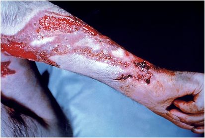
Figure 175.2 Lymphocutaneous sporotrichosis. (CDC/Dr. Lucille K. Georg, CDC Public Health Image Library.)
Pulmonary sporotrichosis
Pulmonary sporotrichosis is a subacute or chronic pneumonitis with cavitation, usually in the upper lobes, clinically indistinguishable from mycobacterial infection or chronic pulmonary histoplasmosis. Almost all patients have underlying chronic obstructive pulmonary disease. They present with productive cough, sometimes with weight loss and increasing dyspnea, but rarely with fever, chills, or sweats. Diagnosis requires isolation of S. schenckii from sputum cultures or its histopathologic recognition in biopsy specimens.
Osteoarticular sporotrichosis
Lesions of deeper tissues may occur in almost any organ, but there is a distinct predilection for the joints, particularly of the extremities, and the long bones adjacent to these joints. The resulting chronic arthritis is often confused with rheumatoid or other chronic inflammatory arthritis, not infrequently for 10 or more years, until destruction of adjacent bone or development of draining fistulas encourage efforts to establish the microbial etiology of the chronic osteomyelitis. Cutaneous or lymphocutaneous lesions are unusual in these patients. The process generally begins in a single joint, but additional joints may be involved successively. The patient usually has pain on motion, and the involved areas may be warm and red. Functional impairment resulting from osteoarticular sporotrichosis can become very severe.
Disseminated sporotrichosis
Sporotrichotic lesions occur infrequently in many other organs such as the eye, the prostate, the oral mucosa, and the larynx, and the clinical manifestations in these patients depend on the organ involved. Involvement of the central nervous system (CNS) and
Related posts:

Stay updated, free articles. Join our Telegram channel

Full access? Get Clinical Tree


