Figure 24.1 Scabies mite Sarcoptes scabiei female. (Courtesy of Dr. Arezki Izri, Université Paris 13 and Department of Parasitology, Hôpital Avicenne, Bobigny, France.)
The definitive diagnosis relies on the identification of mites. Multiple superficial skin samples should be obtained from characteristic lesions by scraping with a scalpel. The specimens are examined under a microscope, looking for mites, eggs, empty eggs, and scybala. Biopsy may help. Failure to find a mite is common and does not rule out scabies.
Therapy
People with scabies and their close physical contacts, even without symptoms, should receive treatment at the same time.
Topical treatment includes 5% permethrin, 10% to 25% benzyl benzoate, esdepalletrine, lindane (withdrawn from the market in most western countries), crotamiton, and precipitated sulfur. Topical scabicides have neurotoxic effects on mites and larvae. Despite the weak level of evidence of randomized controlled trials (RCTs), a recent meta-analysis suggested that topical 5% permethrin is the most effective.
Oral ivermectin interrupts the gamma-aminobutyric acid-induced neurotransmission of many parasites, including mites. However, oral ivermectin is not licensed for scabies in most countries. It is prescribed at 200 µg/kg as a single dose in patients >2 years of age and >15 kg of weight. A second dose is mandatory one to two weeks later due to the lack of ovicidal action of the drug. Ivermectin may be used as first-line therapy, but its higher cost in some countries supports consideration of initial therapy with topical agents. Ivermectin should be routine therapy for patients who have no response to a topical scabicide, and it may be the appropriate first choice for the elderly, patients with generalized eczema, and other patients who may be unable to tolerate or to comply with topical therapy.
Patients should received detailed information about scabies infestation and therapeutic options, including the amount of drug to be used and proper administration. Topical treatment should be applied to the entire skin surface, including the scalp, face (in children and in the elderly), all folds, groin, navel, and external genitalia, as well as the skin under the nails and reapplied again 7 to 14 days after the first treatment. Hands should not be washed during therapy (protection mandatory in infants), otherwise the treatment should be reapplied.
After completion of treatment, patients should use clean clothing and bedding. If possible, potentially contaminated articles should be washed at high temperature (>50°C) or kept in a plastic bag for up to 72 hours, because mites that are separated from the human host will die within this time period. The use of insecticidal products should be restricted to unwashable materials.
Patients should be informed that itching may persist, especially in atopic individuals. After 4 weeks, the cause of itching should be reinvestigated.
Pediculosis capitis
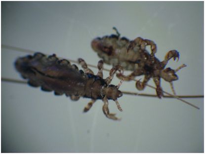
The most common form of louse infestation is caused by the head louse, Pediculus humanus (designated Pediculus humanus capitis in the past to differentiate it from the body louse, formerly designated Pediculus humanus humanus, which has now been found to be genetically identical to the head louse). Since the 1970s, the prevalence has increased in many countries. Head lice predominantly infest schoolchildren (and their mothers) of all socioeconomic groups; transmission occurs through head-to-head contact, with the classroom being the main source of infestation. Active infestation is based on the finding of live lice. The stage of the louse most commonly seen is the nit. Each nit is oval, opaque, and white (about 0.8 × 0.3 mm) and is firmly attached individually to a single hair by the female louse. Nymphs or viable-appearing nits (louse eggs) are located about 1 mm from the scalp surface. Three immature stages (nymphs) precede the formation of the adult louse. Adult and immature lice are wingless and, as in all insects, have six legs. Each leg ends in a claw used for gripping hair. The adult lice are about 2 to 3.5 mm long and are white or cream in color (Figure 24.2).

Figure 24.2 Adult human louse Pediculus humanus. (Courtesy of Dr. Arezki Izri, Université Paris 13 and Department of Parasitology, Hôpital Avicenne, Bobigny, France.)
Infested individuals usually first notice itching of the scalp, most often in the postauricular and occipital regions, but this pruritus occurs in a variable proportion of children. All immatures and adults require blood and, as a result of feeding, produce erythematous, papular lesions that are the cause of the pruritus. Some patients react to louse saliva with urticaria or lymphadenopathy. Secondary bacterial infection may occur as a result of scratching, and concomitant head-louse infestation should always be considered in cases of scalp impetigo or posterior cervical lymph node enlargement in the absence of other lymphadenopathy.
Therapy
Management of head-louse infestation is difficult because good comparative-effectiveness research is still lacking and louse resistance to pyrethroid has emerged. A Cochrane systematic review is in process. DNA sequencing showed that “knockdown resistance” (kdr) to permethrin was linked to a three-point mutation (M815I-T917I-L920F) in the louse voltage-gated sodium channel α-subunit gene, conferring nerve insensitivity. However, genetic resistance might not be predictive of clinical or parasitologic failure. It is recommended to use 1% permethrin or pyrethrin insecticide as first-line therapy. If resistance in the community has been proven or live lice are present 1 day after the completion of treatment, a switch to malathion may be necessary. Other options include wet combing, also called “bug busting,” or treatment with dimethicone or other topical agents (see below), depending on the availability of the agents in the country. All treatments should be applied two times a week apart, because of insufficient ovicidal activity. A recent RCT showed that a single oral dose of ivermectin (400 µg per kilogram of body weight) repeated within 7 days achieved higher louse-free rates on day 15 than 0.5% malathion lotion among patients with difficult-to-treat head lice. The safety of such dosage of ivermectin in patients with head-louse infestation remains unknown, and subsequently should only be used in the case of failure of all other topical treatments (off-label). Topical ivermectin has shown greater efficacy than placebo in a recent RCT and has been approved by the US Food and Drug Administration (see Table 24.1).
Stay updated, free articles. Join our Telegram channel

Full access? Get Clinical Tree


