REPRODUCTIVE EMBRYOLOGY
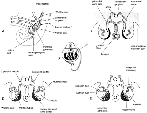
Early in ontogeny, the gonads of both sexes are indifferent and bipotential. Large primordial germ cells are visible in the fourth week among the endodermal cells of the wall of the yolk sac near the origin of the allantois. In the fifth week, these germ cells migrate by ameboid movement along the dorsal mesentery of the hindgut to the gonadal ridges (Fig. 90-9). During the sixth week, the primordial germ cells migrate into the underlying mesenchyme and become incorporated in the primary sex cords (Fig. 90-10, see also Fig. 90-9).
Divergent development begins at ˜40 days of gestation. With the development of the gonad, controlled by a gene or genes regulating testicular differentiation, the translation of gonadal sex into phenotypic sex follows predictably as a function of the type of gonad formed.
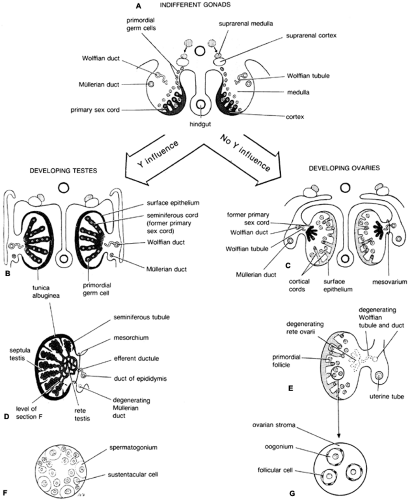
In males, gonaductal differentiation depends on at least two substances secreted by the fetal testes (Fig. 90-11 and Fig. 90-12). The first, testosterone, is produced by the Leydig cells. This hormone stabilizes the wolffian ducts to stimulate the growth and development of the epididymis, vasa deferentia, and seminal vesicles. The second, AMH, is a glycoprotein secreted by the Sertoli cells that acts locally to cause nearly complete regression of the müllerian ducts by 8 weeks of fetal age, before the secretion
of testosterone and the stimulation of the wolffian ducts.39 Studies have established that AMH is similar structurally to transforming growth factor-β (TGF-β) and to ovarian inhibin.39,40 and 41 The structural locus has been localized to the short arm of chromosome 19.42 The receptor for AMH, a serine-threonine kinase with a single transmembrane domain, is expressed in the region around the fetal müllerian duct and in Sertoli and granulosa cells.43,44
of testosterone and the stimulation of the wolffian ducts.39 Studies have established that AMH is similar structurally to transforming growth factor-β (TGF-β) and to ovarian inhibin.39,40 and 41 The structural locus has been localized to the short arm of chromosome 19.42 The receptor for AMH, a serine-threonine kinase with a single transmembrane domain, is expressed in the region around the fetal müllerian duct and in Sertoli and granulosa cells.43,44
Stay updated, free articles. Join our Telegram channel

Full access? Get Clinical Tree


