Regulatory/Suppressor T Cells


Regulatory/Suppressor T Cells


Ethan M. Shevach


INTRODUCTION

T cells are crucial in the immune response because they can function as both effector cells in cell-mediated responses and as helper cells in both humoral and cell-mediated responses. Most biologic systems are subject to complex regulatory controls, and the immune system is not an exception. In addition to T cells that upregulate (help), other populations that downregulate (suppress), the immune response must exist. Once a normal immune response is initiated by antigenic stimulation, mechanisms must be in place to control the magnitude of that response and to terminate it over time. Downregulation should contribute to the homeostatic control of all immune responses serving to limit clonal expansion and effector cell activity in response to any antigenic stimulus. An active mechanism of T-cell suppression is also needed to control potentially pathogenic autoreactive T cells. The primary mechanism that leads to tolerance to self-antigens is thymic deletion of autoreactive T cells, but some autoreactive T cells may escape thymic deletion or recognize antigens that are expressed only extra-thymically. T-cell anergy1 and T-cell ignorance/indifference2 have been proposed as the primary mechanisms used to control autoreactive T cells in the periphery, although these “passive” mechanisms for self-tolerance may not be sufficient to control potentially pathogenic T cells.

It was proposed more than 40 years ago that a distinct subset of T cells is responsible for immune suppression.3 A suppressor T cell is functionally defined as a T cell that inhibits an immune response by influencing the activity of another cell type. Although a strong theoretical basis exists for T cell-mediated suppression, this area of immunologic research has been plagued by controversy. The past decade has seen a resurgence of interest in the concept of T-cell suppression mediated by a distinct subset of T cells that are uniquely equipped to mediate suppressor activity.


HISTORICAL PERSPECTIVE

Gershon and Kondo3,4 first identified suppressor T cells during studies designed to understand the process of “highzone” tolerance. Injection of supraoptimal doses of an antigen including sheep red blood cells (SRBCs) resulted in specific tolerance or nonresponsiveness to subsequent challenge with that antigen. It was believed at that time that the antibody-producing B cell was rendered nonresponsive by exposure to the high concentration of antigen. To investigate whether B-cell tolerance was dependent on the presence of T cells, Gershon and Kondo injected high doses (2.5 × 10¹⁰) of SRBCs into thymectomized (Tx), irradiated, bone marrow-reconstituted mice and then assayed the functional status of B cells from these mice by a secondary challenge with SRBCs in the presence of added thymocytes as a source of T-cell help. Surprisingly, nonresponsiveness as measured by deficient antibody production was only induced in the B cells of animals that had received thymocytes as well as bone marrow cells during the initial exposure to high dose antigen, but not in mice that received bone marrow alone (Fig. 33.1). This result fulfilled Gershon’s prediction that under certain conditions, antigen seen by T cells can induce not only helper and effector cells, but also cells that are able to suppress immune responses. Furthermore, when spleen cells from tolerized animals were transferred into secondary recipients together with normal thymocytes and bone marrow cells, they were capable of suppressing the otherwise competent response of these animals to SRBCs. This suppression, or “infectious tolerance” as it was originally termed, was antigen-specific, as the immune response to an unrelated antigen, horse red blood cells, was not inhibited.4 T cells were necessary for the induction of B-cell tolerance, and these T suppressor cells were assumed to be a distinct cell population with a fully differentiated gene program that allowed them to perform a very specialized function. Other studies5,6,7 during the 1970s supported the existence of T cell-mediated suppression.

Most of the early studies in these models demonstrated that the T cells mediating suppression were distinct from T cells mediating help because the former were cluster of differentiation (CD)8+, while the latter were CD8-. The finding that T suppressor effector cells were CD8+ distinguished them from helper cells, but did not allow them to be distinguished from cytotoxic cells, which are also CD8+. It remained possible that suppressor T cells were actually cytotoxic cells that killed the helper or effector T cells. A cell surface marker that seemed to identify a suppressor cell-specific antigen was discovered in 1976. It was found that an antiserum raised by immunizing the congenic strains B10. A(3R) with cells from B10. A(5R) mice or vice versa gave rise to an antiserum that seemed to react exclusively with suppressor cells.8 CD8+ suppressor cells were also shown to bind antigen in the absence of major histocompatibility complex (MHC) molecules. These experiments suggested that the suppressor effector cells differed from other T cells in that they were capable of binding antigen directly and did not recognize processed antigenic peptides in association with
products of the MHC on the surface of antigen-presenting cells (APCs). Soon after the existence of T cell-mediated suppression was appreciated, some studies suggested that interactions among multiple distinct T-cell subpopulations might be involved. A CD4+ cell was described that appeared to induce CD8+ suppressor cells and was called the suppressor inducer cell. Contrasuppressor T cells had no independent helper, suppressor, or cytotoxic activity on an immune response, but enhanced immune responses by preventing the downregulation mediated by suppressor cells.


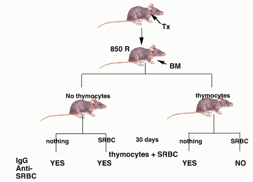
FIG. 33.1. First Demonstration of Suppressor T Cells. Adapted from Gershon and Kondo.3

Research in this area rapidly shifted from studies of the function of intact T cells to studies of their soluble products.9 By the late 1970s, soluble factors produced by T suppressor cells were described by several groups, and cloned T-cell hybridomas that produced such factors were generated. T-cell suppression was regarded as being mediated by numerous soluble antigen-specific and nonspecific factors that comprised a functionally unique network.10,11,12 The cascade involved antigen-specific, I-J-restricted CD4+ suppressor inducer cells (Ts1 cells), CD8+ anti-idiotype-specific cells (Ts2 cells), and CD8+ antigen-specific effector cells (Ts3 cells), whose suppressor function was not restricted by the MHC. Some of these cells were capable of binding directly to immobilized antigen in the absence of MHC molecules. Connectivity in this cellular cascade was mediated by a series of soluble factors: TsF1 was idiotypic and antigen-specific and immunoglobulin (Ig) heavy chain variable (VH) region restricted. TsF2 was anti-idiotypic and required delivery by a macrophage. TsF3 acted totally nonspecifically. TsF1 and TsF2 required APCs, but TsF3 did not. Most of these factors were composed of two polypeptide chains, one of which was capable of binding native antigen and the other which bore a determinant recognized by anti-I-J antibodies.

These elaborate highly convoluted suppressor cell pathways and circuits fell out of favor in the mid-1980s for a number of important reasons. The existence of I-J was called into question by the finding that the region of the MHC complex to which I-J mapped did not contain a gene that could encode a unique I-J polypeptide.13 When the genes encoding the T-cell receptor (TCR) were isolated, they were completely unrelated to the genes encoding Ig heavy chains, thereby calling into question the existence of T-cell factors that expressed Ig V region products. Many of the suppressor T cell hybridomas that produced antigen-specific suppressor factors were found either to have unrearranged genes for the α- or β- chains of the TCR or to have deleted genes for TCR β-chain.14 No studies were ever performed that convincingly characterized at the molecular level any of the suppressor
T cell factors. These studies, together with the inability to identify a marker specific for suppressor T cells and the inability to purify suppressor T cells, raised considerable doubts about the existence of a distinct functional lineage of suppressor T cells.


FIG. 33.2. Depletion of CD4+ CD25+ T Cells Results in Organ-Specific Autoimmunity. Adapted from Nishizuka and Sakakura15 and Sakaguchi et al.21

Two completely different approaches to the demonstration of the importance of regulatory or suppressor T cells in the prevention of organ-specific autoimmunity were also developed in the 1970s. In one, mice that were Tx on the third day of life (d3Tx) were shown to develop organ-specific autoimmune diseases (Fig. 33.2). The specific disease that developed varied with the strain of mouse under study; more than one organ could be involved in a given mouse. Most importantly, autoimmunity was not seen if the mouse was Tx on day 1 or day 7 of life, and disease could be completely prevented if the d3Tx mouse received a thymus transplant between days 10 and 15 of life.15 These observations led to the hypothesis that autoreactive T cells were exported from the thymus during the first 3 days of life and that somewhat later in ontogeny a population of suppressor cells emigrated from the thymus that controlled the autoreactive T cells. Removal of the thymus before the suppressor cells reached the periphery resulted in autoimmune disease. A number of other protocols (Table 33.1) that induced a lymphopenic state also resulted in the development of organ-specific autoimmunity. It was believed that these procedures resulted in a selective depletion of suppressor T cells, while leaving the autoreactive effector populations intact. Subsequent studies demonstrated that the effector cells in this model were CD4+ T cells. The suppressor T cells were also CD4+ cells, and the development of autoimmune disease could be prevented by reconstitution of the d3Tx animals with peripheral CD4+ T cells from normal adult mice.16

A second approach to define the role of regulatory T (Treg) cells in the control of autoimmunity was described by Penhale and coworkers in the 1970s.17,18 They devised a procedure to deplete Tregs from adult animals, while leaving
the helper population responsible for autoantibody production intact. The disease model was autoimmune thyroiditis because circulating antibody to thyroglobulin was believed to play an important pathogenic role. Spontaneous thyroiditis and circulating IgG autoantibodies developed in 60% of rats following the selective depletion of T cells by adult Tx followed by irradiation. Tx was performed between 3 and 5 weeks of age, and the rats were then given four to five repeated doses of 200 rad at 14-day intervals (Fig. 33.3). No evidence of thyroiditis was seen in rats that received only local irradiation to the thyroid region, indicating that irradiation itself did not induce pathologic changes. The conclusion drawn from these studies was that in the normal animal, B cells that recognized thyroid antigens were prohibited from differentiating into autoantibody-producing cells by an active controlling T cell mechanism. It was assumed that the suppressor T-cell population was mediating its functions by acting directly on the B cell and not by regulating other T cells. The active role of T cells in preventing the development of autoimmunity in this model was confirmed by reconstituting, shortly after the final dose of irradiation, the Tx-irradiated mice with lymphoid cells from normal donors. Penhale and colleagues19 also demonstrated that autoimmune diabetes would develop following the Txirradiation protocol in a strain of rats that was normally not susceptible to this disease. Taken together, the d3Tx model in the mouse and the Tx-irradiation model in the rat demonstrated that normally autoreactive helper and suppressor cells may coexist and that certain autoimmune responses are held in check by the equilibrium favoring suppressor activity.


TABLE 33.1 Organ-Specific Autoimmune Disease and Lymphopenia


  • d3Tx


  • Neonatal administration of cyclosporine


  • Thymectomy + repeated low-dose irradiation


  • High-dose fractionated total lymphoid irradiation


  • Adult thymectomy + cyclophosphamide


  • Single TCR a-chain mice


  • Transfer of T cells to T cell-deficient mice


d3Tx, Tx on the third day of life; TCR, T-cell receptor. (Adapted from Sakaguchi and Sakaguchi.16)


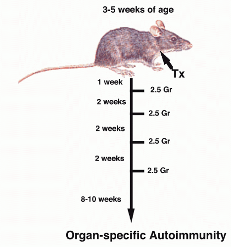
FIG. 33.3. Induction of Organ-Specific Autoimmunity in Rats by Adult Thymectomy and Irradiation. Adapted from Penhale et al.17


IDENTIFICATION OF CD4+ CD25+ FOXP3+ REGULATORY T CELLS

An important extension of this hypothesis was that inhibition of autoreactive T cells by suppressor T cells was not a phenomenon unique to the neonate, but that in the normal adult animal, autoreactive T cells are also under the constant control of the suppressor T cells. If the suppressor lineage was deleted, damaged, or compromised in the adult animal, autoimmune disease might develop. Although a number of studies suggested that the Treg cells in the normal adult animal might be identified by the expression of certain membrane antigens (eg, high levels of CD520), a major advance in our understanding of
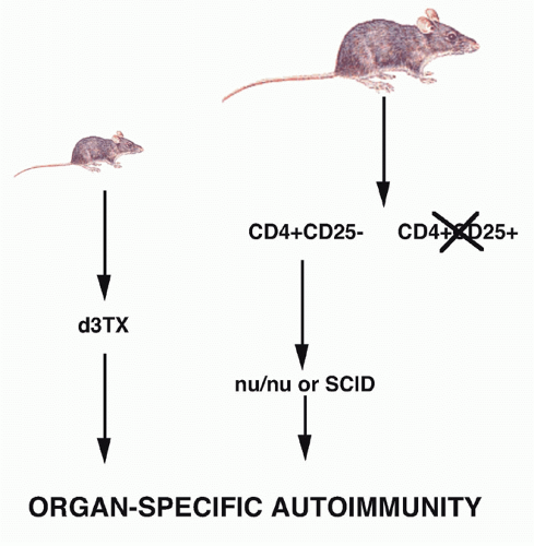
the role of Treg cells was the demonstration by Sakaguchi et al.,21,22 that a minor population of CD4+ T cells (10%) that coexpressed the CD25 antigen (the interleukin [IL]-2R α-chain) appeared to function as Treg cells in the normal adult. When CD25+ T cells were depleted from a population of normal adult CD4+ T cells and the remaining CD4+CD25- T cells transferred to an immunocompromised recipient such as a nu/nu mouse, the recipients developed a spectrum of autoimmune diseases that closely resembled those seen following d3Tx (see Fig. 33.2). Cotransfer of the CD25+ cells prevented the development of autoimmunity. Similarly, the induction of disease post-d3Tx could also be prevented by reconstitution of the animals with CD4+CD25+, but not CD4+CD25-, normal adult T cells before day 14 of life.23 CD8+CD25-T cells alone were not capable of inducing autoimmunity and enhanced the induction of disease induced by CD4+CD25- T cells. These studies solidified the role of the CD4+CD25+ T cells as a major subset of cells that plays a unique role in the regulation of the immune response. The autoimmune diseases induced by depletion of CD4+CD25+ T cells are uniformly accompanied by the development of organ-specific autoantibodies, suggesting that this mode of loss of T-cell tolerance also results in the breakdown of B-cell tolerance as well. It is likely that the activated self-reactive T helper cells provide signals to self-reactive B cells, rescue them from apoptosis, and stimulate autoantibody production.

Powrie and Mason24 were the first to identify cell surface markers that distinguished between regulatory and effector T-cell populations in the rat. When athymic rats were reconstituted with small numbers of CD4+CD45RChigh T cells, they developed a severe wasting disease characterized by extensive mononuclear cell infiltration in the lungs, liver, thyroid, stomach, and pancreas 6 to 10 weeks later. No pathology developed in rats that received unseparated CD4+ T cells or CD45RClow cells. It seemed likely from these studies that the CD45RClow subset controlled the capacity of the RChigh subset to mediate the wasting disease. Fowell and Mason25 directly demonstrated the suppressive effect of the RClow subset in the Tx-irradiation model develped by Penhale. Transfer of RClow CD4+ T cells completely inhibited the development of diabetes and insulitis. RClow T cells from long-term Tx donors could protect as efficiently as cells from normal donors, demonstrating that the Treg cell is long-lived in the periphery. Subsequently, CD4+CD45RBlow subset in the mouse was shown to have regulatory properties similar to the CD4+CD45RClow subset of the rat.26 Taken together, these studies in mouse and rat model systems demonstrated for the first time that two well characterized cell surface antigens (CD25 and CD45RClow/CD45RBlow) could be used to identify suppressor CD4+ T-cell subpopulations present in normal animals. In later studies,27 it was shown that the CD25+ T within the CD4+CD45RBlow population mediated their suppressor activity.

CD4+CD25+ T cells typically represent 5% to 8% of the total population of T cells in the normal mouse lymph node (LN), or 10% to 15% of mouse CD4+ T cells (Fig. 33.4). CD4+CD25+ T cells can also be found in the thymus where they also represent about 5% to 10% of the mature CD4+CD8- population, or 0.5% of mouse thymocytes.21,28 A population with an identical phenotype has been identified in the rat, in human peripheral blood, and in human thymus.29,30,31,32,33,34,35,36 When compared directly to CD4+CD25- T cells, CD4+CD25+ T cells express slightly higher levels of CD5, have a slightly higher proportion of CD62Llow cells, and have a higher proportion of CD69+ cells. They express both intermediate and low levels of CD45RB, and are completely absent from the CD45RBhigh population.37 All of the CD4+CD25+ T cells express the TCR αβreceptor and are NK 1.1 negative. CD25+ and CD25- T cells have a similar distribution of TCR Vα or Vβ specificities.28 One other unique property of CD25+ T cells in both mouse and man is that they are the only nonactivated T-cell population that expresses high levels of the cytotoxic T-lymphocyte antigen (CTLA)-4 antigen intracellularly.27,38,39 The glucocorticoidinduced tumor necrosis factor (TNF)-like receptor (GITR, TNFRSF18) has also been shown to be expressed on the majority of resting CD4+CD25+ T cells and to be expressed at very low levels on CD4+CD25- T cells.40,41 The GITR is upregulated on CD4+CD25- T cells following TCRmediated activation.


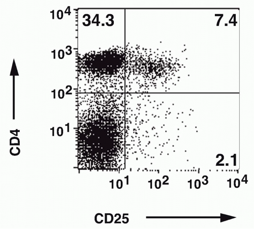
FIG. 33.4. Identification of CD4+ CD25+ T Cells in Normal Mouse Lymph Node. Adapted from Thornton and Shevach.37

A major advance in the study of Tregs was derived from two related experiments of nature. Although CD4+CD25+ T cells were shown to display suppressive properties in multiple disease models, the value of CD25 has a marker is limited as CD25 is highly expressed on both activated CD4 and CD8 T cells, compromising its usefulness in studying Treg cells in settings of immune activation. The X-chromosome encoded forkhead transcription factor,
Foxp3, was identified as the key player in the biology of CD4+CD25+ Tregs.42,43,44,45 Young males with the immune dysregulation polyendocrinopathy enteropathy X-(IPEX) linked syndrome or a mutant mouse strain, scurfy, succumb to similar autoimmune and inflammatory diseases as a result of uncontrolled activation and expansion of CD4+ T cells. Both IPEX and scurfy mice have mutations in a common gene, Foxp3, which encodes a forkheadwinged-helix transcription factor. There is an excellent correlation between expression of Foxp3 and CD25, but a minor population (˜10%) of Foxp3+ cells is CD25- and about 10% of CD25+ cells are Foxp3- (Fig. 33.5); the latter represent activated effector cells. Expression of Foxp3 only in the thymus using a proximal lck-driven transgene does not prevent disease in scurfy mice. Foxp3 expression in peripheral T cells is thus required for maintenance of Treg function. Retroviral-mediated transduction of Foxp3 expression in naïve T cells can convert these cells to a regulatory phenotype functioning in vitro in a manner similar to CD4+CD25+ Tregs. Furthermore, Foxp3-transduced naïve CD25- T cells also manifest suppressor activity in vivo and can inhibit the weight loss, diarrhea, and histologic development of colitis induced by transfer of CD25-CD45RBhigh cells as effectively as Tregs.

Fontenot et al.46 generated Foxp3 deficient (-/-) mice and demonstrated that Foxp3 is specifically required for the thymic development of Tregs. Mixed bone marrow chimeras using wild-type (WT) and Foxp3-/- bone marrow demonstrated that Foxp3-/- T cells behaved normally in the presence of WT Foxp3+ Tregs. Thus, the lethal autoimmune syndrome in Foxp3-/- mice results from a deficiency of Tregs and not from a cell intrinsic defect of Foxp3- T cells. Further study of the role of Foxp3 in Treg function has been greatly facilitated by the development of strains of mice in which a fluorescent protein such as green fluorescent protein (GFP) has been “knocked in” to the Foxp3 locus, permitting ready identification and isolation of Foxp3+ Tregs by cell sorting.47 Studies of Treg development in the thymus of the Foxp3gfp mice demonstrated that less than 0.1% of the CD4+CD8- thymocytes expressed Foxp3 within 12 hours after birth, and that the percentage of Foxp3+ CD4 single positive (SP) thymocytes increased slowly over the following days and reached a plateau of ˜4% at ˜21 days after birth. The largest single day change in the percentage of Foxp3 expressing SP occurred between days 3 and 4. Eighty percent of the Foxp3+ T cells on day 1 were CD4+CD8- SP cells. This result correlates with the early studies on the d3Tx mice that suggested that the generation of Treg cells is delayed relative to generation of nonregulatory CD4+ T cells. Foxp3 was not expressed in nonhematopoietic tissues. Foxp3 expression in peripheral TCR αβ+ T cells irrespective of CD25 expression correlates with suppressor activity.


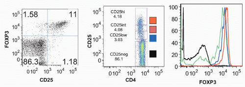
FIG. 33.5. Correlation between CD25 Expression and Foxp3 Expression in Mouse CD4+ Cells.

Direct proof that Foxp3+ T cells maintain control of autoreactive T cells in the periphery has been shown by using a targeting construct encoding the human diptheria toxin receptor fused to sequences encoding GFP and equipped with an internal ribosome entry site into the 3′ untranslated region of Foxp3.48 Complete elimination of Foxp3+ cells was achieved after 7 days of treatment with diptheria toxin. Foxp3 elimination at birth led to a syndrome very similar to that seen in Foxp3-/- mice. Treg cell elimination in adult nonlymphopenic mice resulted in an even more rapid development of terminal autoimmune disease than in neonates. These studies demonstrate that T cells, probably expressing self-reactive TCRs, are targets of continuous Treg-mediated suppression. After Treg-cell ablation, those T cells become activated, produce secreted and membrane bound cytokines, and facilitate dendritic cell (DC) maturation. At day 7 after ablation, there are increased numbers of B cells, macrophages, Gr-1+ granulocytes, natural killer (NK) cells, and a 10-fold increase in the absolute numbers of DCs. Thus, DC dysregulation is a general phenomenon resulting from the absence of Treg cells. Furthermore, Foxp3-independent recessive and dominant tolerance mechanisms established in adult mice are not sufficient to protect mice from fatal autoimmunity after elimination of Treg. Even a partial
decreased level of Foxp3 protein in Treg cells as a result of induced dysregulation of Foxp3 gene transcription results in marked attenuation of Treg suppressor function.49


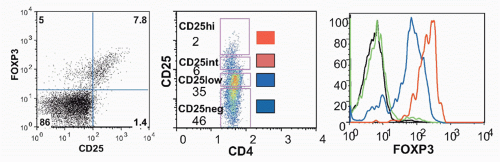
FIG. 33.6. Correlation between CD25 Expression and Foxp3 Expression in Human CD4+ Cells.

Isolation of human Tregs continues to be problematic as one must rely on expression of cell surface antigens. It was reported that among T cells, only the highest expressors of CD25 (˜2% of CD4+ T cells) exerted significant suppressive effects in vitro.29 This observation was substantiated by a comparison of Foxp3 expression with CD25 expression on human CD4+ T cells. Almost all human CD4+ CD25high cells are Foxp3+, but a variable percentage of CD25int cells express lower, but substantial amounts of Foxp3 (Fig. 33.6). Flow cytometric isolation of the CD25high cells does allow one to obtain a population that is almost uniformly Foxp3+, but a substantial subset of Foxp3+ cells present in the CD25int population will be lost. It has been proposed that almost all of human Foxp3+ T cells can be identified as expressing low levels of the IL-7 receptor (CD127) and that CD127low expression can be used to isolate human Treg.50 However, CD127 is downregulated early in the course of T-cell activation, so the CD127low phenotype is also unlikely to be Treg-specific during an ongoing immune or inflammatory response. In addition, only about 40% of the CD127low population is Foxp3+ and even purified CD4+CD127lowCD25+ cells were only 85% to 90% Foxp3+. Nevertheless, the differential expression of CD127 has proven useful for the isolation of human Treg.

Because the number of Foxp3+ T cells is remarkably constant in normal animals in the absence of perturbation of the immune system, they have been frequently referred to as thymic-derived, “naturally occurring” Tregs,51 whereas Foxp3+ T cells that are generated extrathymically are termed “adaptive or induced” Tregs. As both populations of Tregs can exert potent biologic functions in vivo and both are generated during the course of normal T-cell development and differentiation, this nomenclature is not accurate. This chapter will primarily describe the biologic functions of Foxp3+ T cells that will be referred to as Tregs irrespective of their site of generation. In the discussion of some of the papers published prior to the availability of reagents specific for the detection of Foxp3, they are referred to a CD4+CD25+ T cells. More precise definitions of the site of origin will be used where appropriate. The potential regulatory functions of Foxp3- T-cell populations will be summarized in section on Foxp3- Regulatory T cells.


BIOLOGIC PROPERTIES OF FOXP3+ REGULATORY T CELLS


Development of Regulatory T Cells in the Thymus

The potential role of the thymus in the generation of Treg cells was first described several years ago.52 Most studies strongly support the view that the Foxp3+ Treg population is produced in the thymus as a functionally mature distinct T-cell subpopulation. Thymic Foxp3+ Tregs are not derived from peripheral cells that have recirculated from the periphery to the thymus because Tregs are developed in vitro in organ cultures of fetal thymus. Foxp3+ thymocytes are nonresponsive and suppress T-cell activation in vitro in a manner similar to Tregs derived from the periphery.28 The capacity of Foxp3+ T cells to migrate from the thymus to the periphery was documented by injection of fluorescein isothiocyanate intrathymically. The percentage of Foxp3+ T cells within migrants and resident T cells was identical, suggesting that Foxp3+ T cells in the periphery can originate in the thymus.53 Thymectomy at 4 to 5 weeks of age did not modify the number of Foxp3+ T cells in the periphery even when tested 19 months later.

It is widely accepted that conventional CD4+ and CD8+ T cells develop in the thymus by a process of positive and negative selection. Positive selection is mediated by interaction of developing T cells with MHC antigens on thymic cortical epithelium. Negative selection is mediated both by DCs and thymic medullary epithelium. As Treg cells have an antigen-experienced phenotype in the absence of exposure to foreign antigens, it seems logical that their TCRs would be biased toward recognition of self-antigens. A number of studies have suggested that Tregs undergo a unique developmental process during their generation in the thymus. When TCR-transgenic mice bearing a TCR specific for a determinant (S1) derived from influenza hemagglutinin (HA) in association with I-Ed were crossed to mice expressing the HA transgene, the transgenic T cells were not deleted and a
large proportion expressed CD25 and functioned as Tregs.54 Radioresistant elements of the thymus were shown to be both necessary and sufficient for the selection of Foxp3+ T cells in these doubly transgenic mice. Similar results were obtained when TCR transgenic mice on a RAG-/- background were mated to the HA transgenic mice, clearly indicating that thymocytes that can only express a single transgenic TCR can undergo selection to become Tregs. A second TCR transgenic mouse was generated that expressed a variant determinant of HA, but that recognized the S1 determinant with 100-fold less affinity. When these mice were bred to the HA transgenic mouse (S1), they did not have an increased frequency of Foxp3+ T cells. Thus, thymocytes with a low intrinsic affinity for the S1 peptide did not develop into Foxp3+ thymocytes in response to HA. These data are consistent with a model in which selection of Tregs that express a transgenic TCR depends on a high-affinity interaction of the TCR with its ligand. Treg differentiation results from interactions that lie in between the signaling strength required for conventional positive selection on one side and clonal deletion on the other side. It could not be determined from these studies whether this selection process occurs on cortical or medullary epithelial cells.

A number of other models have been proposed55 for Treg differentiation in the thymus. Recent studies favor TCR specificity as playing a dominant role in thymic Treg cell development.56,57 Transgenic mice expressing TCRs derived from Treg cells bore very few thymic Foxp3+ T cells in contrast to TCR-transgenic mice using foreign antigen-specific TCRs. The failure of these mice to develop significant numbers of Foxp3+ T cells suggested that developing T cells with the same antigenic specificity compete for a limited niche for Treg-cell development. In mixed bone marrow chimeras with varying proportions of polyclonal progenitors and Treg cell-derived TCRs, a striking inverse correlation was observed between the percentage of TCR-transgenic cells and the propensity of these cells to give rise to Foxp3+ T cells. When TCR-transgenic cells accounted for 10% of total cells, 1% expressed Foxp3, but when TCR-transgenic cells were only 0.1% of the total cells, 30% expressed Foxp3. This result suggests the existence of a niche that limits Treg cell development. Other studies have demonstrated efficient clonal deletion of CD4+Foxp3- T cells and the complete absence of Foxp3+ T cells in a transgenic mouse expressing a different Treg-derived TCR transgene consistent with the view that Treg-derived TCRs are self-reactive.58

Some studies have raised the possibility that Tregs are generated in the thymic cortex,59 whereas others favor the view that the thymic medulla is the site where Treg precursors mature after contacts with tissue-specific peptides presented by thymic medullary epithelial cells or DCs.60 It is likely that both both DCs and thymic medullary epithelial cells can facilitate Treg development.61 It is still possible that different TCR specificities are selected by each APC subset. AIRE expression in thymic medullary epithelial cells might favor tissue specific antigens in the thymus, but Treg maturation is normal in AIRE-/- mice that have impaired presentation of tissue-specific antigens and in CCR7-/- mice that have defects in thymocyte trafficking. Some peripheral DCs can migrate into the thymus and present peripheral antigens to developing thymocytes. The ability of the different types of APCs to mediate negative selection versus Treg development may also differ.

Events in addition to TCR signaling are also required for the induction of Foxp3+ T cells. CD28 signaling also plays an important cell intrinsic role in the development of Foxp3+ T cells in the thymus.62 CD28 primarily improves the efficiency of Treg cell development rather than enhancing the number of TCRs that can facilitate Treg cell development. CD28 signaling is important for the generation of cytokine responsive Treg cell precursors, as the TCR repertoires of WT and CD28-/- mice were identical.63 The ability of CD28 to support Treg generation does not require an intact phosphoinositide 3-kinase (PI3K)-binding motif as PI3K signaling through Akt and mammalian target of rapamycin (mTOR) will antagonize Treg differentiation.64,65

Cytokines also play a critical role in Treg development. The thymic population of Tregs is only mildly reduced in IL-2-/- or IL-2Rα-/- mice.66 A more dramatic decrease is seen in mice with targeted mutations of IL-2Rγ, JAK3, or signal transducer and activator of transcription (STAT)5, signaling molecules shared by IL-7 and IL-15. Because neither IL-7 or IL-15 deficiency by itself affects the thymic production of Treg cells, it is likely that IL-2 is the principal common γ-chain cytokine required for intrathymic Treg cell development, but that in its absence IL-7 and IL-15 can compensate to a certain extent.67 The development of Foxp3+ thymocytes occurs in two stages. A relatively small CD25+Foxp3- subset of CD4+ SP thymocytes is enriched in precursors of Foxp3+ Treg cells. These Foxp3- Treg precursors require common γ-chain cytokines, but no further TCR stimulation for acquisition of the mature Foxp3+ Treg phenotype. In this “two step model,” the first step is a TCR- and CD28-driven instructive phase, while the second step is the cytokine-driven consolidation phase.68,69 Although multiple cytokines can induce Foxp3+ Treg cell differentiation, they all do so via STAT5 activation. Activated STAT5 is likely to directly bind and regulate the Foxp3 gene at the promoter and at an intronic enhancer region.69 Transgenic mice expressing a constitutively active form of STAT5 have a marked increase in the percentage and total numbers of Foxp3+ Tregs.

While other cytokines that use the γ common chain can substitute for IL-2 for the development of Tregs in the thymus, IL-2 is required for the maintenance and survival of Foxp3+ T cells in the periphery. The numbers of Foxp3+ Treg cells in the periphery of IL-2-/-, IL-2Rα, and IL-2Rβ-/-70 mice are substantially decreased or almost absent.71 Partial or more profound defects both in the number and function of Foxp3+ T cells have been reported in mice deficient for CD80/CD86, CD40,72 CD40L,73 and Stat5a.74 The one common factor that characterizes these mice is that the products of all the deficient genes have important roles in the production or responsiveness to IL-2. Treatment of mice with anti-IL-2 or with CTLA-4Ig to inhibit costimulatory signals also leads to a rapid decline in the number of Foxp3+ T cells.38 As Foxp3+ T cells do not produce IL-2, this deficiency may
be secondary to the capacity of Foxp3- T cells to produce IL-2,75 or to some intrinsic defect in the Foxp3+ T cells in their capacity to respond to IL-2. IL-2Rα-/- T cells in mixed bone marrow chimeras had a substantially decreased competitive fitness compared to WT Foxp3+ Tregs. The conclusion drawn from these studies was that IL-2 is an essential Treg cell growth factor in vivo and that in the absence of IL-2, Treg cells exhibit an impaired metabolic fitness. It appears that Foxp3 expression in Tregs establishes a gene expression program that renders Tregs critically dependent on paracrine IL-2 signalling resulting in repression of IL-2 production and the induction of IL-2Rα. IL-2 is not only required for the survival of Foxp3+ Tregs in the periphery, but also appears to be critical for maintenance of their suppressive functions.76,77,78 Deletion of the proapoptotic factor, Bim, results in the restoration of normal numbers of Foxp3+ T cells in IL-2-/- mice, but Bim-deleted mice still exhibited lethal autoimmunity.78

The potential role of transforming growth factor (TGF)-β in the induction of Foxp3+ T cells during thymic development remains controversial. Liu et al.79 demonstrated that very few Foxp3+ cells could be detected in the thymus of TGF-βRI-/- mice between 3 and 5 days of age. Later in life, the number of Foxp3+ T cells in the thymus increased and this expansion was driven by the production of IL-2. Very similar results were observed in TGF-βRII-/- mice, with a reduced number of Foxp3+ thymocytes at 3 to 5 days of age and increased numbers at 2 to 3 weeks of age.80 The few Foxp3+ T cells in the TGF-βRII-/- mice expressed high levels of caspase and enhanced apoptosis associated with high amounts of the proapoptotic Bcl-2 family members, Bim, Bak, and Bax. Bim deficiency enhanced the number of peripheral Tregs. Thus, TGF-β is probably not essential for the induction of Foxp3 expression during Treg cell development, but the antiapoptotic effects of TGF-β are required for Treg survival.


Transcriptional Regulation of Regulatory T Cell Development

Recent studies of regulatory elements within the Foxp3 locus offer insights into the molecular mechanisms of Foxp3+ T-cell differentiation and maintenance.81 Conserved noncoding deoxyribonucleic acid (DNA) sequence (CNS) elements at the Foxp3 locus encode information defining the size, composition, and stability of the Treg population (Fig. 33.7). CNS3 contains a DNase I hypersensitive site, and the NF-κB family member c-Rel binds to this element and likely facilitates the activity of the Foxp3 promoter. Gene targeting studies reveal an essential role for CNS3 in the induction of Foxp3 expression in the thymus and in the periphery. CNS3-/- mice remain healthy despite an approximately fivefold decrease in Treg cells compared to WT mice. Consistent with the documented binding of c-Rel to CNS3, c-Rel deficiency in thymic precursor cells results in a profound impairment in Treg differentiation.82 In contrast to other CNS regions, CNS3 contains permissive chromatin in double positive (DP) and CD4SP cells that can facilitate binding of c-Rel as a homodimer. This result suggests that c-Rel may be a pioneer transcription factor that opens the Foxp3 locus to other transcription factors. It is likely that that c-Rel binding to CNS3 in cooperation with other transcription factors, including NFAT, CREB, p65, and Smad, facilitates formation of a c-Rel containing enhanceosome at the Foxp3 promoter. Other members of the NF-κB pathway are important for Treg differentiation, as mutations of PKCθ, CARMA1, Bcl-10, TAK1, and IKKβ greatly reduce thymic Treg numbers.83


FIG. 33.7. Transcription Factors Controlling Foxp3 Expression. Adapted from Zheng et al.81 and Ruan et al.82

CNS2 also known as the Treg-specific demethylated region contains a cytosin-phosphatidyl-guanosin (CpG) island whose methylation status is correlated with the stability of Foxp3 expression.84 CNS2 CpGs are demethylated in Foxp3+ T cells, but fully methylated in Foxp3- T cells. While Foxp3 induction in the thymus and periphery was unimpaired in CNS2-/- mice, Treg cells lacking CNS2 progressively lost Foxp3 expression upon division. Thus, CNS2 has a nonredundant function for the maintenance of Foxp3 in the progeny of dividing Treg cells. Foxp3 is also recruited to CNS2 with the assistance of Runx-1 and its co-factor Cbf-β. The binding of the Foxp3-Runx-1-Cbf-β complex confers stability to Foxp3 expression and is also involved in facilitating Foxp3 promoter activity. The recruitment of Foxp3 to CNS2 and the resulting maintenance of Foxp3 expression represent the first description of a feed-forward mechanism enforcing heritable cell lineage destiny. Foxp3 binding to CNS2 occurs after and is dependent on demthylation of CNS2.

CNS1 contains NFAT, Smad3, and STAT5 binding sites. Analysis of mice deficient in CNS1 revealed that thymic differentiation was unaffected, whereas peripheral Foxp3 induction was severely impaired by CNS1 deficiency.81 Deficiency in peripherally generated Treg cells did not result
in detectable tissue specific autoimmunity in CNS1-/- mice until 1 year of age. These results suggest that mechanistic requirements for thymic and peripherally induced Tregs are distinct from Tregs differentiated in the thymus.


T-Cell Receptor Repertoire Analysis of Foxp3+ Regulatory T Cells

As noted previously, several lines of evidence suggest that Treg development hinges upon a particular TCR specificity. Foxp3+ Tregs in general do not develop in TCR-transgenic mice lacking RAG genes, suggesting that only cells with certain TCR specificities can develop into Tregs. Several studies have addressed in detail the TCR repertoire of Foxp3+ Tregs and their antigen specificity.85,86 Hsieh et al.85 directly compared the sequences of the CDR3 region on Treg and conventional T cells and found that they are only partially overlapping. When immunodeficient mice were reconstituted with T cells expressing diverse Vα2 TCR α-chains from Treg or conventional T cells paired with a single Vβ chain, some of the Treg TCR reconstituted T cells were selfreactive, as they induced wasting disease in lymphopenic hosts, but not in conventional recipients. Although 40% of the TCR were strongly self-reactive in vivo, others were only intermittently and moderately self-reactive or nonreactive as measured by T-cell expansion in lymphopenic hosts. Other studies86 have demonstrated a large degree of overlap in TCR usage between Foxp3+ and Foxp3- T cells. The recent experiments56,57,58 demonstrating that TCR self-specificity is required for the intrathymic induction of Foxp3 expression strongly support the view that the TCRs of the Foxp3+ population are predominantly self-specific.

TCR repertoire analysis has also been used to determine the relationship between thymic Foxp3+ Treg cells and Foxp3+ Tregs in the periphery. A comparison of the TCR repertoire of thymic and peripheral Foxp3- and Foxp3+ T cells revealed that the TCRs from Foxp3+CD4+ peripheral T cells resembled those of thymic precursors and differed from TCRs expressed by Foxp3-CD4+ T cells; in addition, the diversity of Foxp3+ TCRs was greater than TCRs on Foxp3-CD4+ naïve T cells.87 These studies suggested that the great majority of peripheral Foxp3+ Tregs derive from thymic precursors and that peripheral conversion from Foxp3- to Foxp3+ is rare. TCR repertoire analysis has also been applied to autoreactive T cells in mice deficient in Foxp3.88 These mice did not exhibit a defect in negative selection. Activated, but not naïve, T cells in Foxp3-/- mice often used TCRs found in the Foxp3+ Treg TCR repertoire of normal mice, suggesting that T cells expressing these selfreactive TCR are not eliminated and might contribute to the pathology associated with Foxp3 deficiency.


Development of Foxp3+ Regulatory T Cells Extrathymically

One important question that must be addressed is whether the thymus is the only site for the generation of Foxp3+ Tregs. A number of studies performed both in vivo and in vitro have demonstrated that under certain conditions Foxp3+ Tregs can be generated extrathymically and that TGF-β is a key component in this process. Administration of peptide antigen to TCR transgenic mice bred onto the RAG-/- background, that lack Tregs, resulted in prolonged expression of CD25 only when the antigen was administered under tolerogenic conditions.89,90 These CD25+ cells had some Treg properties. The mechanism of suppression used by these induced Tregs was not explored. One highly effective method for the induction of Tregs was to expose TCR transgenic T cells on a RAG-/- background in vivo to a continuous supply of subimmunogenic doses of agonist peptides delivered via a mini-osmotic pump. Many of the T cells became Foxp3+ and resembled thymic-derived Tregs in all their phenotypic and functional properties.91 It was postulated from these studies that low doses of peptide presented over relatively long periods of time on nonactivated DCs represented a important mechanism by which Tregs could be generated in the periphery particularly to self-components that do not lead to tolerance in the thymus. Foxp3+ T cells were also induced when mice were treated with peptide agonist ligands targeted to DCs in minute doses by coupling them to antibodies directed to DCs under conditions of suboptimal DC activation.92 Tregs induced with subimmunogenic conditions could subsequently be expanded by delivery of antigen in immunogenic conditions. DC-targeted de novo generation in vivo results in efficient demethylation of conserved CpG motifs within the Treg-specific demethylated region (CNS2) region of the Foxp3 locus, a predictive parameter for long-term stability of induced Foxp3 expression.93

One interpretation of the studies described previously is that weak TCR stimulation is favorable for the peripheral induction of Foxp3 and that robust TCR signaling during Treg cell generation stimulated the Akt-PI3K-mTOR pathway that antagonizes the induction of Foxp364 and results in cellcycle dependent maintenance of a silenced state of the Foxp3 locus.94 More recent studies have examined the role of the strength of the antigenic signal required for the induction of Foxp3+ T cells in the periphery.95 Foxp3+ cells induced by weak agonist peptides did not persist compared to cells generated by a higher affinity ligand. There appear to be qualitative differences between weak and strong signals whereby a few strong signals may be different from many weaker signals. The few strong signals may specifically induce survival signals, while the many weaker signals may induce proapoptotic signals. There also appears to be a unique subpopulation of recent thymic emigrants that are CD25hiCD62Lint CD69+ that are capable of becoming Foxp3+ upon exposure to IL-2 alone in the absence of TGF-β.96

A number of groups97,98,99,100 have presented studies that demonstrate a pivotal role for TGF-β in the generation of Foxp3+ Tregs in vitro. TCR activation in the presence of TGF-β converted naïve T cells into Foxp3+ (Fig. 33.8) cells with an anergic/suppressor phenotype. IL-2 plays a nonredundant role in TGF-β-mediated induction of Foxp3 expression in mouse CD4+ T cells. Once induced, Foxp3 expression was maintained both in vitro and in vivo in the absence of IL-2. Other cytokines utilizing the common γ-chain as part of their receptors were unable to induce Foxp3 expression in IL-2-/- cells. When T cells are activated through the TCR
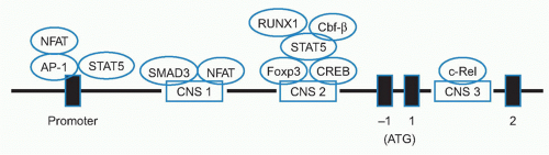
in the presence of TGF-β, both RORγT and Foxp3 are transcribed and interact with each other. In the presence of IL-2, Foxp3 expression dominates leading to suppression of RORγT transcription, promoting Treg development. In contrast, in the presence of IL-6, and low levels of IL-2 the transcription of RORγT is favored which in turn represses Foxp3 transcription with resultant Th17 devlopment.101


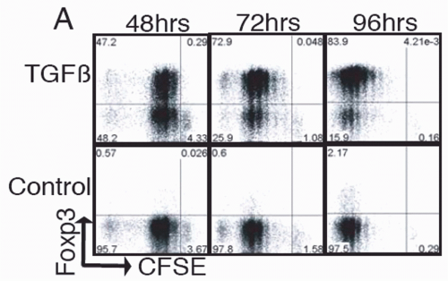
FIG. 33.8. Naïve Foxp3- T Cells were Cultured and Stimulated in the Presence of Transforming Growth Factor-χ. Foxp3 expression was determined by intracellular staining at the indicated times. Adapted from Davidson et al.100

TGF-β-induced Foxp3+ T cells not only exhibit unresponsiveness to TCR stimulation, but also suppress normal CD4+ T-cell activation and Th1 and Th2 cytokine production in vitro. The induction of Foxp3 results in downregulation of Smad7 and renders T cells highly susceptible to the regulatory effects of TGF-β signaling via Smad3/4.98 Foxp3+ T cells induced in vitro can suppress antigen-specific proliferation of CD4+ T cells in vivo and prevent house dust mite-induced mouse asthma.97 One of the major factors preventing induction of Foxp3 expression in vitro is the presence of interferon (IFN)γ with subsequent STAT1 signaling.102 Some studies suggest that TGF-β may also be capable of inducing Foxp3 expression in vivo. Peng et al.103 used an inducible system for the transient induction of TGF-β in the pancreatic islets during the priming phase of diabetes. Approximately 40% to 50% of intraislet CD4+ T cells expressed CD25 and other characteristics of Tregs. However, it is not clear if high levels of TGF-β resulted in the in situ expansion of the small numbers of Tregs already present in the islets or if TGF-β induced the conversion of CD25- T cells to CD25+ suppressors.

The gut appears to be one of the major sites for the generation of Tregs in the periphery, and the gut contains a large number of Foxp3+ Tregs. Mucida et al.104 first demonstrated that repeated antigen feeding induced antigenspecific Foxp3+ Treg cells in the absence of thymic-derived Tregs. Converted cells were rapidly found in the mesenteric LN, Peyer patches, and primarily in the small intestine lamina propria. Gut-associated lymphoid tissue has a number of unique properties that facilitate the generation of Foxp3+ T cells. A vitamin A metabolite, retinoic acid (RA), produced by gut DCs, can selectively induce molecules such as CCR9 and the integrin, α4β7, on both conventional T cells and Treg cells, and these cell surface antigens are involved in directing homing of cells to the gut. DCs from the lamina propria of the small intestine have the unique ability to generate Treg cells in vitro via a mechanism that involves the synergistic action of TGF-β and RA. A subpopulaton (25%) of lamina propria DCs express CD103, and this CD103+ subpopulation was responsible for the induction of Foxp3 in the absence of exogenously added TGF-β. Endogenous TGF-β was responsible for Treg conversion, as anti-TGF-β largely abrogated Treg induction. Addition of RA to splenic DC cultures enhanced Treg conversion, and blockade of nuclear RA signaling decreased converted Tregs, but RA alone is not sufficient for Foxp3 induction. Thus, the capacity of the intestinal immune system to produce TGF-β and RA explains the unique capacity of the gut to induce
tolerance.105,106 In addition to the TGF-β induction pathway, Foxp3+ Tregs can also be induced via PD-L1-PD-1 ligand receptor pathway107 and by environmental factors such as some aryl hydrocarbon receptor agonists.108

One major issue that remains to be addressed is the relative contribution of thymic-derived versus peripherally induced Tregs to the establishment of immune tolerance. Although Tregs are induced in the colitis model from Foxp3- T cells, the induced Tregs are not sufficient to prevent disease.109 Some studies suggest that only the combination of thymic-derived Tregs and in vitro generated Treg cells could fully protect mice from developing inflammatory bowel disease (IBD) induced by naïve T cells from Foxp3-/- donors. When scurfy mice were reconstituted with thymic-derived Tregs alone, a marked inhibition of lethality was observed, but the treated mice still displayed signs of inflammation.110 Surprisingly, the combined treatment with thymic-derived Tregs and conventional Foxp3- T cells, that could differentiate into Foxp3+ T cells, decreased inflammation. In vitro generated Treg cells could substitute for Tregs generated in vivo from Foxp3- conventional cells. One explanation for this result was that the TCR repertoires of the thymic-Tregs and the Tregs induced in the periphery were largely nonoverlapping and that each Treg cell subset is responsible for recognizing different antigens. The major conclusions drawn from this study is that peripherally induced Treg cells are an essential, nonredundant regulatory subset that acts synergistically with thymic-Treg cells to enforce peripheral tolerance.

If a significant percentage of Foxp3+ T cells are generated at peripheral sites, one should expect that the TCR repertoires of peripheral- and thymus-derived Tregs to be different as peripheral conversion would allow for the generation of Treg cells specific for antigens not presented in the thymus such as those derived from commensal bacteria. Lathrop et al.111 found a significant rate of conversion when CD4+Foxp3- T cells were transferred into lymphopenic hosts. There was considerable disparity in TCR usage between converted Foxp3+ and expanded Foxp3- T-cell subsets within the same host. The converted TCRs were found in the normal non-Treg cell subsets because the donor cells for these studies were normal Foxp3- T cells. However, some of these TCRs were also found in the normal Treg subset and made up 5% of the sequences in the normal Treg pool. It appears that some TCRs are able to facilitate both thymic and peripheral Treg cell development. It is likely that tissue-specific antigens influence the local Treg TCR repertoires as several studies suggest that the presence of an organ facilitates Treg-mediated tolerance to that organ.112,113,114,115,116,117,118

Peripherally induced Tregs may differ markedly from thymic-Tregs in terms of TCR repertoire expression, stability, suppressor mechanisms, and potential suitability for in vitro expansion and cellular therapy. Thus far, no cell surface antigen has been identified that allows one to reliably distinguish thymic-derived Tregs from Tregs induced in peripheral sites. Some studies have suggested that expression of Helios, a member of the Ikaros transcription factor family, distinguishes thymic Tregs from peripheral Tregs.119 Helios was expressed intracellulary in 100% of Foxp3+ mouse thymocytes, but is only expressed by ˜70% of Foxp3+ T cells in peripheral lymphoid tissues in both mouse and man (Fig. 33.9). Foxp3+ T cells induced in vivo by oral tolerance, following transfer of Foxp3- T cells to RAG-/- mice, or following induction by reconstitution of germ-free mice with bacteria,120,121 were primarily Helios-. These studies suggest that a significant proportion of Foxp3+ Tregs are generared extrathymically. Further studies are needed to validate the use of Helios expression as a marker for thymic-derived Tregs. Major differences in the composition of the Treg pool may also exist between man and mouse. A subpopulation of human Tregs was highly proliferative in vivo with a doubling time of 8 days.122 However, these cells were also susceptible to apoptosis and had critically short telomeres and low telomerase activity raising the possibility that they must be produced from another population source. Heteroduplex analysis of TCR from memory T cells and Tregs suggested that they are closely clonally related raising the possibility that a significant proportion of human Tregs are not thymus derived, but are generated from rapidly dividing highly differentiated memory CD4+ T cells.


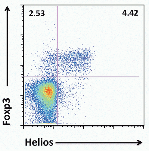
FIG. 33.9. Approximately 70% of Mouse Foxp3+ T Cells Express Helios. Adapted from Thornton et al.119


Analysis of Gene Expression by Foxp3+ Regulatory T Cells

Both DNA microarray technology and serial analysis of gene expression technology have been used to compare patterns of gene expression between different cell types.40,123,124 These technologies have also been applied to compare the patterns of gene expression in Tregs with Foxp3- T cells and other cell types with regulatory functions. One major goal of this approach is to determine whether Tregs simply represent a population of previously activated T cells or whether they display a unique pattern of gene expression that is correlated
with their functional properties. Most of the results are consistent with the latter possibility. Some genes are differentially expressed between the resting Foxp3- T cells and Tregs, while others are differentially expressed following activation.


TABLE 33.2 Genes Selectively Activated in CD4+CD25+ T Cells


  • Signaling: COS, SOCS-1, SOCS-2, SLAP-130


  • Secreted molecules: IL-10, IL-17, Enkephalin, ETA-1, ECM-1, MIP-1a, MIP-1b


  • Cell surface molecules: CD2, OX40, CD25, CD122, GPCR83, GITR, Ly-6, Galectin-1, Thy-1


(Adapted from Hill et al.125)


A comparison of a number of these studies has permitted the identification of groups of genes that are relatively Treg specific and allow one to begin to define a distinct Treg molecular signature (Table 33.2). The “signature” includes both genes encoding certain cell-surface receptors, and genes encoding a wider array of transcription factors and other intracellular proteins.125 Using a Treg to T conventional fold ratio of 1.5/1 or 1/1.5, 603 genes were differentially expressed, with 407 overexpressed and 196 underexpressed in Tregs. The vast majority of the signature’s differential elements were already present in the thymus. Thus, most of the Treg cell signature is already specified at an early stage of lineage commitment, independent of peripheral influences. Many of the Treg signature genes also identify activation antigens expressed on activated conventional T cells including CTLA-4, GITR, and other members of the TNF-receptor superfamily that are expressed by activated effector cells. Some, including neuropilin, GITR, CD38, and CD5, appear to be shared with Foxp3-anergic T cells that do not exert suppressive functions.126 On balance, the studies on global gene expression indicate that Tregs express a unique pattern of gene expression that may contribute to their survival and anergic state.

A Foxp3 chromatin immunoprecipitation method has been used to analyze the direct targets of Foxp3.127,128 About 700 to 1000 genes contain Foxp3 binding regions, and many of these genes are up- or downregulated in Foxp3+ T cells, confirming that Foxp3 can function as both a transcriptional activator and repressor. However, Foxp3 target genes comprise only a small portion of Foxp3-dependent gene expression, suggesting that Foxp3 regulates a large part of its transcriptional program by acting on other transcription factors (Table 33.3). It is likely that some of genes encoding molecules that could potentially mediate suppressor function (eg, IL-10, granzyme B) are secondary Foxp3 targets, as they are not found in the group of genes that are direct Foxp3 targets.


TABLE 33.3 Transcription Factors Bound and Regulated by Foxp3


Prdm1


Irf6


CREM


Helios


(Adapted from Zheng et al.127 and Marson et al.128)


One novel approach to understand the role of Foxp3 in directing Treg function is to engineer a mouse expressing the coding sequence of GFP knocked into the Foxp3 locus together with disruption of Foxp3 protein expression.129,130 Surprisingly, the GFP+Foxp3- T cells in these mice shared many phenotypic features with their GFP+Foxp3+ counterparts. The former are what might be termed Treg “wannabees,” as they develop in normal mice and respond to signals required to induce Foxp3 expression. The GFP+Foxp3- cells were unable to produce IL-2 and effector cytokines, expressed elevated levels of CD25, were relatively nonresponsive to TCR stimulation, but lacked suppressor activity. Treg “wannabees” expressed some (including transcriptional activity at the Foxp3 locus), but not all, of the genes of the Treg signature. Thus, several of the characteristics of the Treg cell lineage can be selected and persist in the absence of Foxp3. Foxp3 does not function as the “master regulator” of the Treg lineage, but stabilizes or enhances certain Treg characteristics, particularly its immunoregulatory activities including suppression. Foxp3 is also not sufficient to elicit the full Treg signature after transfection or induction by TGF-β. Treg “wannabees” do not become conventional effector cells and transfer of these cells into RAG-/- recipients did not induce the scurfy phenotype.88,131 It appears that WT mice have a population of self-reactive non-Tregs that express the Treg self-reactive TCR repertoire, which are not subject to negative selection but can potently induce autoimmune disease in the scurfy mouse.

Studies of the Treg “wannabee” mice indicate that certain Treg cell phenotypic properties including transcription of the Foxp3 locus and expression of some the Treg signature genes can be independent of Foxp3. One candidate family of genes that can regulate these Foxp3-independent Treg properties are the Foxo (Forkhead-box-O) family of transcription factors that play critical functions in the control of diverse cellular responses. Mice with a T cell-specific deletion of Foxo1 have defects in T-cell survival as well as defects in expression of the IL-7 receptor α-chain and CD62L.132 Foxo1 is required for the inhibition of T-cell activation and Foxo1-/- mice develop a mild autoimmune disease. Foxo3 plays a role in DC function.132 Mice with a deletion of both Foxo1 and Foxo3 develop a lethal inflammatory disease associated with a compromised Treg-cell differentiation. Foxo1 and Foxo3 function in a cell intrinsic manner to suppress IFNγ and IL-17 production. Foxo1/Foxo3-/- Treg cells fail to protect lymphopenic mice from autoimmunity induced by scurfy T cells or from colitis induced by Foxp3- T cells.131 Mixed bone marrow chimeras containing bone marrow from Scurfy mice and Foxo1/Foxo3-/- developed a disease as severe as that observed with scurfy bone marrow alone; this was associated with fewer thymic and splenic Treg cells. Thus, it appears that Foxo proteins control the expression of Foxp3 and a subset of Treg cell-associated genes needed for suppressing inflammatory cytokine production in Treg cells. In chromatin-immunoprecipitation assays using antibody
to Foxo1 and Foxo3, Treg cells were selectively enriched in genomic fragments containing the CNS1 and CNS3 regions, but not the CNS2 region of the Foxp3 promoter. Foxo transcription factors constitute one element of higher order control132,133 of Treg development and function.


SUPPRESSOR MECHANISMS UTILIZED BY FOXP3+ REGULATORY T CELLS


In Vitro Studies

An in vitro model system has been established for the analysis of Treg-cell function that allows rapid assays and that may offer some insights to the mechanism of action of Treg function in vivo.37,134,135,136 This approach also allows a comparison of the requirements for activation (costimulation, antigen concentration) of Foxp3+ T cells compared to Foxp3- T cells. Purified Foxp3+ T cells were completely unresponsive to high concentrations of IL-2 alone, to stimulation with plate-bound or soluble anti-CD3, or to the combination of anti-CD3 and anti-CD28. They could be induced to proliferate when stimulated with the combination of anti-CD3 and IL-2. The most striking property of the Foxp3+ T cells is their ability to suppress proliferative responses of both CD4+Foxp3- and CD8+ Foxp3- T cells (Fig. 33.10). The Foxp3+ T cells must be activated via their TCR to suppress. No suppression was seen when Foxp3+ T cells were separated by semipermeable membrane from the CD25- T cells. This demonstrates that cell contact between CD25+ and CD25- T cells is required. Neutralization of the suppressor cytokines IL-4, IL-10, and TGF-β individually or in combination also had no effect on the Treg-mediated suppression. Similarly, Foxp3+ T cells from mice deficient in IL-4, IL-10, or TGF-β were fully competent suppressors. Indo-1-loaded Foxp3+ T cells did not flux calcium in response to TCR stimulation,123 suggesting that they have a block in proximal signaling similar to that seen in T cells rendered anergic in vitro.


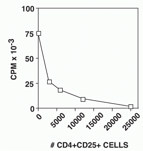
FIG. 33.10. CD4+ CD25+ T Cells Suppress the Proliferative Response of CD4+CD25-T cells. Graded numbers of CD4+CD25+ T cells were mixed with 5 × 10⁴ CD4+CD25- T cells, T-depleted spleen cells, and soluble anti-CD3. Proliferation was measured after 72 hours. Adapted from Thornton and Shevach EM.37

Most studies have demonstrated that Foxp3+ T cells mediate suppression by inhibiting the induction of IL-2 messenger ribonucleic acid (mRNA) and mRNA for other effector cytokines in the responder Foxp3- T cells. Suppression can be abrogated by the addition of IL-2, thereby circumventing the block. Although IL-2 gene transcription was inhibited in the presence or absence of exogenous IL-2, the addition of anti-CD28 overcomes suppression by potently stimulating the production of endogenous IL-2 and overriding the suppressive effects of the Foxp3+ cells. Surprisingly, transcription of IL-2 mRNA was also restored in the cocultures in the presence of anti-IL-2. These results suggest that Tregs do not suppress the initial activation of Foxp3- T cells, but mediate their suppressive effects following production of IL-2 by the responder cells resulting in both the expansion of the Treg and induction/enhancement of their suppressor functions.77 Foxp3+ T cells do not directly mediate the death of the responders, but induce a cell-cycle arrest at the G1-S phase of the cell cycle. Such a cell-cycle arrest is often followed by cell death; it is difficult to recover significant numbers of viable cells when the suppressors and responders are cocultured for periods longer than 48 hours. Suppression of T-cell proliferation is the exclusive property of Foxp3+ T cells isolated from normal animals. The induction of CD25 expression by stimulating CD25- T cells via the TCR, in the absence of TGF-β, does not render the stimulated cells suppressive.

The role of IL-2 consumption in the suppressive function of Foxp3+ T cells is controversial. Treg cells express all three components of the high affinity IL-2R complex (CD25, CD122, and CD132), and one study137 has suggested that Tregs may compete with Foxp3- T cells for IL-2 and thereby inhibit the proliferation of Foxp3- T cells secondary to the induction of a form of apoptosis dependent on the proapoptotic factor Bim. In a hybrid system in which human Treg cells were shown to efficiently suppress mouse Foxp3- T cells, the addition of an antihuman CD25 that blocks IL-2 binding had no effect on the function of the human Treg.138 Taken together with the data that Foxp3+ Tregs inhibit the production of IL-2, it is highly unlikely that Tregs function as IL-2 “sinks,”139 at least in vitro. IL-2 consumption might play a role in Treg suppression in vivo. However, WT Tregs suppress autoimmunity in IL-2Rβ-/- mice where all other cells in the recipient are nonresponsive to IL-2 directly demonstrating that effective suppression is not simply due to depriving IL-2 growth promoting signals to autoreactive T cells by Treg consumption of this cytokine.140

Foxp3+ T cells can be easily propagated in vitro for 3 to 14 days by stimulation initially with anti-CD3 and IL-2 and then expansion in IL-2 alone.136 It has also been possible to clone murine Foxp3+ T cells by repeated stimulation with anti-CD3 and IL-2.41 Following 7 to 14 days of activation and
culture in IL-2, activated Foxp3+ T cells remain nonresponsive and cannot be induced to proliferate when restimulated via their TCR in the absence of IL-2. The activated Foxp3+ T cells have more potent suppressor activity on a per cell basis (three- to fourfold) than freshly explanted Foxp3+ T cells. Foxp3+ T cells that appear to be antigen-specific can be identified in mice that express a transgenic TCR. In general, the percentage of Foxp3+ cells is reduced in TCR transgenic mice (3% to 5% compared to 10% in normal animals). The expression of the transgenic TCR α-chain is a convenient tool that allows activation of the Foxp3+ T cells with peptide/MHC rather than with anti-CD3.136 When Foxp3+ T cells from TCR-transgenic mice are activated with their peptide/MHC ligand and then expanded in vitro in IL-2, the activated suppressors are subsequently capable of suppressing the proliferative responses of fresh Foxp3- T cells from mice that express a different transgenic TCR. There is no MHC restriction in the interaction of the activated suppressors and the Foxp3- responders. Therefore, the suppressor effector function of activated Foxp3+ T cells in vitro is completely nonspecific (Table 33.4).

Treg-mediated suppression is highly sensitive to antigenic stimulation. For example, when Foxp3+ and Foxp3- T cells are prepared from the same TCR-transgenic mouse and stimulated with a specific peptide, the antigen concentration required to stimulate the Foxp3+ T cells to suppress is 10- to 100-fold lower than that required for triggering the proliferation of the Foxp3- T-cell population.134 The partially activated phenotype of Foxp3+ T cells combined with their enhanced sensitivity to antigen stimulation suggests that they are highly differentiated in their function and are ready to mediate their suppressive functions immediately upon encounter with their target antigens. Their capacity to rapidly suppress responses in vitro suggests that they have been continuously stimulated by self-antigens in the normal physiologic state and continuously exert some degree of suppression in vivo.

Foxp3+ T cells suppress the proliferative responses of CD8+ T cells in a manner similar to that seen with CD4+ responders.141 Marked suppression of the effector cytokine, IFNγ is also seen in the presence of Foxp3+ T cells. While suppression of the proliferation of CD4+ responders by Foxp3+ T cells can be completely reversed by the addition of IL-2 or anti-CD28, the suppression of CD8+ T-cell responses is not reversed by the addition of IL-2 or anti-CD28. The failure of IL-2 to abrogate suppression is secondary to a failure of full upregulation of the expression of CD25 on the responder CD8+ T cells. Foxp3+ T cells thereby prevent responses mediated by CD8+ T cells both by inhibiting their ability to produce IL-2 and by inhibiting their ability to respond to IL-2, thus disrupting CD4+ T cell help for CD8+ T cells.


TABLE 33.4 The Suppressor Effector Function of Activated CD4+ CD25+ T Cells is Completely Nonspecific


First Culture


Second Culture (TCR Tg CD4+CD25- T cells Specific For)


% Suppression


CD4+CD35+ from HA TCR Tg stimulated with HA126-138 + IL-2 3 to 7 days


I-Ad + HA126-138


99


I-Ed + HA110-119


99


I-Ek + PCC88-104


95


I-Au + MBPAc1-11


91


HA, hemagglutinin; TCR, T-cell receptor; Tg, transgenic.


CD4+CD25+ T cells from mice expressing a TCR transgene specific for HA126-138 were cultured with peptide, T-depleted spleen cells, and IL-2. They were then washed and mixed with CD4+CD25-T cells from mice expressing the same or different TCR transgenes and then stimulated with the appropriate peptide. (Adapted from Thornton and Shevach.136)


In general, murine Tregs failed to inhibit responses induced by plate-bound anti-CD3, but readily inhibited responses induced by soluble anti-CD3.37 This finding raised the possibility that the cellular target of the Treg was the APC rather than the responder T cell, as responses to platebound anti-CD3 are relatively APC-independent, but other studies demonstrated that Foxp3+ T cells can mediate suppression in vitro via a T-T cell interaction and that the APC is not directly required for delivery of the suppressive signal.141 Similar conclusions were drawn in other studies in which Foxp3+ T cells were shown to suppress responses of either mouse142 or human.33 Foxp3- T cells induced by anti-CD3 coupled to beads in the absence of APCs and can efficiently directly target responder T cells. It remains unclear whether the mechanisms utilized by Tregs to suppress the proliferation of Foxp3- T cells in the presence or absence of APCs are the same or whether assays of Treg function under these distinct conditions measure distinct components of Treg-mediated suppressor function.


Molecular Analysis of Regulatory T Cell Suppression and Anergy

Recent studies have attempted to define the biochemical and molecular pathways that mediate the anergic or nonresponsive state of Foxp3+ Tregs.143,144 A number of studies have shown that Tregs exhibit diminished responses of both proximal and distal signalling pathways both in absolute amplitude and in duration when compared to the responses of CD25- T cells. Tregs have reduced phosphorylation of the TCR ζ-chain, reduced recruitment of ZAP-70, and reduced phosphorylation of SLP-76. Defects were seen in the activity of PLC-γ1 and in signals downstream of this enzyme including calcium mobilization, NFAT, NF-κB, and Ras-ERK-AP-1
activation. The diminished activity of PLC-γ1 in Tregs is likely to contribute, not only to the delay and reduced appearance of NFAT in the nucleus, but also to the attenuated activation of the PKC- and Ras-ERK-directed pathways as well. Other studies145 have suggested that the proximal promoter of the IL-2 gene fails to undergo chromatin remodeling and remains in a closed chromatin configuration.

Despite the widely recognized importance of IL-2 in Treg homeostasis, very little is known about the intracellular mechanisms that regulate IL-2R signaling in Tregs. Indeed, a defining characteristic of Tregs is their inability to expand in vitro upon stimulation with IL-2 alone despite expression of all three chains of the IL-2 receptor. It has been shown that JAK/STAT-dependent signaling is intact in Tregs stimulated with IL-2, but that downstream mediators of PI3K are not activated.146 The nonresponsiveness of Tregs is associated with elevated levels of PTEN, a phosphoinositol 3,4,5-triphosphatase that catalyzes the reverse reaction of PI3K, negatively regulating the activation of downstream signaling pathways. In conventional T cells, the level of PTEN is downregulated after T-cell activation, but PTEN remains highly expressed in Tregs. A critical role of PTEN, in the nonresponsiveness of Tregs to IL-2, was established by studying PTEN-/- mice.147 PTEN-/- Tregs readily proliferated after stimulation with IL-2 alone in vitro and exhibited enhanced peripheral turnover in vivo. They retained their ability to suppress effector T-cell responses both in vitro and in vivo. Forced expression of PTEN in recently activated non-Tregs inhibited their ability to expand in response to IL-2 confirming the ability of this lipid phosphatase to negatively regulate IL-2-dependent proliferation.

As anticipated from their heightened expression of PTEN, Tregs have a defect in the activation of the PI3K-AKT pathway indicated by a reduction in AKT phosphorylation.64 Thymic-derived Tregs with established and stable Foxp3 levels were resistant to the effects of constitutively activated AKT, while activated AKT inhibits the induction of Foxp3 expression in Foxp3- T cells both in vitro and in vivo. IL-6 may block the differentiation of Tregs by activating an AKTdependent pathway. The repression of Foxp3 expression by active AKT required its kinase activity and was partly counteracted by rapamycin treatment placing mTOR as a downstream target of AKT. mTORC1 is more sensitive to rapamycin treatment, so it is likely that AKT is exerting part of its effect via mTORC1. Conversely, inhibition of the PI3K-AKT-mTOR pathway results in de novo expression of Foxp3 in a TGF-β-independent manner.148

The role of Foxp3 in directly controlling the activation of Tregs has been extensively studied. Many of the genes regulated by Foxp3 are also target genes for NFAT. The IL-2 and IL-4 genes are activated by NFAT and repressed by Foxp3, while the CD25 and CTLA-4 genes are upregulated by both NFAT and Foxp3. It is likely that Foxp3 represses IL-2 expression by competing with NFAT for binding to DNA. Foxp3 may also repress NFAT-driven cytokine transcription by sequestering NFAT away from DNA. Examination of the crystal structure of an NFAT:FOXP2:DNA complex revealed an extensive protein-protein interaction interface between NFAT and Foxp2.149 Structure guided mutations of Foxp3, predicted to disrupt its interaction with NFAT, interfere in a graded manner with the ability of Foxp3 to repress expression of IL-2 and upregulate Treg markers CTLA-4 and CD25. Thus, NFAT can switch its transcription partner from AP-1 that drives T-cell activation to Foxp3 that programs Treg function. Foxp3 also has been shown to interact with the transcription factor AML1/Runx1 that is required for normal hematopoiesis, as well as activation of IL-2 and IFNγ gene expression.150 The physical interaction of Foxp3 with RUNX1 suppresses IL-2 and IFNγ production and induces Treg cell-associated molecules such as CD25, CTLA-4, and GITR. Treg-cell deficiency of Cbf-β, a cofactor for all RUNX proteins, attenuated expression of Foxp3 and resulted in high expression of IL-4. The RUNX1-Cbf-β heterodimer is indispensable for optimal expression of Foxp3, maintenance of suppressive function, and suppression of T effector responses.151 Foxp3 can also directly interact with c-JUN/AP-1 in Treg cells, sequestering activated AP-1 in the nucleus, altering the subnuclear distribution of activated AP-1, and disrupting chromatin binding of activated AP-1.152 Foxp3 can form a transcriptional repressor complex with several posttranslational modification enzymes such as the histone deacetylases HDAC1, HDAC7, and HDAC9, and histone acetyltransferase TIP60.153

Other transcription factors including Eos, a member of the Ikaros family, have been shown to play critical roles in Foxp3 dependent gene silencing in Tregs.154 Silencing of Eos in Tregs abrogates their ability to suppress in vitro immune reponses and endows them with partial effector function, as they are capable of inducing IBD in vivo. Knockdown of Eos reversed Foxp3-mediated suppression of IL-2 production. Ninety percent of the genes suppressed by Foxp3 were no longer downregulated when Eos was knocked down in Tregs, while ninety-five percent of the Foxp3 upregulated genes were unaltered by Eos knockdown. One other gene that appears to be important in the regulation of Foxp3 suppressor function is the genome organizer SATB1, which is one of the genes most repressed in human and mouse Treg cells.155 Foxp3 binds directly to the SATB1 locus in Treg cells and prevents SATB1 transcription. Foxp3+ T cells overexpressing SATB1, despite expressing normal levels of Foxp3, lost their suppressive activity and produced T effector cytokines, suggesting that downregulation of SATB1 in Treg cells was necessary for a stable suppressive phenotype. Thus, Treg-cell function not only depends on the induction of Foxp3-dependent genes that mediate suppressor function, but also on the specific repression of molecules such as SATB1 that prevent T effector function

Microribonucleic acids (miRNAs) also act as posttranslational regulators of Treg-cell phenotype, as mice with a deletion of either DICER or DROSHA, RNAaseIII enzymes necessary for miRNA maturation, convert Treg cells to Th1 or Th2 cells.156 Conditional deletion of DICER in Treg cells confirmed that miRNA are critical for the homeostatic potential of Treg cells, as well as Treg-cell function in inflammation.157 miRNA-155 is essential for the competitive fitness of Treg cells by modulating SOCS-1 expression thus regulating the response of STAT5 to limiting amounts of IL-2.158 In contrast, miRNA-146a is important in the suppression
pathogenic Th1 responses by downregulating STAT-1 expression in Treg cells.159

Much less is known about the molecular/biochemical signals generated in the responder cell following interaction with Foxp3+ Tregs. Detailed time course studies in vitro have shown that suppression is established after 6 to 12 hours of contact with Tregs, and that responders are refractory to suppression after 12 hours of activation. As suggested by the requirements for IL-2 to activate suppressor function,77 the IL-2 response of the responders is normal for the first 6 hours of the coculture and then abruptly terminates. Costimulation with anti-CD28 generated resistance to suppression by stabilizing IL-2 transcripts enabling the responder T cells to counterbalance transcriptional down regulation by Tregs.160 Tregs do not inhibit the early induction of CD69 or CD25 and viability of responder cells was excellent at 36 hours. Gene expression analysis in responder T cells using a microarray approach has shown that a set of genes expressed after culture with Tregs was distinct from genes induced in anergized T cells, T cells deprived of IL-2, or T cells treated with TGF-β.161 Hopefully, further detailed studies of these genes will elucidate the specific pathways involved in Treg-mediated inhibition.


Production of Suppressor Cytokines by Regulatory T Cells

Most of the studies with either human or mouse Tregs that have studied suppression of T-cell activation in vitro have failed to identify a soluble suppressor cytokine. It is difficult to rule out the involvement of a cytokine that acts over short distances or a cell-bound cytokine. Nakamura et al.162 raised the possibility that TGF-β produced by Tregs and then bound to their cell surface by an as yet uncharacterized receptor might mediate suppression in a cell contact-dependent fashion. In their studies, TGF-β was detected on the surface of resting and activated CD25+ T cells, and suppression could be reversed by high concentrations of anti-TGF-β monocloncal antibodies (mAbs). They postulated that latent (inactive) TGF-β bound to the cell surface of activated Tregs is delivered directly to responder CD25- T cells and is then locally converted to its active form. In contrast to these studies, Piccirillo et al.163 were unable to show a requirement for either the production of TGF-β or responsiveness to TGF-β in Treg-mediated suppression. CD25- T cells from Smad3-/- and from mice expressing a dominant negative form of the TGF-β receptor II (TGFβRII), that are completely resistant to the immunosuppressive effects of TGF-β were readily suppressed by Treg cells from WT mice (Table 33.5). Tregs from TGF-β-/- mice were as efficient as Tregs from WT mice in mediating suppression of WT CD25- T cells. High concentrations of anti-TGF-β did not reverse suppression, nor did anti-TGF-β or a soluble form of the TGF-βRII inhibit suppression mediated by activated CD25+ T cells.

An alternative possibility is that TGF-β plays a role in the enhancement or maintenance of Foxp3+ Treg suppressor activity. Indeed, Marie et al.164 have demonstrated that peripheral TGFβ1-/- Tregs expressed diminished levels of Foxp3. These studies suggested that TGF-β is required for maintenance of Foxp3 expression in peripheral Tregs, that the source of the TGF-β is not the Treg, and that paracrine production of TGF-β by APCs is needed for optimal Treg survival and suppressor function. Further evidence for the role of TGF-β in the maintenance of Tregs was derived from studies of mice with T cell-specific deletion of TGF-βRII.165 In these mice, TGF-β was not required for induction of Foxp3 expression in the thymus and for the thymic development of Treg cells. In fact, TGF-βRII-/- mice have an increase in the percentage and number of Foxp3+ T cells in the thymus and increased BrdU uptake, suggesting that TGF-β normally inhibits the proliferation of thymic Tregs. Increased Treg proliferation was also seen in the periphery of TGF-βRII-/- mice, but these mice have a decrease in the percentage of Foxp3+ Tregs in the periphery, consistent with the possibility that TGF-β is essential for their survival in the periphery. When mixed bone marrow chimeras between TGF-βRII-/- and WT mice were generated, the knock out Foxp3- T cells demonstrated complete resistance to suppression by WT Tregs. This result suggests that there is a defect in a cell intrinsic mechanism of TGF-β-mediated control of T-cell reactivity in the TGF-βRII-/- T cells.


TABLE 33.5 CD4+ CD25+ Suppressor Function Occurs Independently of Transforming Growth Factor-β


CD4+CD25+


CD4+CD25-


% Suppression


Wild-type


Wild-type


Yes


Wild-type


Wild-type + anti-TGF-β


Yes


Wild-type


SMAD3-/-


Yes


SMAD3-/-


Wild-type


Yes


Wild-type


DNTGFβRII


Yes


TGF, transforming growth factor.


CD4+CD25+ T cells were mixed with CD4+CD25- T cells in the presence of T-depleted spleen cells and soluble anti-CD3. Proliferation was measured at 72 hours. (Adapted from Piccirillo et al.163)


The TGF-β gene has been deleted in activated T cells and Treg cells or in Treg cells alone to determine the exact source of TGF-β for regulating T-cell tolerance and differentiation. Abrogation of TGF-β1 in activated T cells and Treg cells, but not in Treg cells alone, protected mice from experimental autoimmune encephalomyelitis (EAE) and was associated with compromised Th17 differentiation.166 Th17 cells were the main producers of TGF-β1 in vitro and in vivo. TGF-β1 produced by Treg cells is not essential for Th17 differentiation in the EAE model.167 Mice with a specific deletion of TGF-β1 in Tregs showed no signs of inflammation even at the age of 9 months. These mice had increased frequencies of Foxp3+ T cells in peripheral and mesenteric nodes, but not in the spleen, with no increase in conventional CD4+ or CD8+ T cells. These results demonstrate that TGF-β produced by Treg cells alone is specifically required for inhibiting Treg-cell proliferation.

The potential role of TGF-β as a mediator of Treg suppression must be reevaluated, as recent studies168,169,170 have
identified GARP (LRRC32) as the cell surface receptor for latent TGF-β. Although the GARP/Latent TGF-β complex is also expressed by platelets, within the immune system, GARP expression is most often observed on activated Foxp3+ Tregs (Fig. 33.11). However, the function of the GARP/latent TGF-β complex on Tregs has remained elusive. Tregs that lacked the surface expression of LAP following treatment with TGF-β siRNA or the expression of GARP and LAP following treatment with GARP siRNA were somewhat less suppressive than Tregs treated with the control siRNA in the in vitro suppression assay. The roles for secreted or cell surface associated TGF-β as major mediators of the in vitro or in vivo suppressive functions or other functions of Tregs remains to be further explored.

TGF-β does appear to play a major role in the in Treg-NK cell interactions.171 Tregs were shown to inhibit NKG2D-mediated cytolysis in vitro largely by a TGF-β-dependent, IL-10-independent mechanism. Similarly, Tregs directly inhibited NKG2D-mediated NK cell cytotoxicity in vivo, resulting in suppression of NK cell-mediated tumor rejection.172 Other studies have shown that human Tregs could also directly inhibit NK cell effector functions by a TGF-β-dependent mechanism with a concomitant downregulation of NKG2D receptors on the NK cell surface.173 NK cell cytotoxicity was restored in the presence of Tregs when the cocultures were performed in the presence of cytokines known to activate NK cells such as IL-2, IL-4, IL-7, and IL-12. Curiously, in both of these studies, Treg cells did not induce a global inhibition of NK-activating receptors, but only an inhibition of NKG2D. In other experiments, depletion of Tregs resulted in enhanced proliferation of NK cells as well as their cytotoxic potential. The role of Tregs in the regulation of NKcell function has also been studied in a bone marrow graft rejection model.174 One explanation for the susceptibility of NK cells to Treg control is that uninhibited NK cells might be a danger to the host in autoimmune diseases.

Only gold members can continue reading. Log In or Register to continue

Stay updated, free articles. Join our Telegram channel

Aug 29, 2016 | Posted by in IMMUNOLOGY | Comments Off on Regulatory/Suppressor T Cells

Full access? Get Clinical Tree

Get Clinical Tree app for offline access