PET tracer
Pooled detection rate for PET/CT
Correlation between detection rate and serum calcitonin values
Prognostic value
Tolerance
Radiation dose
Radionuclide production
Comparative costs
Comparative availability
Theranostic value
FDG
59 %
Yes
Yes
No serious undesirable effects
0.019 mSv/MBq
by cyclotron
+
+++
No
FDOPA
72 %
Yes
Uncertain
No serious undesirable effects
0.025 mSv/MBq
by cyclotron
+++
+
No
Gallium-68-somatostatin analogues
60 %
Yes
Uncertain
No serious undesirable effects
0.0167-0.023 mSv/MBq
by generator
++
+
Yes
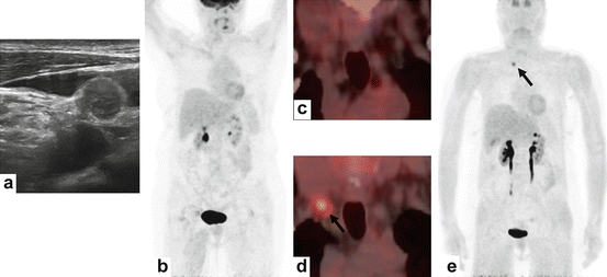
Fig. 18.1
A 54-year-old female affected by MTC with evidence of increasing serum calcitonin levels (44 pg/mL) after thyroidectomy and cervical lymphadenectomy. Serum CEA was normal. Neck ultrasonography showed a small right laterocervical (IV level) lymph node suspicious for recurrence (a). Maximum intensity projection FDG-PET image (b) and coronal FDG-PET/CT image (c) did not show any abnormal tracer uptake. On the other hand, maximum intensity projection FDOPA-PET image (e) and coronal FDOPA-PET/CT image (d) correctly detected the recurrence of disease (black arrow)
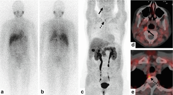
Fig. 18.2
A 44-year-old female affected by MTC with evidence of increasing serum calcitonin levels (80 pg/mL) after thyroidectomy and cervical lymphadenectomy. Serum CEA was normal. Neck ultrasonography (not shown) did not detect suspicious lymph nodal recurrence. Radioiodinated MIBG scintigraphy in anterior (a) and posterior (b) view did not demonstrate any abnormal tracer uptake. On the other hand, maximum intensity projection FDOPA-PET image (c) and axial FDOPA-PET/CT images (d, e) correctly detected disease recurrence showing tracer uptake corresponding to small retropharyngeal (black arrow) and paraesophageal (dotted arrow) lymph nodes
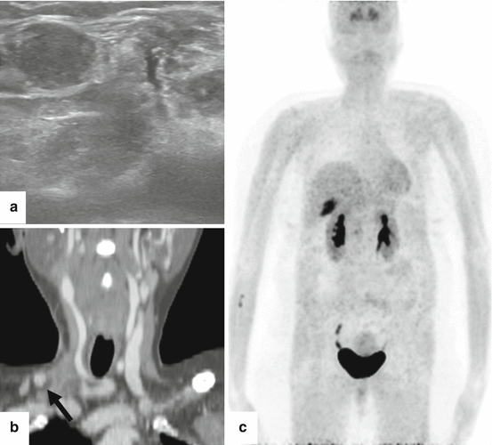
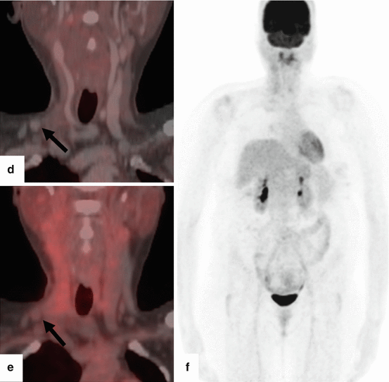
Fig. 18.3
A 48-year-old female affected by MTC with persisting high serum calcitonin levels (39 pg/mL) after thyroidectomy and right laterocervical lymphadenectomy for lymph nodal metastases. Serum CEA was normal. Neck ultrasonography (a) showed a right laterocervical (IV level) lymph node suspicious for metastasis adjacent to a scar tissue. Coronal contrast-enhanced CT (b) detected a 12 mm right laterocervical lymph node with contrast enhancement (black arrow). Neither FDOPA-PET/CT (c, d) nor FDG-PET/CT (e, f) demonstrate any abnormal tracer uptake corresponding to the right laterocervical lymph node (black arrows). Histopathology on this lymph node showed a MTC metastasis
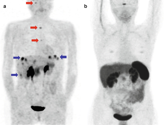
Fig. 18.4
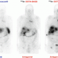
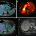
A 47-year-old female affected by MTC with evidence of increasing serum calcitonin levels (280 pg/mL) after thyroidectomy and cervical lymphadenectomy. FDOPA-PET (a) showed multiple areas of abnormal tracer uptake corresponding to bone (red arrows) and liver metastases (blue arrows). On the other hand, somatostatin receptor PET was negative (b)
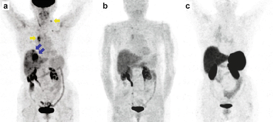
Fig. 18.5
A 80-year-old female affected by MTC with evidence of increasing serum calcitonin (480 pg/mL) and CEA (322 ng/mL) levels after thyroidectomy and cervical lymphadenectomy. FDG-PET (a) showed multiple areas of abnormal tracer uptake corresponding to lymph nodal (yellow arrows) and liver metastases (blue arrows). FDOPA-PET (b) and somatostatin receptor PET (c) demonstrated only minimal abnormalities
Comparative analyses between FDOPA and FDG have shown better results with FDOPA in terms of sensitivity and specificity and a complementary role of the two radiopharmaceuticals in the assessment of recurrent MTC [46, 50, 63–65]. The different behaviour of FDOPA and FDG in recurrent MTC can be explained by their different uptake mechanisms that, in turn, reflect the different metabolic pathways of NET cells, including MTC cells. FDOPA is a marker of amino acid decarboxylation that is a feature of the neuroendocrine origin of MTC. So, it can be assumed that a higher FDOPA uptake is related to a higher degree of cell differentiation, whereas a higher FDG uptake is related to a high-proliferative activity and a poor differentiation [46, 50, 63–65]. Although FDOPA-PET/CT has less prognostic value compared to FDG, it can more accurately assess the extent of the disease in patients with residual/recurrent MTC [46, 50, 63].
Comparative analyses between somatostatin analogues labelled with Gallium-68 and FDG have shown the complementary role of these PET tracers in recurrent MTC without statistically significant difference in terms of detection rate of MTC lesions [32, 50, 56, 58, 61].
To date, only one multicentric study compared FDOPA, FDG and somatostatin analogues labelled with Gallium-68 demonstrating that FDOPA-PET/CT is the most useful functional imaging method for detecting recurrent MTC lesions in patients with increased serum calcitonin levels, performing better than FDG and somatostatin analogues labelled with Gallium-68 and leading a change in the patient management in a significant percentage of cases [50].
According to the injected activity of radiopharmaceutical and the volume of the body explored by low dose CT, the total effective dose from FDG, FDOPA or somatostatin receptor PET/CT varies between 13 and 15 mSv, and the radiation dose is very similar for all these radiopharmaceuticals [66]. The actual effective dose is currently decreasing with a trend to reduce the injected activity of radiopharmaceuticals by using time of flight PET/CT [66].
While FDG and FDOPA may be prepared industrially (for PET centres without an on-site cyclotron) and delivered ready to use, for labelling somatostatin analogues with Gallium-68, both Germanium-68/Gallium-68 generator and lyophilised peptides are needed [66]. The easy synthesis process or radiolabelled somatostatin analogues is an advantage supporting their clinical use as PET tracers [54, 55].
FDOPA is the most expensive PET tracer for evaluating recurrent MTC, mainly for its difficult synthesis, but it is questionable whether its first-line indication in recurrent or metastatic MTC will increase the total costs of diagnostic workup. In fact, FDOPA-PET/CT allowing the most precise diagnostic information among other functional imaging techniques and sometimes providing additional informational compared to conventional morphological imaging in recurrent or metastatic MTC may permit avoiding further examinations, savings which may compensate for its price [66]. Unfortunately no cost-effectiveness studies are currently available for the diagnostic workup of recurrent or metastatic MTC by using radionuclide imaging.
The limited availability of FDOPA and somatostatin analogues labelled with Gallium-68 compared to FDG is probably not a major drawback in case of a rare cancer such as MTC, a limited number of specialised centres being able to match the demand [66].
18.4.3 Proposed Algorithm About the Use of Radionuclide Imaging Methods in MTC
The role of radionuclide imaging in staging MTC before treatment seems to be limited compared to conventional morphological imaging [1].
In suspected recurrent MTC based on rising tumor markers levels after thyroidectomy, radionuclide imaging methods may provide additional information compared to conventional imaging if serum calcitonin is >150 pg/mL. In particular, PET/CT with different tracers may change the patient management in a significant percentage of cases [43, 67].
FDOPA-PET/CT should be used as first radionuclide imaging method in recurrent MTC showing significant diagnostic performance in patients with higher serum calcitonin levels and shorter serum calcitonin doubling time. In negative cases, FDG-PET/CT should be the next radionuclide imaging method, in particular if calcitonin and CEA levels are rapidly rising or a more aggressive disease is suspected. PET/CT with somatostatin analogues labelled with Gallium-68 could be performed when neither FDOPA- nor FDG-PET/CT are conclusive or in selecting patients suitable for therapy with cold or radiolabelled somatostatin analogues. Bone scintigraphy could complement FDG-PET/CT if FDOPA is not available [66].
Conflict of Interest
The authors declare that they have no conflicts of interest.
References
1.
Wells Jr SA, Asa SL, Dralle H, Elisei R, Evans DB, Gagel RF, et al. Revised American Thyroid Association guidelines for the management of medullary thyroid carcinoma. Thyroid. 2015;25:567–610.CrossRefPubMedPubMedCentral
2.
3.
4.
5.
Marsh DJ, Learoyd DL, Andrew SD, Krishnan L, Pojer R, Richardson AL, et al. Somatic mutations in the RET proto-oncogene in sporadic medullary thyroid carcinoma. Clin Endocrinol. 1996;44:249–57.CrossRef
6.
Boichard A, Croux L, Al Ghuzlan A, Broutin S, Dupuy C, Leboulleux S, et al. Somatic RAS mutations occur in a large proportion of sporadic RET-negative medullary thyroid carcinomas and extend to a previously unidentified exon. J Clin Endocrinol Metab. 2012;97:E2031–5.CrossRefPubMedPubMedCentral

