Radiologic Imaging Concerns
Jeffrey A. Bennett
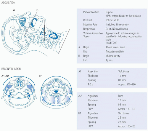
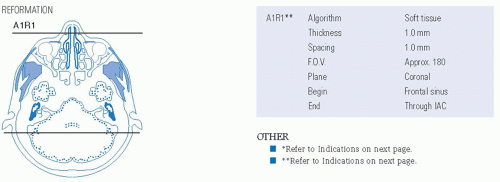
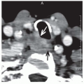
The optimal diagnostic imaging evaluation for patients presenting with cervical lymph node metastases from an unknown source is still controversial. The purpose of imaging is to help identify potential sites to pay special attention to panendoscopy with directed biopsies. Currently, it is recommended that patients receive a contrast-enhanced CT scan or MRI of the head and neck, and FDG-PET/CT should be considered.1 Suggested CT and MRI protocols are given in Figures 15-2 and 15-3. The location of the cervical lymph nodes is important as patients with lymphadenopathy primarily in levels II and III will most often have a primary in the tonsillar fossa or the tongue base,2 whereas patients with low neck lymphadenopathy, primarily in level IV and the supraclavicular fossa, will often have a primary tumor below the clavicles.3 CT or MRI may identify a submucosal mass that could not be detected on physical exam or endoscopy. Alternatively, a small tumor located deep in the
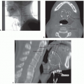
crypts of lymphatic tissue in the nasopharynx, the palatine tonsils, or the tongue base may produce asymmetry that could guide the endoscopist to a location to biopsy (Fig. 15-4). The problem arises though, that normal lymphatic tissue is quite asymmetric, especially in the palatine tonsils and the lingual tonsil, and so a better approach is to use the asymmetry to guide a search for deep invasion into the parapharyngeal space or the tongue musculature, which is a clear sign that cancer is present (Fig. 15-5).
crypts of lymphatic tissue in the nasopharynx, the palatine tonsils, or the tongue base may produce asymmetry that could guide the endoscopist to a location to biopsy (Fig. 15-4). The problem arises though, that normal lymphatic tissue is quite asymmetric, especially in the palatine tonsils and the lingual tonsil, and so a better approach is to use the asymmetry to guide a search for deep invasion into the parapharyngeal space or the tongue musculature, which is a clear sign that cancer is present (Fig. 15-5).
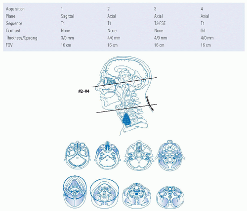 FIGURE 15-3. Protocol for MRI of the unknown primary.
Stay updated, free articles. Join our Telegram channel
Full access? Get Clinical Tree
 Get Clinical Tree app for offline access

|
