Radiation Therapy Technique
Louis B. Harrison
Rudolph Woode
James Dolan
Kenneth Hu
The most common sites treated with radiation therapy include the lip, the floor of the mouth, and the oral tongue. Lesions of the buccal mucosa, retromolar trigone are treated with techniques similar to those used for the tonsil, which are discussed in Chapter 17C. For oral cavity tumors, primary surgery is the most common approach. Radiation therapy is generally used as an adjuvant treatment in the postoperative setting. Primary RT can be used in certain situations, and brachytherapy is an important component.
Lesions of the upper lip are uncommon; the treatment techniques described hereafter apply primarily to the lower lip, but can be modified to treat the upper lip. Patients with superficial, well-differentiated squamous cell carcinoma of the lip have a low risk of cancer in the neck nodes and may be irradiated with brachytherapy alone or external beam irradiation alone. A short course of external beam irradiation precedes the interstitial implant, if the lesion is too thick to be adequately encompassed in a single-plane implant (> 1 cm). External beam
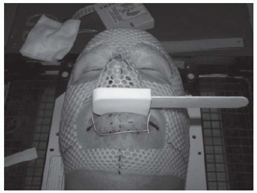
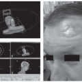
irradiation is usually done with local electron field, although photons can sometimes be used. Figure 16-32 shows a patient with squamous cell cancer of the lower lip, and the field setup for electrons.
irradiation is usually done with local electron field, although photons can sometimes be used. Figure 16-32 shows a patient with squamous cell cancer of the lower lip, and the field setup for electrons.
Interstitial irradiation may be accomplished with iridium-192 (192Ir) using the plastic tube technique. These procedures can usually be done under local anesthesia. Figure 16-33 shows a patient who had an 192Ir implant for a squamous cell cancer of the lower lip.
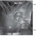
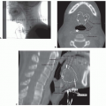
Small cancers of the floor of the mouth or the oral tongue can be treated with an interstitial implant alone. This is usually accomplished with an 192Ir implant afterloaded into plastic tubes. This is done under general anesthesia, in the operating room. Sometimes, a tracheostomy is performed, as a temporary protection of the airway, especially if the posterior portion of the oral tongue is involved. The tracheostomy protects the airway in the advent of the tongue swelling or bleeding. The afterloading catheters are implanted using a looping technique. The catheters enter and exit via the submental approach. Figure 16-34 shows an intraoperative photograph of an oral tongue implant and highlights the looping technique. Figure 16-35 shows AP and lateral images of an oral tongue implant, with isodose curves.
Stay updated, free articles. Join our Telegram channel

Full access? Get Clinical Tree


