Prognostic Factors in Patients with Head and Neck Cancer


Prognostic Factors in Patients with Head and Neck Cancer


Sung Kim

Benjamin D. Smith

Bruce G. Haffty


INTRODUCTION

Treatment of head and neck cancer requires accurate risk stratification in order to determine the type and extent of therapy and the expected clinical outcome. Physical exam, diagnostic imaging studies, and pathologic review enable the clinician to determine the size and extent of the primary tumor, the status of cervical lymph nodes, and the likelihood of distant metastatic disease, thus generating an accurate tumor, lymph node, metastasis (TNM) stage for each patient. In addition to TNM staging, other clinical and pathologic factors not routinely incorporated into the staging system have been shown to influence response to therapy and eventual outcome. These factors may be categorized as follows: (a) prognostic factors related to the primary tumor, (b) prognostic factors related to the cervical lymph nodes, (c) prognostic factors related to patient demographics, and (d) prognostic factors related to the patient’s general medical condition.

In addition to clinical and pathologic factors, recent interest has focused on identifying molecular factors that may influence clinical outcome. These molecular markers not only provide useful prognostic information, but also serve as targets for novel pharmacotherapies that antagonize cellular proliferation by interfering with specific cellular processes. Molecular factors that influence tumor behavior fall into several broad categories, including proto-oncogenes, tumor suppressor genes, growth factors, immune-related factors, loss of heterozygosity at various genetic loci, total cellular DNA content, and parameters related to the kinetics of in vivo tumor growth. Finally, the correlation of human papilloma virus (HPV) has been crucial to furthering our understanding of this new and important subset of cancers. This will be discussed in detail in Chapter 12.

When considering prognostic factors in head and neck cancer, several specific outcomes merit attention. In addition to diseasefree and overall survival, the relationship between a specific prognostic factor and local recurrence, regional recurrence, and distant metastatic disease must be considered. Factors specifically related to local recurrence may necessitate wider surgical resection, external beam radiotherapy dose escalation or altered fractionation, or the use of a brachytherapy boost to the primary tumor bed. Factors specifically related to regional recurrence may prompt prophylactic neck dissection or radiotherapy in the clinically NO neck or determine the type of neck dissection selected in patients with clinically positive cervical lymph nodes. Finally, factors predictive of distant metastases may prompt more aggressive screening for concomitant distant metastases before local-regional treatment1 and influence the decision to offer systemic therapy.


FACTORS RELATED TO THE PRIMARY TUMOR


Tumor Dimensions

Tumor size and extent of invasion determine clinical and pathologic T stage for head and neck squamous cell carcinomas (HNSCC). T stage is determined clinically by measuring the maximal surface diameter of a mucosal neoplasm or pathologically by measuring the maximal cross-sectional diameter of a resected tumor. Because tumors arising from mucosal surfaces do not conform to spherical geometry, the maximal tumor diameter does not perfectly correlate with either tumor depth or tumor volume.

As the primary determinant of T stage, maximum tumor diameter has traditionally been considered an important risk factor for the presence of concomitant nodal metastases, local recurrence, and poor survival. For example, in two studies conducted on a total of 603 patients with HNSCC, Magnano et al. found that T stage was a consistent, independent predictor of pathologically positive cervical lymph nodes.2,3 In addition, pathologic maximal tumor diameter has been shown to predict local recurrence in tumors arising from the lower lip,4 oral cavity and oropharynx,5 and larynx.6 Finally, most studies,5,7,8,9,10 though not all,11 have shown a univariate association between either clinical or pathologic tumor diameter and survival.

One limitation of using tumor size as a prognostic determinant was highlighted by Moore et al., who stratified 155 patients with oral SCC based on surface diameter of the primary tumor.7 Eighty-four percent of patients with tumors <2 cm survived disease free for 3 years, compared with 52% of patients with tumors larger than 2 cm. However, no significant differences in survival existed between tumors with surface diameters in the following three groups: 2.1 to 3 cm, 3.1 to 4 cm, and >4 cm. Thus, although a gross trend exists between surface diameter and survival, this trend does not follow a simple dose-response relationship. In a follow-up study conducted on 151 patients with oral and oropharyngeal SCC, Moore et al. found that tumor thickness was a more consistent predictor of concomitant nodal metastasis and 3 year disease free survival than tumor surface diameter.12


The correlation between tumor thickness and risk of concomitant cervical lymph node metastases has been confirmed in multiple studies.4,13,14,15,16,17,18,19,20 The critical thickness, above which the risk of nodal metastasis increases substantially, varies depending upon the specific anatomic site involved, perhaps due to variation in density of lymphatic channels.21 Nevertheless, for most anatomic subsites, the critical thickness ranges from 3 to 5 mm depending upon the specific study. For example, a recent study of 105 patients with clinically node negative oral cavity tumors reported that the risk of occult nodal metastasis was 10% if the tumor thickness was <5 mm, compared to 46% if the tumor thickness was >5 mm.14 Furthermore, in a series of 76 patients with carcinoma of the tongue who all received neck dissection, Woolgar demonstrated that the mean reconstructed thickness of tumors with pathologically positive cervical lymph nodes was 19 mm, compared to 10 mm in patients without metastases.22 Tumors with a large mucosal surface area but minimal invasion were not at increased risk of nodal metastases. In such cases, clinical and pathologic T stage may overestimate the likelihood of concomitant nodal disease.

In addition to consideration of maximal tumor diameter and thickness, utilization of computed tomography (CT) enables the calculation of total tumor volume. In a series of 63 patients with supraglottic primaries treated with primary irradiation, Mancuso et al. found that tumor volume calculated from pretreatment CT scans was an independent predictor of local control, with local control rates of 89% in tumors <6 cm3 versus 52% for tumors >6 cm3.23 Furthermore, in a study of 103 patients with supraglottic SCC, tumor volume as calculated from pretreatment CT was a stronger predictor of local-regional failure than clinical T stage in multivariate analysis.24 Hence, calculation of tumor volume using diagnostic imaging provides important prognostic information that can supplement traditional clinical staging for tumors of the larynx. In contrast, T stage, rather than tumor volume estimated from diagnostic imaging studies, appears to be a better predictor of local control for tumors of the oropharynx and hypopharynx treated with definitive radiation.25


Margin Status

The presence of residual carcinoma at the margins of surgical resection is an important risk factor for local recurrence in HNSCC. Although the presence of positive margins may indicate an error in surgical judgment at the time of resection, it may also imply a more biologically aggressive tumor that extends microscopically through the muscle bundles, submucosal lymphatics, and perineural spaces.26 The precise definition of “positive margins” varies depending on the study and may include invasive tumor involving the initial surgical margin, invasive tumor involving the final surgical margin, invasive tumor approaching within 5 mm of the final surgical margin, carcinoma in situ involving the final surgical margin, or dysplasia involving the final surgical margin. Guidelines used in the United Kingdom define “negative” margins as invasive tumor more than 5 mm away from the surgical margin, “close” margins as invasive tumor within 1 to 5 mm from the surgical margin, and “positive” margins as tumor <1 mm from the surgical margin.21

In a review of resections for head and neck cancer performed at Memorial-Sloan Kettering Cancer Center, Looser et al. classified 1,775 cases according to final margin status.27 After excluding patients with gross residual disease, they identified 62 patients with microscopically positive margins, defined as either cancer within 0.5 cm of the margin, marked atypia or premalignant changes in the margin, carcinoma in situ in the margin, or invasive carcinoma in the margin. Interestingly, patients in all four of these groups experienced increased local recurrence rates, ranging from 64% in patients with invasive cancer at the margin to 85% for patients with carcinoma in situ at the margin, compared to a local recurrence rate of 32% in patients with negative margins.

Many other studies have confirmed the prognostic importance of margin status in HNSCC. The presence of positive margins has been shown to predict local or local-regional recurrence for the following sites: lower lip,4 oral cavity,28,29,30 buccal mucosa,31 oral tongue,13,32 oral and oropharyngeal tongue,33,34 base of tongue,35 oral cavity and oropharynx,36 larynx,37,38 and all sites combined.39,40,41,42,43 Furthermore, the presence of positive margins predicts poor overall survival in univariate analysis for the following sites: oral cavity,30 oral tongue,44 oral and oropharyngeal tongue,34 oral cavity and oropharynx,45 base of tongue,35 supraglottic larynx,46 larynx,8,38,47 and all sites combined.39,40,48,49 Whether or not margin status is an independent predictor of survival remains somewhat controversial, with two studies confirming an independent association,38,49 but six others failing to find an association.5,8,9,39,46,50 Margin status has not been shown to predict regional or distant recurrent disease.

Given the negative prognostic implications of positive margins, Byers et al. demonstrated that use of intraoperative frozen sections can identify those patients with initially positive margins and thus allow resection of additional tissue to remove residual carcinoma and reduce the risk of local recurrence.48 In this cohort of 216 patients with SCC arising from the oral cavity, oropharynx, and hypopharynx, patients with initially positive margins on frozen section received additional resection to achieve final negative margins. Local recurrence in this group was 13%, compared with a local recurrence rate of 12% in patients with negative margins on initial frozen section. Patients with positive final margins suffered from an 80% local recurrence rate. Hence, use of intraoperative frozen section analysis of surgical margins may result in a clinically significant reduction in local recurrence.

However, in a follow-up study of 268 patients with SCC of the tongue, those patients with initially positive margins that were ultimately rendered negative experienced an increased risk of local recurrence and death compared to patients with initially negative frozen section margins.26 Thus, the authors recommended use of postoperative radiotherapy in all patients with initially positive frozen section margins. One additional concern regarding the use of intraoperative frozen sections to determine margin status is potential inaccuracy, with one study reporting a 14% (7/49) false negative rate for oral cancer.51

For those patients with persistently positive margins, postoperative external beam radiotherapy remains an important component of therapy. However, even with combined modality therapy, most investigators,31,39,42,52 although not all,53 report an increased incidence of local recurrence in those patients with positive margins. One important factor influencing local control in patients with positive margins is the dose of postoperative radiotherapy, as Zelefsky et al. noted a 7-year local control rate of 92% in patients who received >60 Gy, compared with 44% in patients receiving <60 Gy.53 In addition, interest has grown in using postoperative brachytherapy, either alone or following postoperative external beam radiotherapy, to improve local control in patients with positive margins.54,55,56,57 Beitler et al. reported 29 patients with microscopically close or positive margins after curative surgery treated with postoperative external beam radiotherapy to a median dose of 60 Gy followed by125 I permanent implant designed to deliver a cumulative lifetime dose of 120 to 160 Gy to the high-risk target volume.57 This treatment strategy resulted in a 92% 2-year actuarial local control rate and clearly merits further study. Finally, growing evidence from two large randomized trials indicates that the addition of cisplatin (100 mg/m2 on days 1, 22, and 43) to postoperative radiation improves local-regional control and perhaps overall survival for patients with positive surgical margins.58,59,60


Malignancy Grading

Pathologists have long recognized the potential prognostic significance of cellular morphology in squamous cell carcinoma. In 1920, Broders proposed a four-tiered grading system for carcinoma of the lip that was based on the proportion of the neoplasm resembling normal squamous epithelium.61 This grading system provides an easily assessed scheme for assessing tumor differentiation and roughly correlates with prognosis, as poorly differentiated tumors are more likely to recur and reduce survi val.30,46,62,63,64,65,66,67,68 Broders’ grading system has been criticized, however, for its subjectivity and for its failure to consistently predict survival in multivariate modeling.9,30,46,69,70 The lack of independent significance of Broders’ grading system may be due to the association between higher grade (more poorly differentiated] and advanced stage.71

In 1973, Jakobsson proposed a semiquantitative grading system that considered not only histologic parameters of the tumor cell population but also the host-tumor interface.72 Parameters describing the tumor cell population included structure and growth of the neoplasm, degree of keratinization, nuclear pleomorphism, and the frequency of mitoses. The tumor-host interface was assessed for mode of invasion, degree or stage of invasion, vascular invasion, and lymphoplasmacytic cellular response. When this classification system was applied to a series of 42 laryngeal SCCs treated with radiotherapy, tumors with high malignancy grade were more likely to recur and result in death with disease. However, in a subsequent multivariate analysis conducted on 77 patients with oropharyngeal cancer, Crissman et al. found that invasion pattern was the only histologic parameter that independently predicted survival.73 None of the other histologic parameters, either independently or as a composite score, were significant predictors of outcome.

In 1987, Anneroth et al. reviewed efforts to devise a malignancy grading system and proposed that grading consist of six morphologic features: degree of keratinization, nuclear polymorphism, number of mitoses, pattern of invasion, stage of invasion, and lymphoplasmacytic infiltration.74 In 1989, Bryne et al. applied Anneroth’s grading system to only the most anaplastic fields in the most invasive areas of the tumor (Table 3.1).75 In two cohorts of 68 and 61 patients with oral SCC, Bryne’s invasive cell grading was a highly significant, independent, and reproducible predictor of survival.75,76,77 Those patients with a total malignancy score between 5 and 10 experienced a 57% 5-year survival, compared to a 19% 5-year survival in patients with malignancy scores >10. In contrast, Broders’ grading system failed to correlate with survival in both cohorts. The authors concluded that histologically invasive regions might be responsible for the clinical behavior of HNSCC. Further study of invasive cell grading (ICG) conducted by other groups has shown that a high ICG score strongly predicts the presence of occult cervical metastases and extracapsular extension (ECE).17,22,78 Due to the strong correlation between ICG and concomitant nodal metastases, this factor should be considered in decisions regarding elective treatment of the clinically negative neck.


TABLE 3.1 Invasive Cell Grading System


Score


Morphologic Feature


1


2


3


4


Degree of keratinization


Highly keratinized (>50% of the cells)


Moderately keratinized (20%-50% of the cells)


Minimal keratinization (5%-20% of the cells)


No keratinization (0%-5% of the cells)


Nuclear polymorphism


Little nuclear polymorphism (>75% mature cells)


Moderately abundant nuclear polymorphism (50%-75% mature cells)


Abundant nuclear polymorphism (25%-50% mature cells)


Extreme nuclear polymorphism (0%-25% mature cells)


Number of mitoses (high-power field)


0-1


2-3


4-5


>5


Pattern of invasion


Pushing, well-delineated infiltrating borders


Infiltrating, solid cords, bands, and/or strands


Small groups or cords of infiltrating cells


Marked and widespread cellular dissociation in small groups and/or in single cells (n < 15)


Lymphoplasmocytic infiltration


Marked


Moderate


Slight


None


Source: From Bryne, M., Koppang HS, Lilleng R,et al., New malignancy grading is a better prognostic indicator than Broders’grading in ora squamous cell carcinomas. J Oral Pathol Med. 1989;18(8):432-437, with permission.


In addition to assessment of malignancy grade, many studies have attempted to determine which individual histologic parameters contribute most strongly to prognosis. Several studies suggest that pattern of invasion may predict outcome independent of other histologic parameters. Tumors that infiltrate in small groups or cords of cells, or infiltrate with marked cellular dissociation, may behave more aggressively. For example, Spiro et al. found that oral tongue SCCs with a high-grade pattern of invasion were more likely to present with concomitant nodal metastases, develop distant metastases, and result in death.32 In addition, for oral carcinoma that invades the mandible, highgrade invasion pattern increases the rate of mandibular margin positivity and local recurrence and results in a fourfold increased risk of death with disease in multivariate analysis.79 The correlation of aggressive invasion pattern with poor localregional control4,33,80,81,82,83 and survival10,45 has been noted in several other studies.

In addition to pattern of invasion, lymphoplasmacytic infiltration of the tumor bed has been considered as a potential independent marker of prognosis. In a study of 396 patients with HNSCC, multivariate analysis revealed that the presence of a lymphocytic tumor infiltrate decreased the risk of concomitant
cervical lymph node metastases, whereas a plasmacytic infiltrate increased the risk.2 In addition, several studies have reported a correlation between lymphocyte infiltration and improved localregional control.81,84,85

Basaloid SCC (BSCC) is a rare variant of SCC that has historically been thought to be chemo- and radiosensitive, with similar-to-better loco regional control than “typical” SCC, but with higher risk of distant metastases. Soriano reported a case control series of BSCC versus SCC treated with various modalities and reported that loco regional recurrence caused death in only 15% of the BSCC patients versus 51% of the SCC patients. Distant metastasis, however, occurred in 45% versus 7%, respectively.86 Thariat reported a case control series of radiated patients with BSCC versus poorly differentiated (PSCC) or well-differentiated SCC (WSCC). The loco-regional control for BSCC, PSCC, and WSCC was similar at 81%, 86%, and 82%, respectively; distant metastases free survival was worse for BSCC at 71%, 86%, and 86%, respectively, but not significantly so.87

Why the seeming inconsistency that BSCC is considered an aggressive variant of SCC, yet HPV-associated SCC is more commonly BSCC but has a better prognosis? It may be that BSCC is a more heterogeneous disease than previously recognized. Certainly the demographics of the BSCC patient are evolving: whereas the typical BSCC patient has been thought to be in his 60s or 70s, with history of frequent, intense tobacco use and alcohol use, recent cases have also affected young women without a significant tobacco or alcohol history, suggesting a causal link between BSCC and HPV-associated SCC.88 Therefore, BSCC may have a dual nature similar to SCC, depending on factors such as HPV status and tobacco and alcohol use.


Perineural Invasion

Infiltration of perineural spaces occurs in up to 52%89 of HNSCCs and was first noted to influence surgical and adjuvant treatment strategies by Ballantyne et al. in 1963.90 Perineural invasion (PNI) may result in dysphagia secondary to involvement of the vagal trunk91 or pain and paresthesias along the territories of the glossopharyngeal or trigeminal nerves.91,92 CT findings suggestive of PNI include obliteration of the fat within the pterygopalatine fossa, enlargement of neural foramina, and increased enhancement in the region of Meckel’s cave.93 In addition, a study of 48 patients with oral tongue, base of tongue, or floor of mouth SCC utilized CT to identify the presence of vascular and/or PNI with a sensitivity of 88% and a specificity of 83% using criteria including “aggressive” tumor margins, invasion of the sublingual space, and direct adjacency to the lingual vasculature.94

PNI may be mediated by the presence of nerve cell adhesion molecule (N-CAM) on the surface of squamous carcinoma cells, which engages in homophilic binding with N-CAM expressed in neural and perineural tissues. In two studies of 76 and 66 patients with HNSCC, expression of N-CAM on the surface of neoplastic cells was significantly associated with PNI detected on review of pathologic sections.95,96

Numerous clinical studies have identified PNI as an important predictor of poor prognosis. In tumors arising from the lower lip,4,97,98 oral tongue,99 oral cavity,20,89,100,101 larynx,102 and all sites combined,2,3 the presence of PNI is associated with a higher risk of metastasis to regional lymph nodes. Furthermore, following definitive treatment of HNSCC, the presence of PNI in the primary tumor is associated with poor local control,4,89,103,104 regional control,64 local-regional control,81,84,105,106 cause-specific survival,89,105 and overall survival.44,105 PNI has been associated with an increased risk of distant metastasis in some,107 though not all,108 studies.

The association between PNI and local recurrence may result from either centrifugal or centripetal propagation of malignant cells along perineural spaces and away from the primary tumor. Most primary tumors will only disseminate up to 2 cm along the perineural space,109 although PNI 12 cm from the primary tumor has been reported.90 As a result, PNI may allow malignant cells to evade surgical excision or radiotherapy and result in local recurrence. In addition, the association between PNI and regional recurrence implies that these tumors may be more biologically aggressive. The association between perineural invasion and tumor aneuploidy, a known marker of poor prognosis, lends support to this hypothesis.110

Despite the clear importance of PNI, the percentage of mucosal HNSCCs positive for PNI varies widely in the literature, from 5 %104 to 52 %.89 This discrepancy may result from a tendency to identify PNI only when large, named nerves are involved. However, although PNI of small, unnamed nerves may not result in clinical symptoms, the relationship between PNI and prognosis appears to be independent of nerve diameter.89 Hence, all HNSCC pathologic specimens should be closely examined for PNI, even in nerves <1 mm in diameter.


Vascular Invasion

Vascular invasion, defined as the presence of neoplastic epithelium within an endothelial-lined channel, occurs in over 50% of HNSCCs.5,22,111 As with tumor depth and pattern of invasion, the presence of vascular invasion may identify tumors with an aggressive biologic nature due to their ability to invade normal anatomic structures. Indeed, vascular invasion has been shown to correlate with the presence of concomitant cervical metastases in both univariate20,22,45,103,111 and multivariate models.2,3,99,112 In addition, the presence of vascular invasion in the primary tumor specimen has been noted to increase the risk for subsequent local regional recurrence in oral cavity, oropharyngeal, and laryngeal SCC treated surgically.5,64,103 However, in a large series of 226 patients treated with surgery and postoperative radiotherapy, vascular invasion did not correlate with local-regional control, suggesting that postoperative radiotherapy may mitigate the poor prognosis associated with vascular invasion.106 Vascular invasion has also been associated with increased risk of distant metastatic disease.5,69


Imaging

18F-fluorodeoxyglucose (FDG) PET scan is a functional test that combines a positron-emitting nuclide with a glucose molecule, thereby revealing areas of increased glucose metabolism. PET/CT scans are commonly performed prior to therapy to determine extent of disease, and afterward to assess response and need for further treatment.

The utility of a posttreatment PET has been well studied. Isles performed a meta-analysis of radiated patients who received posttreatment FDG PET scans. The weighted mean pooled sensitivity, specificity, PPV, and NPV of posttreatment PET for the primary site was 94%, 82%, 75%, and 95%, respectively. At the neck, the corresponding numbers were 74%, 88%, 49%, and 96%, respectively. They reported that sensitivity was greater for scans performed 10 weeks or longer after completion of therapy.113 Gupta also performed a meta-analysis; his corresponding numbers for the primary site were 79.9%, 87.5%, 58.6%, and 95.1%, respectively, and for the neck 72.7%, 87.6%, 52.1%, and 94.5%, respectively. Scans performed >12 weeks after therapy had moderately higher diagnostic accuracy.114

Moeller prospectively compared posttreatment PET/CT with contrast-enhanced CT in radiated patients. Patients were imaged with PET/CT and CT prior to and 8 weeks after radiation.
Though postradiation maximum SUVs were significantly higher in nonresponders compared with responders, the positive and negative predictive values for PET/CT were similar to CT alone. In order to improve the PPV of the postradiation scans, they stratified patients into high-risk (HPV negative, nonoropharynx primary, tobacco history) and low-risk categories. In the highrisk patients, PET/CT outperformed CT alone, especially in terms of accurately detecting residual nodal disease, where the PPV for PET/CT was twice that of CT alone (75% vs. 37.5%).115

Whether pretreatment PET/CT reveals loco-regional versus metastatic disease is clearly prognostic, but the predictive value of pretreatment SUV in localized disease is controversial. Moeller found no significant difference in the pretreatment SUVmax at the primary site or lymph nodes between responders and nonresponders.115 Higgins came to a similar conclusion, but did report that increased pretreatment SUVmean at the primary site correlated with disease-free survival.116 Imsande found that SUVmax at the primary site correlated with overall survival, whereas Demirci found that disease-free survival correlated with nodal (but not primary site) SUVmax.117,118 Tang119 evaluated metabolic tumor volume (MTV) on pretreatment PET-CT and concluded that MTV was an independent predictor of outcomes. Specifically, MTV > 17 cm3 was associated with a greater risk of both disease progression and death. Lim120 also reported that primary tumor MTV was predictive of local failure, distant metastases, and overall survival.

In conclusion, a negative posttreatment PET/CT that is appropriately timed portends a favorable prognosis and may be especially helpful in higher-risk patients. A positive result is less reliable, probably due to the possibility of a false positive due to residual inflammation or infection.


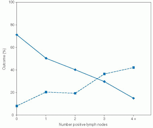
FIGURE 3-1. Number of positive cervical lymph nodes as a predictor of distant metastasis and 5-year overall survival in a cohort of 914 patients undergoing lymph node dissection for a primary head and neck squamous cell carcinoma.

Source: From Mamelle G, Pampurik J, Luboinski B, et al. Lymph node prognostic factors in head and neck squamous cell carcinomas. Am J Surg. 1994;168:494-498, with permission.


FACTORS RELATED TO THE CERVICAL LYMPH NODES


Number of Positive Lymph Nodes

The number, size, and location of positive cervical lymph nodes define the N stage for HNSCC and provide important information regarding prognosis and selection of treatment. The number of cervical lymph nodes histologically positive for squamous cell carcinoma provides one of the simplest, and perhaps most important, prognostic markers in head and neck cancer. Mamelle et al. retrospectively reviewed 914 patients who received cervical lymph node dissection as a component of initial therapy for oral cavity, oropharyngeal, hypopharyngeal, and laryngeal SCC.121 Patients with palpable lymph nodes >3 cm underwent radical lymph node dissection, and all other patients underwent selective neck dissection on sentinel nodes with immediate pathologic evaluation. Those patients with positive sentinel nodes then received ipsilateral modified radical neck dissection and contralateral selective neck dissection. Thus, all patients, regardless of their clinical N stage, received pathologic evaluation of their cervical lymph nodes. Those patients with positive cervical lymph nodes received 50 Gy to the neck with a 15 Gy boost in areas of extracapsular spread. In this study, lymph node number exhibited a strong dose-response correlation with distant metastasis and survival (Fig. 3-1). In multivariate analysis stratified by tumor site and patient age, number of positive nodes was a significant, independent predictor of survival. Furthermore, although extracapsular spread and node location (upper vs. middle vs. lower neck) were significant predictors of survival in univariate analysis, after controlling for number of positive lymph nodes,
they lost their prognostic significance. Thus, with this particular treatment algorithm, the number of positive nodes emerged as an important, independent predictor of prognosis.

The importance of positive cervical nodes has been noted in many other studies and appears to correlate with risk of regional recurrence and distant metastasis. Studies have demonstrated a correlation between increasing number of positive nodes and regional recurrence in patients with advanced SCC who receive postoperative radiotherapy,122 patients who do not receive radiotherapy,64 patients with oral cavity30 and hypopharyngeal or lateral epilaryngeal primary tumors,123 and patients who receive neck dissection for mucosal HNSCC from any site.124,125 Furthermore, number of positive nodes clearly predicts risk of distant metastatic disease for all sites of HNSCC.11,108,121,126,127,128,129,130 Number of positive nodes predicts both regional recurrence64 and distant recurrence,129 even after controlling for other prognostic variables in multivariate analysis.

Finally, the number of positive cervical lymph nodes consistently correlates with survival in univariate analysis for all major sites of HNSCC.9,30,62,122,123,126,131,132,133,134,135 The relative importance of ECE versus number of positive nodes remains somewhat controversial. In addition to the Mamelle et al. study,121 Moe et al. found the number of positive nodes to predict poor survival independent of ECE in 159 patients with advanced laryngeal cancer.11 However, cohorts of 136 patients with clinically node positive oral cavity cancer63 and 281 patients with supraglottic cancer46 found that ECE, not number of positive nodes, was an independent predictor of poor survival. Differences in treatment modalities and stratification of pathologic variables may explain some of the differences noted in these studies. Regardless, patients with either multiple positive lymph nodes or ECE are at higher risk for recurrence and should be considered for aggressive therapy.


Extracapsular Extension

ECE occurs in roughly 60% of patients with positive cervical nodes and is of paramount importance in predicting patient outcomes.136,137 A recent study of 337 patients reported a strong association between the presence of ECE and clinical N stage, with ECE present in 10.5% (18/171) of clinical NO patients, 35% (26/75) of clinical N1 patients, 55% (35/64) of clinical N2 patients, and 74% (20/27) of clinical N3 patients.135 The extent of ECE can be stratified into the following three levels based on the morphology of the involved cervical lymph nodes: (a) macroscopic extracapsular spread with involvement of adjacent anatomic structures such as the internal jugular vein or skeletal muscle, (b) macroscopic extracapsular spread confined to the perinodal fibro-adipose tissue, and (c) microscopic extracapsular spread.22 A recent study of 96 dissected cervical lymph nodes with ECE measured the maximum distance from the capsule border to the farthest extent of the tumor.138 The mean extension was 2.2 mm, and in 96% of lymph nodes the maximum extension was <5 mm. Rarely, the extension measured up to 9 mm. These findings have important implications for target delineation in the setting of intensity modulated radiation therapy to treat lymph nodes at high risk for ECE.

As a general marker of tumor invasiveness and biologic aggressiveness, some authors have proposed that the presence of ECE in cervical lymph nodes may predict recurrence at the primary site. This hypothesis remains unresolved, as two studies conducted on a total of 207 patients with laryngeal cancer noted a significant association between ECE and local recurrence,139,140 but two other studies conducted on a total of 392 HNSCC patients failed to confirm this association.123,136

Regardless of its relationship with local recurrence, ECE is a significant determinant of prognosis due to its association with increased risk of recurrence in the neck and distant metastatic disease. For example, in a review of 284 patients with HNSCC who received neck dissection as a component of initial therapy, the presence of gross or microscopic ECE tripled the risk of neck recurrence in multivariate modeling.64 In addition, patients with gross ECE were 1.5 times more likely to develop regional recurrence when compared to patients with microscopic ECE, although this trend did not reach statistical significance. Many other series have confirmed this relationship for the following sites: oral tongue,22 oral cavity and oropharynx,5,29 hypopharynx and lateral epilarynx,123 pyriform sinus,9 supraglottic larynx,139,141 larynx,140,142,143 and all sites combined.70,122,124,125,144 The association between ECE and regional recurrence is independent of other prognostic variables in HNSCC.64,125,142

The presence of ECE also increases the risk of distant metastatic disease. In a review of 281 patients who received a neck dissection as a component of initial therapy for HNSCC, the presence of ECE tripled the risk of distant metastasis as the first site of failure (19.1% in ECE positive patients vs. 6.7% in ECE negative patients).127 These findings have been reproduced in several large studies.108,126,128,135,145 Currently, it is unclear whether the relationship between ECE and distant metastatic disease is independent of the number, size, and location of positive cervical lymph nodes.

In addition, ECE is a significant predictor of poor disease-free, cause-specific, and overall survival. In a retrospective study of 281 patients with supraglottic SCC treated with surgery with or without postoperative radiotherapy, pathologic NO patients experienced a 5-year overall survival of 65%, compared to 49% in node-positive patients without ECE and 20% in node-positive patients with ECE.46 In a multivariate model, the presence of ECE conferred the highest risk of death, resulting in a sixfold increased risk of death when compared with node-negative patients. In contrast, patients with positive nodes but no ECE experienced a threefold increased risk of death. This independent association between ECE and poor cause-specific or overall survival has been confirmed for the following sites: oral cavity,63 oral cavity and oropharynx,5 supraglottic larynx,62 larynx and hypopharynx,143 and all sites combined.49,125,142

The importance of ECE as compared to other pathologic factors was underscored in a prospective randomized clinical trial conducted by Peters et al. that evaluated radiation dose in the postoperative setting.146 In this trial, patients with ECE experienced significantly higher local-regional recurrence rates than patients without ECE. In addition, in the subset of patients without ECE, only those patients with four or more adverse risk factors experienced a local-regional recurrence rate comparable to those patients with ECE. Adverse factors examined included oral cavity primary, close or positive margins, PNI, two or more positive nodes, largest node >3 cm, Zubrod score ≥2, and delay in starting radiotherapy. As a result, the study authors concluded that ECE is the dominant pathologic risk factor in HNSCC. Furthermore, this study showed that, in patients with ECE, localregional control increased from 52% to 74% as dose increased from 57.6 to 63 Gy. No such dose-response was evident for patients without ECE, suggesting that tumors with ECE require a higher dose of postoperative radiation to achieve adequate localregional control.

Due to the poor prognosis associated with ECE, several investigators have explored the potential benefit of postoperative chemoradiation in this patient population. The first randomized data to support a benefit of chemoradiation was reported by Bachaud et al. in 1996.147 This trial included 83 patients with histologically documented ECE treated with surgery and postoperative radiation with or without concurrent cisplatin (50 mg given weekly with radiation for seven to nine cycles). The addition of cisplatin lowered the risk of local-regional recurrence, from 41% in the radiation alone group down to 23% in the chemoradiation
group. Subsequently, large multicenter trials conducted by the Radiation Therapy and Oncology Group (RTOG) and the European Organization for the Research and Treatment of Cancer (EORTC) randomized patients with high-risk, resected head and neck cancer to receive postoperative radiation or postoperative radiation with concurrent cisplatin (100 mg/m2 given on days 1, 22, and 43).58,59 A post hoc pooled analysis revealed that the addition of cisplatin was specifically beneficial for patients with ECE, lowering the relative risk of local-regional recurrence by 50% and also improving overall survival.60 The updated data on the RTOG 9501 Intergroup Phase III trial has failed to show an overall benefit to the addition of chemotherapy, with a median follow-up of 9.4 years.148 However, the subgroups with positive resection margins or extracapsular spread did have improved loco regional control with the addition of chemotherapy.


Node Location

The anatomic location of positive cervical lymph nodes has been classically described by dividing the neck into five anatomic levels.149 Level I refers to nodes within the submandibular and submental triangles. Levels II, III, and IV refer to the chain of nodes along the upper, middle, and lower third of the jugular vein, respectively. Level V refers to nodes along the spinal accessory nerve and within the posterior cervical triangle. Using these categories, Mamelle et al. defined sentinel nodes as those nodal groups that provide the primary lymphatic drainage for a particular site within the head and neck.121 Thus, sentinel nodes for oral cavity tumors were defined as levels I, II, and III; and sentinel nodes for oropharyngeal, hypopharyngeal, and laryngeal tumors were defined as levels II and III. Applying this classification to cervical node pathologic specimens from 914 HNSCC patients, Mamelle et al. found that the presence of nodal metastases outside the sentinel node region independently decreased 5-year survival by more than 50% and nearly doubled the rate of distant metastasis. Furthermore, node location inside versus outside of the sentinel area predicted survival independent of number of positive nodes and extracapsular spread. The increased risk of regional recurrence, distant recurrence, and death associated with positive low cervical lymph nodes has been noted in several other studies of patients with HN-SCC.11,36,123,129,131,132,134,150,151 Setton152 reported that low-lying cervical metastases (levels IV, VB) were independent predictors of distant metastasis. Finally, de Bree et al. found that 33% of HNSCC patients with low jugular lymph node metastases showed evidence of concomitant distant metastatic disease on preoperative CT of the thorax.1 Clearly, patients with positive lymph nodes outside of the sentinel node regions merit aggressive preoperative screening for distant disease and are at increased risk of treatment failure and death following aggressive local-regional therapy.


Node Size

The diameter of the largest metastatic cervical lymph node contributes to the assessment of N stage in HNSCC and may correspond with total tumor burden. In a review of 250 radical neck dissection specimens, Carter et al. found that pathologic nodal size >2 cm correlated with increased risk for regional recurrence.125 However, several other studies have failed to determine a relationship between node size assessed clinically or pathologically and either regional recurrence or local-regional recurrence.36,38,124 In contrast, Mamelle et al. found that nodal size increased the risk of distant metastases, with patients having nodal diameter <3 cm experiencing a distant metastasis rate of 22%, compared with 35% for patients with nodal diameter between 3 cm and 6 cm and 49% for patients with nodal diameter >6 cm.121 Furthermore, in a multivariate analysis of clinical parameters only, node size was a significant predictor of poor overall survival. In contrast, other studies have not found an independent relationship between pathologically assessed nodal size and survival when other relevant pathologic variables are considered.9,38,125,143 Thus, the diameter of the largest positive cervical lymph node may serve as a helpful clinical predictor of outcome, but may not exert an independent prognostic when other pathologic factors are considered.

Only gold members can continue reading. Log In or Register to continue

Stay updated, free articles. Join our Telegram channel

Jun 21, 2016 | Posted by in ONCOLOGY | Comments Off on Prognostic Factors in Patients with Head and Neck Cancer

Full access? Get Clinical Tree

Get Clinical Tree app for offline access