Primary Considerations in Managing the Older Patient with Foot Problems: Introduction
Diseases and disorders of foot and their related structures in the older patient represent a significant health concern. The immobility that results from local foot conditions and the focal complications of systemic diseases have a significant impact on individuals’ ability to maintain their independence, retain a quality of life, and become financial concerns for society in general. Two important factors involved in the older patient’s ability to remain as a vital part of society are a keen mind and the ability to retain their mobility through ambulation.
The human foot is both a static and a mobile organ of function. It provides support for the body at rest and during propulsion and ambulation. It supports our ability to walk upright, which is a specific characteristic of modern humans. The ability to remain mobile and functional in society is a key activity of daily living and may well be the primary catalyst to independence for the older population. The loss of the ability to walk owing to some foot problem or change not only produces physical limitations but also has a significant impact on the patient’s mental, social, and economic status. On average, 90% of the adult population older than 65 years will demonstrate some evidence of foot pain that alters independent activity. Foot and related complications also represent a significant factor for many hospital admissions, especially related to complications associated with diabetic and vascular ulceration and infection.
The goal of this chapter is to provide the geriatric health-care provider with a foundation of knowledge about the aging foot in health and disease. There is an extensive and unique comprehensive podiatric assessment and a medical vocabulary associated with the field of podiatry. An example of a comprehensive assessment and a list of podiatric terms are provided as appendices available in the online version of this text.
Etiological and Epidemiological Factors
There are multiple factors that contribute to the etiology of foot problems in older patients. The primary factors include the aging process itself, years of use and abuse, repetitive stress, neglect, foot deformity, and the presence of multiple chronic diseases. Other significant factors include the degree of ambulation, limitation of activity, prior institutionalization, as well as prior and improper self-care. Foot problems are also affected by impaired vision, obesity, the inability to bend, and peripheral sensory loss. Local foot factors include altered biomechanics and pathomechanics, soft-tissue atrophy and plantar fat pad displacement, limited joint mobility, and contractures. Discomfort, pain (podalgia), ambulatory dysfunction, and pain when walking (pododynia dysbasia) become distressing and disabling.
In addition to factors of the host, agent factors that contribute to the development of foot conditions include hard flat surfaces for ambulation, trauma, foot-covering material and fabrication, foreign bodies, and foot-to-shoe-last incompatibilities. Environmental factors that affect the foot include customs and shoe styles; low income; adequate care and referral; nutrition; poor foot health education; cultural barriers; physical changes such as climate, flooring materials, and covering; the health-care system and insurance limitations; and the lack of school foot health programs.
Anatomy of the Foot and Changes Related to Age
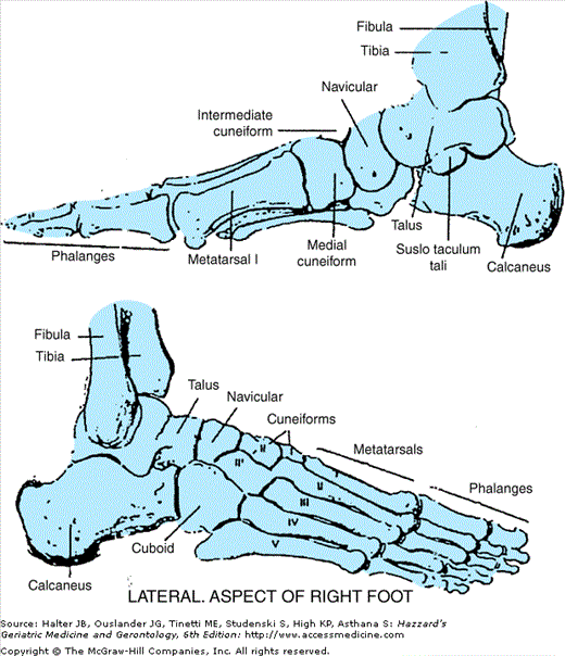
Figure 122-1 illustrates the major anatomic components of the foot. The human foot is an organ that is static and mobile. It provides support for the body at rest and during propulsion and ambulation. The human foot from a morphological standpoint is basically a modified rectangle that is also three dimensional.
The human foot is complex. Each foot has 26 bones (together constituting one-fourth of the body’s total), ossicles, ligaments, muscles, tendons, arteries, veins, nerves, and its covering of skin. The foot normally bears weight on a triangular base, with the long sides being the inner and outer longitudinal arches and the metatarsal arch consisting of the bias along the five metatarsal heads. Weight is transferred from the calcaneus to the fifth and first metatarsal heads initially.
The talus, with the tibia and fibula, comprises the ankle joint. The talus has no muscular attachments and thus with the tendons and muscles, which parallel it, controls activity. The talus articulates with the calcaneus. The Achilles tendon attaches to the superior-posterior segment and is a significant structure to foot function. The cuboid is anterior to the calcaneus and is rectangular in shape, which adds to the structural support to the foot. The navicular is anterior to the talus and is coin shaped, which also provides muscular attachments to aid in function and stabilization. Anterior to the navicular and lateral to the cuboid are three wedge-shaped bones called the first, second, and third (medial, middle, and lateral) cuneiforms. These help provide stability to the foot. These bones comprise the “rear foot.” The arches do not fall. The foot does rotate, pronate, supinate, invert, evert, and elongate and changes structurally in relation to Wolff’s law (bone is deposited and resorbed in accordance with the stresses placed upon it) and Davis’s law (soft tissue models itself along its imposed demands). The mid-foot consists of the remaining five tarsal bones, that is, the navicular, cuboid, and three cunieforms.
Anterior to the cuneiforms and cuboid bones are the five metatarsals. Each metatarsal consists of a base, a shaft, and a head. These are anatomically classed as long bones. Anterior to the metatarsals are the phalanges. Each of the lesser toes contains three phalanges. The first or great toe comprises two phalanges. The metatarsals and phalanges represent the forefoot.
Each osseous structure attaches by ligaments to another to form the joints of the foot. Their functional role is to maintain proximity by keeping the segments of bone articulating with each other. Their properties are strength and rigidity with limited elasticity, which permit the joints to maintain some degree of motion or movement. The basic change that occurs in aging bone is a loss of calcium (osteopenia and/or osteoporosis) and, in foot ligaments, the transition from elasticity to rigidity. The lateral longitudinal arch is formed by the calcaneus, cuboid, fourth, and fifth metatarsals. It is the lower structural arch. The medial longitudinal arch is formed by the calcaneus, talus, navicular, three cuneiforms, and the first, second, and third metatarsals. It is higher with its summit at the head of the talus and navicular. The pitch of the calcaneus (the angle formed by the plantar surface of the calcaneus and plane of support) determines, to a great degree, the height of that segment. The transverse arches of the foot include the posterior metatarsal arch, formed by the bases of the metatarsals and is reasonably firm, and the anterior metatarsal arch, formed by the five metatarsal heads, is flexible, and flattens during gait phases, with weight bearing, pronation, and supination.
The most predominant and significant membranous covering of the foot is known as the plantar fascia. It attaches to the plantar posterior segment of the calcaneus or tuberosity and fans out anteriorly to the metatarsal area. The plantar fascia covers all of the foot muscles, all of the ligaments, and all of the bones on the plantar aspect of the foot. It acts as a spring to aid in function as well as offers protection to plantar structures. It is a very elastic or flexible mass in youth, which becomes wasted and rigid with age. The dorsum of the foot does not have a strong fascial attachment. Most of the structures are subcutaneous.
The muscles that control the major functions of the foot have their origin in the leg. The posterior muscles include the tibialis posterior, flexor digitorum longus, flexor hallucis longus, plantaris, and calcaneal or Achilles tendon (formed by the gastrocnemius and soleus muscles). The lateral muscles include the peroneus longus and peroneus brevis. The anterior muscles include the tibialis anterior, extensor hallucis longus, and extensor digitorum longus. As individuals age and chronic disease becomes evident, there is a decrease in muscle strength that can result in ambulatory dysfunction, imbalance, foot drop, and a higher risk for falls.
The muscle on the dorsum of the foot is the extensor digitorum brevis. The muscles on the plantar surface of the foot are covered by the plantar fascia and are organized in four layers from superficial to deep. The muscles of the deepest fourth layer include the dorsal interossei (four), plantar interossei (four), and the tendons of the peroneus longus and tibialis posterior muscles. It is the atrophy of the interossei muscles that is a major factor in the development of hammertoes in the aging patient.
The nerves that control the foot include the lateral sural cutaneous, superficial and deep fibular (peroneal), medial dorsal cutaneous, dorsal digital, tibial, lateral calcaneal, medial plantar, lateral plantar, and saphenous nerves.
The arterial supply to the foot follows the femoral artery, to the popliteal artery, and divides to become the anterior and posterior tibial arteries. The anterior tibial artery becomes the dorsalis pedis artery on the dorsum of the foot, whose terminal branches are the dorsal metatarsal and dorsal digital arteries. The posterior tibial artery becomes the medial and lateral plantar arteries in the foot. The lateral calcaneal artery supplies the lateral segment of the calcaneal area of the foot. The venous return includes the dorsal venous arch, plantar cutaneous venous plexus, medial and lateral plantar veins, and the greater and lesser saphenous veins.
The skin of the foot is made up of fitted, flexible, elastic inner dermis, covered by a much less sensitive outer epidermis. The skin varies in thickness from one-half a millimeter (as in the eyelid) to 4 or 5 mm, in the sole of the foot. The skin of the foot undergoes many changes during the aging process. It becomes thinner, even parchment like; loses its elasticity, usually atrophies; and loses hair. It loses its hydration or water content because there is generally less perspiration and lubrication. The skin loses its suppleness, becomes brittle and dry, and injures easily. This condition, accompanied by a diminished blood supply, can be quite serious.
The normal toenail includes the nail matrix, nail bed, nail plate, lunula, eponychium, cuticle, and nail folds. The nail plate is a sheet of keratin and is important in clinical medicine, as it reflects the health events of the previous months. The nail plate grows slowly forward until it breaks free of the nail bed at the free edge. The tissue below the free edge is termed the hyponychium. The matrix is the area under the proximal nail fold, cuticle, and lunula. The arterial supply to the nail bed stems from small arterial branches from the dorsal and plantar anastomoses. Nail growth is affected by age, trauma, chemicals, environment, and disease.
The skin is one of the first structures to demonstrate change. There is a loss of hair below the knee and on the dorsum of the foot and toes. Atrophy follows with the skin appearing parchment like and xerotic. Brownish pigmentations are common and related to the deposition of hemosiderin. Hyperkeratotic lesions are associated with keratin dysfunction (hypertrophy and hyperplasia), with a residual to repetitive pressure, with atrophy of the subcutaneous soft tissue, and/or as space replacement as the body attempts to adjust to the changing stress placed on the foot.
Toenails undergo degenerative trophic changes (onychopathy), thickening (onychauxis and onychogryphosis), and/or longitudinal ridging (onychorrhexis) related to repeated microtrauma, disease, and nutritional impairment. Deformities of the toenails become more pronounced and complicated by xerotic changes in the periungual nail folds as onychophosis (hyperkeratosis) and tinea unguium (onychomycosis). These conditions are usually long-standing, chronic, and very common in the elderly and, in the case of onychomycosis, present a constant focus of infection.
Progressive loss of muscle mass and atrophy of soft tissue decreases function and a lack of activity that increases the susceptibility of the foot to injury, so that even minor trauma can result in a fracture and a marked limitation of activity. Atrophy of the interossei is a precipitating factor in the development of digital contractures (hammer toes), metatarsal prolapse, atrophy, and displacement of the plantar fat pad.
Peripheral vascular disease and arterial insufficiency produce trophic changes, rest pain, intermittent claudication, coldness, and color variations, such as rubor and cyanosis. There may be a loss of sensation and other changes related to neuropathy. The presence of hemorrhage subungually or in subhyperkeratotic tissue indicates marked vascular disease.
Clinical Assessment and Medicare Considerations
The podiatrist provides clinical management based on important principles of podogeriatric assessment; recognizing primary changes in the aging foot; identifying complications related to systemic diseases and, in particular, diabetes mellitus and peripheral arterial occlusive disease; risk stratification; understanding Medicare reimbursement policies, serving as the patient’s advocate; and being knowledgeable about primary management procedures. Given the many risk factors for foot diseases and conditions in older adults, specialized foot care is often required. Reasons to refer to a podiatrist for specialized foot care are listed in Table 122-1.
|
The Pennsylvania Department of Health’s Diabetes Prevention and Control Program working with Temple University’s School of Podiatric Medicine’s Department of Community Health, Aging, and Health Policy and the Pennsylvania Diabetes Academy developed a Comprehensive Clinical Podogeriatric and Chronic Diseases Assessment Protocol (Helfand Index). The format provides a methodology to assess foot problems associated with aging and chronic diseases, stratify those patients most at risk to develop complications, maintain a surveillance instrument related to patient care, and provide a means to evaluate data for outcome measurement, which would be valid, accurate, consistent, and relevant. The protocol instrument served as an adjunct to develop prevention and management programs for individual patients and their communities and as a component for comprehensive geriatric assessment. The protocol’s application includes elements of primary, secondary, and tertiary prevention and stresses a need for patient and professional education as well as the need for appropriate multidisciplinary patient management.
As a part of the current Medicare regulations for coverage, there are a number of diseases and disorders that have been categorized as special risk conditions that qualify for coverage for “routine foot care.” In Medicare terms, primary foot care refers to the débridement of hyperkeratotic lesions (heloma and tyloma) and hypertrophic onychodystrophy. Those patients who demonstrate neurosensory and vascular deficits and who present with clinical findings represent an exemption to the exclusion and become eligible for coverage, within utilization guidelines. Medicare has indicated that although not intended as a comprehensive list, the following metabolic, neurological, and peripheral vascular diseases commonly represent the underlying conditions that might justify coverage for “routine foot care.” Table 122-2 lists the primary diseases and disorders that meet current Medicare requirements for routine foot care. This list is not comprehensive but illustrates major conditions.
Amyotrophic lateral sclerosis (ALS) |
Arteriosclerosis obliterans (A.S.O., arteriosclerosis of the extremities, and occlusive peripheral arteriosclerosis) |
Arteritis of the feet |
Buerger’s disease (thromboangiitis obliterans) |
Chronic indurated cellulitis |
Chronic thrombophlebitis* |
Chronic venous insufficiency |
Diabetes mellitus* |
Intractable edema—secondary to a specific disease (e.g., congestive heart failure, kidney disease, and hypothyroidism) |
Lymphedema—secondary to a specific disease (e.g., Milroy’s disease and malignancy) |
Peripheral neuropathies involving the feet |
Associated with malnutrition and vitamin deficiency* |
Malnutrition (general and pellagra) |
Alcoholism |
Malabsorption (celiac disease and tropical sprue) |
Pernicious anemia |
Associated with carcinoma* |
Associated with diabetes mellitus* |
Associated with drugs and toxins* |
Associated with multiple sclerosis* |
Associated with uremia (chronic renal disease)* |
Associated with traumatic injury |
Associated with leprosy or neurosyphilis |
Associated with hereditary disorders |
Hereditary sensory radicular neuropathy |
Angiokeratoma corporis diffusum (Fabry’s) |
Amyloid neuropathy |
Peripheral vascular disease |
Raynaud’s disease |
The podiatrist obtains a history of present illness and past medical history and screens for risk conditions. The foot examination includes a dermatological, an orthopedic, a vascular, and a neurological examination. An assessment is summarized briefly here.
The basic dermatologic evaluation of the foot includes, but is not limited to, the following: focal hyperkeratatotic lesions, onychauxis, bacterial infection, ulceration, onychomycosis, onychodystrophy, onychocryptosis, cyanosis, xerosis, tinea pedis, verruca, hematoma, rubor, discoloration, and preulcerative changes.
Coverage for the débridement of onychomycosis also includes documentation of mycosis/dystrophy causing secondary infection and/or pain, which results or would result in marked limitation of ambulation. The specific clinical findings include discoloration, hypertrophy, subungual debris, onycholysis, secondary infection, limitation of ambulation, and pain. In addition, evidence of dystrophy, onychodysplasia, onychauxis, and onychogryphosis are also frequently noted.
The initial foot orthopedic assessment includes an evaluation of the primary deformities related to biomechanical and pathomechanical changes that commonly cause pain and contribute to ambulatory dysfunction. The foot type, muscle strength, and ranges of motion should be noted.
There should be some identification of gait and movement changes, including the use of mobility aids and if the patient is able to reach and see their feet. An evaluation of footwear is also performed. (See section “Footwear and Orthoses”.)
As part of the assessment, the patient is evaluated for biomechanical risk for foot ulcers. Factors that increase pressure and stress should be considered. Those factors include body mass, gait (particularly antalgic), ambulatory speed, evidence of repetitive micro tissue trauma, change in weight diffusion, and weight dispersion. Imbalance is the force on the foot that produces abnormal stresses. These factors include force (alteration in physical condition, either shape or position), compression stress (one force moves toward another), tensile stress (a pulling away of one part against another), shearing stress (a sliding or gliding of one part on the other), friction (the force needed to overcome resistance and usually associated with a sheering stress), elasticity (weight diffusion and weight dispersion), and fluid pressure (soft-tissue adaptation and conformity to stress).
The primary peripheral vascular assessment evaluation should include coldness, trophic changes, and palpation of the dorsalis pedis and posterior tibial pulses. If pulses are not present, the popliteal and femoral pulses should be identified. A history of night cramps, intermittent claudication and deep venous thrombosis, edema, atrophy, varicosities, and atrophy should be noted. Any amputation or partial amputation should be noted and the etiology identified, particularly related to diabetes mellitus, arterial occlusion, and/or infection.
The primary neurological assessment should include the following: Achilles, patellar, and superficial plantar reflexes; vibratory sense (pallesthesia); response to a loss of protective sensation using a 5.07 (10 g) nylon monofilament at bilateral and multiple sites; sharp, dull, and light touch reaction; joint position; burning; two-point discrimination; and related findings. The response to heat, cold, pain, and two-point discrimination are also important considerations. A Tip Therm can measure temperature sensation.
For Medicare purposes, the assessment for the evaluation and management of a diabetic patient with diabetic sensory neuropathy, resulting in a loss of protective sensation must include the criteria listed in Table 122-3.
|
The final segment of the assessment process is providing an initial impression and/or diagnosis and indicated referrals for management to include podiatric referral, medical specialty referral, patient education, special footwear, orthotics, vascular studies, clinical laboratory referrals, radiographic and other imaging studies, prescriptions as indicated for the management of diseases, and disorders of the foot and its related structures.
All of the assessment components including the focal diseases and disorders of the foot, its related structures, and the pedal manifestations and complications of acute and chronic diseases represent the multiple covariants involved in managing and treating older patients with foot conditions. One mandatory legal key to management includes the requirement for licensure by the individual providing and prescribing indicated medical and surgical care components.
Primary Management—Guidelines and Principles
The management of foot problems in the older patient requires a review of their etiology, symptoms, signs, and clinical manifestations, including related diagnostic studies. The covariant complications, sequelae, relevant treatment, prognosis, and the overall management of the geriatric patient should reflect a reasonable approach that will reduce pain, improve the functional capacity of the patient, maintain that restored function, and provide for the comfort of the patient in activities of daily living and permit the individual to live life to the end of life. Management strategies include radiographs and other imaging studies, nonsteroidal anti-inflammatory drugs, local steroid injections, the use of physical modalities, exercise, and shoe modifications.
