© Springer Science+Business Media New York 2015
Terry F. Davies (ed.)A Case-Based Guide to Clinical Endocrinology10.1007/978-1-4939-2059-4_2121. Primary Aldosteronism
(1)
Endocrine and Diabetes Specialists of CT, 112 Quarry Road, Ste 250, Trumbull, CT 06611, USA
(2)
Division of Endocrinology, Diabetes and Bone Diseases, Department of Medicine, Icahn School of Medicine at Mount Sinai Hospital, 1 Gustave L. Levy Place, 1055, New York, NY 10029, USA
Keywords
Primary aldosteronismHypertensionAldosteroneReninAldosterone/renin ratioAdrenal venous samplingCase Description
A 50-year-old woman was referred to endocrinology by her primary care physician for complaints of fatigue and hair loss. Her past medical history was notable for hypertension diagnosed 1 year prior with several blood pressure recordings of 160/100. Her blood pressure (BP) was effectively managed with the angiotensin-converting enzyme inhibitor (ACEi) ramipril 10 mg daily and the β-blocker bisoprolol 10 mg daily. She reported a family history of essential hypertension in both parents and two of her siblings.
On physical examination she was a thin woman with no stigmata of Cushing’s disease. Her blood pressure was 140/80 with pulse 72. She had mild diffuse scalp hair thinning. She had no thyromegaly or palpable nodules. Lungs were clear to auscultation and cardiac exam was normal. Abdomen was soft, and there was no peripheral edema.
Laboratory evaluation revealed a serum sodium of 140 mEq/L, potassium 4.1 mEq/L, and creatinine 0.8 mg/dL. Plasma aldosterone concentration (PAC) was 18 ng/dL with plasma renin activity (PRA) 0.23 ng/mL/h (0.25–5.82). The aldosterone/renin ratio (ARR) was 78 ng/dL per ng/L/h, suggesting primary aldosteronism (PA) as the underlying cause of her hypertension.
The patient underwent confirmatory testing with a 2 L intravenous saline infusion. Pre-infusion PAC was 16 ng/dL, post-infusion was 11 ng/dL, indicating lack of appropriate suppression and confirming the diagnosis of PA.
CT abdomen was performed and demonstrated thickening in the medial limb of the left adrenal, but no adenoma. She then underwent adrenal venous sampling (AVS), which did not demonstrate lateralization, suggesting bilateral adrenal hyperplasia (BAH) as the underlying pathophysiologic mechanism driving her PA.
She was started on the mineralocorticoid receptor antagonist spironolactone at a dose of 25 mg daily and ramipril was stopped. She tolerated spironolactone without side effects. Bisoprolol was subsequently discontinued and spironolactone was ultimately titrated to 100 mg daily, resulting in control of her blood pressure.
How Does PA Cause Hypertension?
Aldosterone exerts its main effect at the distal tubule of the nephron where it binds to the mineralocorticoid receptor (MR) and promotes gene expression of epithelium sodium channels (ENaC). Increased ENaC expression results in sodium and water absorption, volume expansion with renin suppression, reflexive vasoconstriction, and ultimately hypertension in states of aldosterone excess, such as PA [1].
What Is the Prevalence of PA in Hypertensive Patients Without Hypokalemia?
Jerome Conn described the initial patient with PA in 1954, a young woman with intractable hypertension, tetany, and hypokalemia in which bilateral adrenalectomy was planned in order to control her symptoms. Fortunately, an adrenal adenoma was identified and resected in the operating room, sparing the patient from bilateral adrenalectomy, and resolving her metabolic abnormalities, tetany, and hypertension [2].
Classical teaching recommends screening patients for PA only if they have hypertension and hypokalemia. And, in fact, patients with concomitant hypertension and potassium less than 3.2 mEq/L have a 11-fold increase in the incidence of PA compared to normokalemic (potassium greater than 3.5 mEq/L) patients [3]. However, recent prevalence studies have demonstrated only a 9–37 % incidence of hypokalemia in patients with documented PA, indicating the absence of hypokalemia should not be used as exclusion criteria for PA [4].
Current guidelines recommend screening all patients with moderate or severe hypertension, spontaneous or diuretic-induced hypokalemia, or hypertension with an adrenal adenoma [5].
What Is the Prevalence of Primary Aldosteronism in Outpatient Hypertensive Patients?
After 10 years of studying and treating patients with PA, Conn postulated the prevalence in hypertensive patients to be high, potentially 20 %. He later modified this estimate to 10 %, however, a 1967 study by Fishman et al. suggested disease presence in <1 % of hypertensive patients screened [6], a statistic that debunked the estimates of Conn and has persisted in the medical literature until recently.
Since the widespread use and validation of the ARR in the early 1990s, worldwide prevalence studies in patients with hypertension have demonstrated a much higher prevalence, closer to Conn’s original estimates. Prevalence studies in Australia, Singapore, and Italy demonstrated a prevalence of 5–12 % [7–9]. Even higher prevalence rates have been described in specific populations, such as patients with resistant hypertension (20 %), diabetes and hypertension (13–14 %), and hypertension with obstructive sleep apnea (34 %) [10–16].
Why Is Early Screening and Detection Beneficial?
In addition to its role in salt and water homeostasis, aldosterone has effects at the local tissue level, mediating expression of tissue growth factors and collagen, and increasing oxidative stress and endothelial dysfunction. Ultimately, this results in the deposition of collagen within arterial walls, leading to increased stiffness and increased intima to media thickness [17].
Independent of blood pressure control, aldosterone excess in patients with PA results in left ventricular remodeling and hypertrophy [18–20]. In a retrospective study of patients with PA, Milliez et al. demonstrated in increased risk of cardiovascular events including nonfatal myocardial infarction (MI), cerebrovascular accidents, and atrial fibrillation independent of blood pressure control [21]. These finding were subsequently recapitulated in a prospective study by Catena et al., in which coronary artery disease, cerebrovascular disease, and sustained arrhythmias were more prevalent in patients with untreated PA compared to age, sex, and blood pressure-matched controls [22]. Aldosterone excess has also been implicated in obstructive sleep apnea, insulin resistance and the metabolic syndrome, osteoporosis, and renal insufficiency.
What Is the Best Screening Test for Primary Aldosteronism?
The aldosterone–renin ratio (ARR) is the most sensitive and specific screening tool for PA. It relies on the known pathophysiology of PA, in which aldosterone levels are disproportionately high in comparison to renin concentration. Screening with ARR is generally accepted as positive if ARR exceeds 20 ng/dL per ng/mL/h and PAC exceeds 10 ng/dL [23]. Aldosterone elevation is crucial to interpreting the ARR, as false positives can occur if renin is sufficiently suppressed, as seen in patients with low-renin hypertension.
The ARR is affected by most anti-hypertensive medications; however, it can be reliably interpreted if patients are on any agent except the MR-receptor antagonists (spironolactone and eplerenone) with the knowledge of how these agents affect the ratio [5]. For example, a patient with essential hypertension on an ACEi or angiotensin receptor blocker (ARB) would be expected to have elevated renin with compensatory decrease in aldosterone due to downstream inhibition of the renin–angiotensin system. If the patient had PA, aldosterone synthesis and release would escape regulation by the renin–angiotensin system and would be elevated. Furthermore, elevated aldosterone and volume expansion would suppress renin levels despite the expected renin elevation with ACEi or ARB use.
Patients with suspected PA on MR-receptor antagonist require a 6-week medication washout prior to screening. Alternative medications that do not significantly affect the ARR include the nondihydropyridine calcium channel blockers (verapamil and diltiazem), hydralazine, or the peripheral α-blockers (prazosin, doxazosin, or terazosin) [5, 23].
Finally, even in the setting of a negative ARR screen, knowledge of renin and aldosterone levels are useful tools in selecting appropriate antihypertensive therapy for any patient. For instance, an agent that interferes with the renin–angiotensin system such as an ACEi or ARB may be ineffective in patients with low-renin hypertension.
How Is the Diagnosis Confirmed?
Confirmatory testing must be performed in all patients who screen positive with the ARR for PA. Current clinical guidelines support confirmation with oral sodium loading, saline infusion, fludrocortisone suppression, or captopril challenge testing. Using any of the four modalities, failure to suppress aldosterone secretion is diagnostic of PA. There is no consensus on the optimal test due to insufficient evidence [5]; therefore, the selection of a confirmatory test should be based on patient preference and institution capabilities.
What Is the Molecular Pathophysiology of Primary Aldosteronism?
Aldosterone synthesis and secretion is mediated by renin-dependent angiotensin II. Hyperkalemia and, to a lesser extent, adrenocorticotroph hormone (ACTH) also stimulate aldosterone secretion. PA is a high aldosterone, low renin state resulting from constitutive aldosterone synthesis and release from one or both adrenal glands.
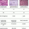
The most common etiology of aldosterone excess, accounting for two-thirds of cases, is bilateral adrenal hyperplasia (BAH). Aldosterone-producing adenomas (APAs) account for one-third of cases. Unilateral hyperplasia, adrenocortical carcinoma, ectopic aldosterone production, and familial hyperaldosteronism 1, 2, and 3 (FH 1, 2, 3) are rare causes.
The underlying molecular changes that result in PA are largely unknown; however, recent descriptions of the germline mutations underlying FH 3 has yielded some insight into sporadic disease. The index family with FH 3 presented with severe hypertension and hypokalemia during childhood requiring bilateral adrenalectomies for management [24]. Pathologic analysis of the adrenals demonstrated massive hyperplasia. The underlying genetic mutation was located to the KCNJ5 gene, which encodes a component of the Kir3.4 potassium channel. Abnormal function of Kir3.4 decreases its selectivity for potassium and allows sodium entry into the cell, prolonged depolarization, and resultant constitutive aldosterone synthesis and release [25]. Several mutations within the KCNJ5 sequence have been identified, resulting in variable phenotypes. Further, genetic analysis of presumed sporadic APAs has demonstrated somatic mutations in KCNJ5 suggesting acquired genetic abnormalities have a significant contributing factor in the development of PA [25–30].
Once PA Is Confirmed Should You Proceed with Subtype Differentiation?
Once the diagnosis of PA is confirmed, it is crucial to adapt a patient-centered approach in the final diagnostic steps, taking into consideration the age, comorbid conditions, and treatment goals of the patient. Abdominal computed tomography (CT) is typically the first localization study performed once the diagnosis of PA is confirmed. CT enables the identification of adrenal enlargement, adenomas, or rarely carcinomas [5]. In patients who ultimately do not desire or would not be candidates for surgery, the requirement for any localization study is questionable. Current guidelines recommend CT in all patients to exclude the possibility of an adrenocortical carcinoma (ACC) [5]. However, in practice, ACC is very rare, and also typically cosecretes other adrenocortical hormones. In patients with mild disease or long-standing hypertension desiring medical therapy, our practice is to not pursue CT.
Adrenal venous sampling (AVS) is an invasive diagnostic test used for localization that must be pursued in all patients in which surgery is the planned treatment modality. CT alone is insufficient for presurgical localization as concordance between CT and AVS is approximately 50 %, placing 25 % of patients at risk for unilateral adrenalectomy of the wrong side.
AVS technique and interpretation varies by institution. In our center, the bilateral adrenal veins are accessed via femoral puncture and catheterization, and ACTH is infused via peripheral IV. ACTH-stimulated adrenal vein and inferior vena cava (IVC) cortisol levels are compared to confirm proper catheter placement in the adrenal veins. Adrenal vein aldosterone values are then corrected for cortisol to account for venous dilution by the phrenic vein. The bilateral cortisol-corrected aldosterone values are compared, and a 4:1 ratio favoring one side is suggestive of unilateral disease. More importantly however, adrenal vein aldosterone secretion must be suppressed from the contralateral side to confirm the diagnosis of unilateral disease.
AVS is 95 % sensitive and 100 % specific for localizing unilateral APAs, however, success of this procedure and complication rate is practitioner dependent, mainly due to the technical difficulty of catheterizing the right adrenal vein [31, 32]. Because of these risks, AVS should only be pursued for patients in whom surgical intervention would be considered.
What Is the Preferred Treatment for Each Subtype?
The majority of patients with PA have BAH which is amenable to medical therapy with MR-antagonists such as spironolactone or eplerenone. Spironolactone at doses of 25–400 mg daily is the initial agent of choice and results in a 25 % systolic blood pressure reduction and a 22 % diastolic reduction [5]. Spironolactone is inexpensive and may decrease polypharmacy. In a study of patients with PA and uncontrolled hypertension, addition of spironolactone improved blood pressure to goal in 50 % of patients studied and was the sole agent required to maintain control in 50 % of these responders [33].
In addition to its actions at the MR, spironolactone has antagonistic properties at the androgen receptor and agonistic properties at the progesterone receptor, which result in its main side effects of gynecomastia in men and mastodynia in women. Although these side effects are dose- dependent, patients with PA often require high doses, and as a result eplerenone may be a more tolerable agent [5].
Eplerenone is a selective MR antagonist and, as a result, lacks the progesterone receptor-stimulating and anti-androgen properties of spironolactone that result in its side effects [34]. However, in a head-to-head trial comparing effectiveness of the two agents, spironolactone resulted in more potent blood pressure reduction [35].
Amiloride and triamterene target blood pressure at the level of the ENaC receptor, which is increased as a result of aldosterone action at the distal renal tubule. These agents are less potent than MR antagonists in achieving blood pressure control in patients with PA and should be reserved for second-line use.
Surgery should be pursued in all patients with APAs in which the benefit outweighs the risk of unilateral adrenalectomy. Laparoscopic unilateral adrenalectomy is the preferred procedure. Postoperatively, BP and hypokalemia improve in 100 % of patients [5], and myocardial fibrosis improves within 1 year [36]. However, long-term hypertension cure rate is only 30–60 % [37–39]. Persistent postoperative hypertension is likely related to underlying essential hypertension and correlates to duration of disease, increased creatinine, older age, family history of essential hypertension, and use of more than two antihypertensive agents preoperatively [37, 39]. Patients with APAs who are not surgical candidates due to comorbid conditions or those who are unlikely to be cured can be managed medically, although higher doses of MR antagonists are typically required.
Related posts:

Stay updated, free articles. Join our Telegram channel

Full access? Get Clinical Tree


