Fig. 24.1
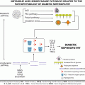
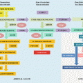
Mechanisms of bone resorption. The stromal cells or osteoblast releases RANKL and osteoprotegerin. RANKL will bind to RANK on the surface of the osteoclast precursor, leading to fusion and differentiation of this cell into mature osteoclasts, which in turn release cathepsin K. When there is increased production of osteoprotegerin, such as in estrogen deficiency, the osteoprotegerin binds to RANKL inhibiting the formation of mature osteoclast

Fig. 24.2
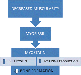
Mechanisms of bone formation. The osteocyte, under the action of PTH, mechanical strain, and NPY, releases sclerostin, which will block the binding of proteins Wnt and Dickkopf to osteoblast receptors, especially the LRP-5
The two major risk determinants for developing osteoporosis are peak bone mass and rate of bone loss. Risk factors that influence these determinants should be evaluated in all postmenopausal women in order to properly estimate the threat of fractures, exclude secondary causes of osteoporosis, identify modifiable risk factors, and determine the appropriate drug therapy for each case [4].
The main risk factors for osteoporosis are listed in Table 24.1.
Table 24.1
Risk factors for osteoporosis
Family history of osteoporosis | Cushing syndrome, and use of corticosteroids |
Advancement of age | Chronic renal failure |
Female gender | Celiac disease |
Sedentary lifestyle | Hyperthyroidism |
Malnutrition | Primary hyperparathyroidism |
Low calcium and vitamin D intake | Multiple myeloma |
Diabetes mellitusa | Time of menopause |
Smoking | Low body weight |
Alcoholism | Obesityb |
Personal history of fractures | Deficiency of GH and IGF-1 |
Delayed puberty and/or hypogonadism | Vitamin D deficiency |
Prolonged immobilization | Depression |
FRAX
FRAX™ is a tool available online (http://www.shef.ac.uk/FRAX/index.htm) which was developed by the World Health Organization. It is employed to gather independent risk factors for osteoporotic fractures involving individuals over 50 years of age, in order to quantify the probability of fracture of the femoral neck or other major osteoporotic fractures (vertebral, hip, forearm, and humerus) over the following 10 years.
The variables considered include gender, age, BMI, personal history of fractures after the age of 40, family history of hip fractures, smoking, excessive alcohol consumption, rheumatoid arthritis, use of glucose-corticoids, or other secondary causes of osteoporosis [5].
Using population samples from Europe, North America, Asia, and Australia, country-specific data was compiled, allowing calculations based on regional differences. When FRAX is associated with bone mineral density (BMD) testing, it is a more useful tool for predicting fracture risk than the use of either FRAX or BMD alone.
Although its use is not routinely indicated for patients who are already being treated for osteoporosis, a recent study demonstrated that FRAX may be a useful tool for assessing fracture risk in these patients, pointing to the need to either continue or discontinue medication [6].
Diagnosis
Clinical History and Physical Examination
Through a clinical history and thorough physical examinations it is possible to detect secondary causes of osteoporosis and risk factors for osteoporosis. In the clinical evaluation of the patient, weight, height, family history of osteoporosis, age, race, nutritional status, calcium and vitamin D intake, thoracic–lumbar pain (chronic or acute), decreased stature, chest deformities, medication use (current and previous), menstrual cycles, time of menopause, history of fractures, and lifestyle habits (smoking, alcohol consumption, and physical activity) should all be taken into consideration.
Most patients with osteoporosis are asymptomatic until the onset of clinical fractures. Vertebral fractures can result in loss of height and/or kyphosis, and local pain, due mainly to the shortening and contracture of the para-spinal musculature caused by the reduction of vertebral height [7]. However, most vertebral micro-fractures are asymptomatic.
Postmenopausal women should also be questioned regarding clinical factors associated with an increased risk of falls, including history of falls, fainting, muscle weakness, problems with coordination and balance, difficulty walking, arthritis in the lower limbs, peripheral neuropathy, and decreased visual acuity.
Laboratory Evaluation
Laboratory tests are important, primarily to exclude secondary causes of osteoporosis. The initial evaluation should include the following: CBC, VSH, 24-h calciuria, calcium, albumin, phosphorus, transaminases, alkaline phosphatase, protein electrophoresis, renal function, thyroid function, PTH, and 25 (OH) vitamin D. If any secondary cause is clinically suspected and/or bone loss greater than expected for the age, investigations should be extended to include cortisol after 1 mg of dexamethasone, anti-gliadin and anti-endomysial antibodies, bone marrow study, serum iron, and ferritin (for suspected hemochromatosis).
Bone Markers
Bone markers are substances released during bone remodeling processes, which can be assayed in serum or urine, and provide dynamic assessment of the activity of the skeleton.
Markers for resorption as well as formation may be utilized (Table 24.2).
Table 24.2
Bone formation and resorption markers
Formation markers | Resorption markers |
|---|---|
Alkaline phosphatase | Telopeptides of collagen cross-links |
Osteocalcin | Amino-terminal amino-NTX (N-telopeptide) |
Pro-peptides of type I collagen | Carboxy-terminal—CTX (C-telopeptide) |
– Amino-terminal (PINP) | Pyridinolines |
– Carboxy-terminal (PICP) | Hydroxyproline |
– Osteocalcin (OCN) | Tartrate-resistant phosphatase acid |
These markers should not be used single-handedly for the diagnosis of osteoporosis, nor even to determine which patients require treatment, but they can be useful in predicting bone loss. Studies show that the higher the level of markers, the greater the decrease in bone mass in subsequent years if treatment is not instituted.
The best and most validated use of bone markers is to monitor treatment. Anti-resorptive therapy is associated with the reduction of all resorption markers after 3 months, remaining at these reduced levels while the patient is in treatment. In cases where the response is inadequate and treatment compliance by the patient has been confirmed, the possibility of changing medication or increasing the dosage should be evaluated.
Imaging
Plain Radiography
Plain radiographs demonstrate low sensitivity for the diagnosis of osteoporosis because they show an alteration only when a bone loss of at least 30 % already exists. They can be useful for diagnosing fractures, or specialized diagnosis involving other diseases that can affect bone, such as multiple myeloma, osteomalacia, and bone metastases.
Since most vertebral fractures are asymptomatic, several techniques have been studied with the aim of objectively recognizing subclinical vertebral deformities by measuring the height of the vertebral bodies (called morph-metric fractures). The semi-quantitative score permits a percentage differential evaluation of the anterior, middle and posterior heights of the vertebral bodies, in order to effectively assess the severity of vertebral fractures [8].
0 Degree—No fracture exists
1st Degree—Mild fracture—reduction ranging from 20 to 25 % of the vertebral height
2nd Degree—Moderate fracture—reduction ranging from 25 to 40 % of the vertebral height
3rd Degree—Severe fracture—reduction > 40 % of the vertebral height
Bone Densitometry
Osteoporosis can be diagnosed before the onset of clinical fractures by means of noninvasive methods for determining bone mineral density (BMD), which is the best single predictor of fracture risk [9]. The most accurate noninvasive method is bone densitometry, and the most widely used measure of absorption is dual energy X-ray absorptiometry (DXA), which measures the area density (grams of mineral per square centimeter of bone; g/cm2). It can be used at central (lumbar spine, and hip) or peripheral (distal radius, heel, and phalanges) sites; however, only central sites are used for diagnosis and monitoring response to treatment.
The World Health Organization (WHO) has defined the diagnosis of low bone mass and osteoporosis, based on the number of standard deviations (SD) below mean BMD detected in normal young adults of the same sex (T-score) [10] (Table 24.3).
Table 24.3
Definition of osteoporosis by the WHO criteria
WHO classification | T-score |
|---|---|
Normal | To −1.0 DP |
Osteopenia | −1.0 to −2.5 DP |
Osteoporosis | < −2.5 DP |
The BMD of osteoporotic patients may also be compared with that of a population of corresponding age (Z-score). A Z-score below −2.0 SD is considered below the expected range for the age group [11], and in these cases should be investigated for secondary causes of osteoporosis.
Since all postmenopausal women are at risk of developing osteoporosis, it would be ideal to evaluate the BMD of all of them. As a way of limiting costs, the International Society of Clinical Densitometry (ISCD) suggests screening for osteoporosis in women over 65 years of age; those with a history of fractures after minimal or no trauma; in early menopause; with prolonged use of corticosteroids; osteopenia evidenced by plain radiography; a maternal history of osteoporosis or fracture, loss of height or thoracic kyphosis; underweight (BMI < 19), secondary causes; and the use of medications associated with bone loss [11].
Quantitative Computerized Tomography
This is a technique that measures volumetric density (g/cm3) at the lumbar spine and peripheral sites using specialized software and standard computerized tomography equipment. It is able to distinguish cortical and trabecular bone compartments and predict fracture risk, as well as DXA, but has a high cost along with limited availability and increased radiation exposure, being used mainly in clinical research.
Ultrasonography
This evaluates the heel bone and the proximal tibia, is practical and inexpensive, and is useful as a method for screening the population at risk for osteoporosis.
Bone Quality
The concept of bone quality has been widely used to justify the occurrence of clinical events not explained by the evaluation of BMD alone. Bone quality takes into consideration the composition and structure of bone, contributing to bone strength regardless of density. Several factors interact to form bone quality, such as bone turnover, geometry, micro-architecture, mineralization; micro-aggressions, and components of the mineral and bone matrix [12].
The evaluation of bone turnover may be conducted through bone marker evaluation and biopsies performed on bone marked with tetracycline [13]. New techniques for bone quality assessment have been developed, such as high-resolution magnetic resonance imaging, and high-resolution peripheral quantitative computerized tomography. However, these costly techniques have yet to become readily available.
Treatment
Indication
Many guidelines have been published concerning the management of osteoporosis, in which treatment decisions are based primarily on the results of BMD in combination with patient characteristics.
The FRAX approach developed by the WHO plays a crucial role in guiding treatment recommendations for the management of osteoporosis [14].
The National Osteoporosis Foundation (NOF) recommends treatment of postmenopausal women (and men 50 years or older) with a history of vertebral or hip fracture or osteoporosis based on the measurement of BMD (T score of −2.5 or less), as well as postmenopausal women with osteopenia, (BMD T score between −1.0 and −2.5) associated with a 3 % or greater likelihood of hip fracture within 10 years, or a 20 % or greater likelihood of osteoporotic fracture calculated by the FRAX approach [15].
The ideal optimal duration of pharmacological treatment for postmenopausal osteoporosis remains controversial. The decision to continue or discontinue therapy should be based on the history and fracture risk, balanced with the risks and benefits of the medication.
Non-pharmacological Treatment
There are three main components in the non-pharmacologic therapy of osteoporosis: diet, exercise, and the cessation of smoking. In addition, the patient should also avoid drugs that increase bone loss, such as glucose-corticoids.
Calcium/Vitamin D
An optimum diet for the treatment of osteoporosis includes an adequate amount of calories (to prevent malnutrition), along with calcium and vitamin D. Postmenopausal women should have an adequate intake of elemental calcium in divided doses, totaling 1,000–1,200 mg/day [16].
A recent study with 31,022 patients showed that vitamin D supplementation leads to a less than significant reduction of 10 % in the risk of hip fracture (hazard ratio, HR 0.90, CI 95 % 0.80–1.01) and a 7 % reduction in the risk of non-vertebral fractures (HR 0.93, CI 95 % 0.87–0.99) when compared with a control group. When intake levels were differentiated, fracture risk reduction was demonstrated only at the highest level of intake (median, 800 IU per day), with a 30 % reduction in the risk of hip fracture (HR 0.70; CI 95 % 0.58–0.86) and a 14 % reduction in the risk of any type of non-vertebral fracture (HR 0.86, CI 95 % 0.76–0.96) [17].
Physical Exercise
Women with osteoporosis should perform physical exercise for at least 30 min three times a week, since exercise has been associated with a reduced risk of hip fracture in older women [18].
A recent meta-analysis of 43 random clinical trials with 4,320 postmenopausal women showed a significant positive effect of exercise on the BMD of the lumbar spine and trochanter. The most effective type of exercise for femoral neck BMD was resistance training using progressive force. A combined program that included more than one type of exercise was the most efficient for lumbar spine BMD [19].
Pharmacological Treatment
There are several medications that can be used in the treatment of osteoporosis. The main medications employed are reviewed below (Table 24.4).
Table 24.4
Reduction in fracture incidence
Drugs | Vertebral fracture | Non-vertebral fracture | Hip fracture |
|---|---|---|---|
Zolendronate | + | + | + |
Risendronate | + | + | + |
Alendronate | + | + | + |
Strontium | + | + | +a |
Estrogen | + | + | + |
Teriparatide | + | + | − |
Calcitriol | + | − | − |
Ibandronate | + | + | +a |
Raloxifen | + | − | − |
PTH 1-84 | + | − | − |
Calcitonin | + | − | − |
Denosumab | + | + | + |
Estrogens
Several placebo-controlled, randomized studies, including the WHI study and the postmenopausal estrogen/progesterone intervention (PEPI) study have established that decreases in BMD are attenuated by estrogen, resulting in a lower risk of fracture [20, 21].
In the WHI study, estrogen-progestin therapy was associated with significant reductions in hip fractures (OR 0.7, CI 95 % 0.4–1.0 unadjusted; less than five hip fractures per 10,000 person-years), along with vertebral and other osteoporotic fractures (OR 0.7, CI 95 % 0.4–1.0 unadjusted, and OR 0.8, CI 95 % 0.7–0.9, respectively) [20]. A similar risk reduction for hip fractures was shown using estrogens alone (OR 0.61, 95 % CI 0.41–0.91), as were reductions for vertebral fractures (OR 0.62, 95 % CI 0.42–0.93) [22].
In a forthcoming sample study, the Million Women Study, current users in postmenopausal therapy were shown to have a significantly lower risk of any fracture when compared to nonusers (RR 0.62, CI 95 % 0.58–0.66) [23].
The coadministration of a progestogen, cyclically or continuously, to prevent endometrial hyperplasia does not impair the beneficial effects of estrogen [21].
However, estrogen-progestin therapy is no longer a front-line approach for the treatment of osteoporosis in postmenopausal women, owing to the increased risk of breast cancer, venous thromboembolism, stroke, and perhaps also coronary disease [20].
Tibolone
Tibolone is a synthetic steroid whose metabolites have estrogenic, androgenic, and progestogenic properties. It is used to treat osteoporosis in some countries. In postmenopausal women with osteoporosis, tibolone use has produced a 5–12 % increase in lumbar spine BMD within 2 years. However, despite the fact that the LIFT study has reported a reduced risk of vertebral and non-vertebral fractures through the use of tibolone, it was stopped at an early stage because of the unacceptable risk of cerebral stroke [24]. This casts doubt on the drug’s safety.
Calcitonin
Using calcitonin for the treatment of osteoporosis, a study that included 5 years of follow-up with 1,255 women with T-scores of less than −2 (lumbar spine and at least one vertebral fracture), randomly assigned either a placebo or doses of 100, 200 or 400 IU/day of intranasal calcitonin. A small and inconsistent beneficial effect on the vertebral BMD from nasal calcitonin treatment was found, and included a reduction in the risk of vertebral fractures [25].
Data on the effect of calcitonin in locations other than the spinal column are conflicting.
A recent meta-analysis with heterogeneous results, using a limited number of patients, showed calcitonin to be of benefit for the short-term relief of acute pain (less than 10 days) in patients who have suffered a vertebral fracture. In contrast, calcitonin has not proved to be effective for patients with chronic pain (over 3 months) [26].
SERMs (Selective Modulators of Estrogen Receptors)
Selective modulators of estrogen receptors (SERMs) bind with high affinity to the estrogen receptor, having agonist and antagonist properties that vary, depending on the target organ.
Raloxifene is a SERM effective in the treatment of established osteoporosis, which increases BMD in both the lumbar spine and the hip [27–30], and reduces the risk of vertebral fractures [28]. It also appears to reduce the risk of breast cancer without stimulating endometrial hyperplasia or vaginal bleeding, but does seem to increase the risk of venous thromboembolism (VTE) [27]. In addition, there are studies that refer to an increased risk of fatal cardiovascular accidents (CVA) [27, 31]. Although serum concentrations of low density lipoprotein (LDL) cholesterol and total cholesterol decrease, there seems to be no change in the risk of coronary cardiac disease [27].
However, despite the fact that raloxifene reduces vertebral fracture risk in postmenopausal women, it is not clear whether there is a reduction in non-vertebral fractures, and therefore seems to be a less potent anti-resorptive agent than alendronate or estrogen [32, 33].
Moreover, unlike the bisphosphonates, SERMs do not appear to have a long-lasting effect on the skeleton, and have no residual beneficial effects on BMD after discontinuation of treatment.
In a recent study, raloxifene was shown to decrease the mortality rate from all causes, mainly due to the reduction in non-cardiovascular and non-oncological deaths owing to a mechanism that has yet to be clarified [34].
Tamoxifen, a SERM used most commonly for the treatment of estrogen-dependent breast cancer, also affords some protection against bone loss in postmenopausal women, and can be used to treat osteoporosis by reducing fracture rates [35].
Bazedoxifene has also decreased the incidence of new vertebral fractures, but not non-vertebral ones, with common adverse effects that include hot flashes, cramps, low rates of endometrial hyperplasia, cancer, polyps, and slightly higher rates of DVT, effects somewhat similar to those of raloxifene [36].
Lasofoxifene, like raloxifene, reduces the incidence of vertebral fractures, but also increases the risk of thromboembolic events, hot flushes, and cramps in the legs. After 5 years of use, lasofoxifene has also been shown to be associated with a decrease in non-vertebral fractures, an effect that raloxifene has not shown. However, none of the SERMs reduce the risk of hip fractures [37].
Bisphosphonates
Bisphosphonates are synthetic analogues of pyrophosphate in which the oxygen bridge is replaced by a carbon atom [38]. They suppress bone resorption mediated by osteoclasts through a mechanism different from other anti-resorptive agents, binding to hydroxyapatite on bone surfaces, particularly those undergoing active resorption. When the osteoclasts begin to reabsorb bone that is impregnated with bisphosphonate, the bisphosphonate released during resorption impairs the ability of osteoclasts to form the wrinkled edge needed to adhere to the bone surface, thereby producing the protons required to continue bone resorption. In addition, they also reduce the activity of the osteoclasts, compromising the development of osteoclast progenitors, along with the recruitment and promotion of apoptosis of the osteoclasts. There also appears to be a beneficial effect on the osteoblasts [39].
Bisphosphonates can be administered orally (alendronate, risedronate, ibandronate) or intravenously (zoledronic acid at a dose of 5 mg every 12 months, and ibandronate in a dose of 3 mg every 3 months) [38, 40–43]. They avidly bind to bone minerals, especially to trabecular bone, with a high degree of specificity [44]. However, oral absorption is low (0.6–1.5 % of the administered dose). Approximately 40–60 % of the dose is distributed in the bone, the remainder being excreted unchanged in the urine without substantial metabolism [38].
Oral bisphosphonates should be taken once a week after fasting, (alendronate in a dose of 70 mg, and risendronate in a 150 mg dose), once a month (ibandronate in a dose of 70 mg or risendronate in a 150 mg dose), or on 2 consecutive days, once a month (risendronate in a dose of 75 mg). The patient must remain upright for at least 30 min after taking the drug in order to minimize gastroesophageal reflux and enhance absorption. Afterwards, food, medications, and other liquids should be avoided for at least 30–45 min [40].
Oral and intravenous bisphosphonates are contraindicated in patients who have had previous allergic reactions to any bisphosphonate, or creatinine clearance estimated at 35 ml/min or less, vitamin D deficiency (serum 25 hydroxy-vitamin D less than 30 ng/ml), osteomalacia or hypocalcemia [40].
Oral bisphosphonates are also contraindicated in patients with impaired swallowing, or esophageal disorders such as achalasia, esophageal varices, severe gastroesophageal reflux, or those who are unable to sit for at least 30 min after taking the medication [40].
An acute phase reaction (fever, myalgia, bone pain and weakness) occurs in 20 % of patients after an initial intravenous infusion of bisphosphonate and, in a very small number of patients, during oral therapy. Erosive esophagitis, ulceration, and bleeding have been associated with daily oral therapy using alendronate or risedronate, but seldom occur with the current regimes (not daily). Heartburn, chest pain, hoarseness, and irritation of the vocal cords can occur with weekly (alendronate or risedronate) or monthly therapy (ibandronate or risedronate) [40].
Osteonecrosis of the jaw is a rare but serious complication of long-term therapy that can appear spontaneously, or after dental surgery. Case reports suggest that atypical fractures of the femur (subtrochanteric and mid-diaphyseal portions) may also occur during prolonged therapy [40, 45].
There are no known interactions between bisphosphonates and other drugs. Evidence of treatment failure with patients adhering properly to a treatment regime indicates the need to change from orally administered bisphosphonate to intravenous zoledronic or another class of drugs, such as anabolic agents (e.g., teriparatide) [40].
Bisphosphonates suppress biochemical indices of bone resorption by around 50 % in a month, significantly reducing the incidence of vertebral, and non-vertebral fractures, including femoral fractures in patients with osteoporosis within a few months after the start of therapy [44].
BMD increases modestly by around 2–6 % during the first year of treatment. In the lumbar spine, it continues to increase slowly for several years, but in the femur, it reaches a plateau after about 2 years. Therapy preserves bone, but does not increase bone volume, or restore the bone structure [44].
Stay updated, free articles. Join our Telegram channel

Full access? Get Clinical Tree


