Definition
Atherosclerotic cardiovascular disease is the most significant health problem in the United States, as heart and cerebrovascular diseases are leading causes of mortality and peripheral vascular disease is a leading cause of morbidity in elderly people. Peripheral arterial disease (PAD) is the most typical form of peripheral vascular disease. PAD is characterized by a partial or complete failure of the arterial system to deliver oxygenated blood to peripheral tissue. Atherosclerosis is, by far, the most common etiology of PAD. However, several other processes can lead to the clinical syndrome (Table 80-1). Although lesions (and symptoms) of PAD can occur in both the upper and the lower extremities, these are much more common in the lower extremity, which is the focus of this chapter.
Epidemiology
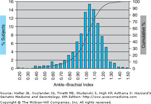
The ankle–brachial blood pressure index (ABI), defined as the systolic blood pressure measured at the ankle divided by the systolic blood pressure measured in the arm during supine rest, is the most widely used quantitative measure to identify subjects with PAD and to determine PAD severity. The ABI varies widely in the general population and is generally normally distributed with a long tail of low values (Figure 80-1). The prevalence of PAD is highly dependent on the exact ABI cutpoint used to detect inadequate peripheral circulation. The definition of an abnormal ABI has ranged between <0.80 and <0.97, with a value of ≤0.90 generally considered to be the best reference standard.
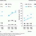
Figure 80-1.
Ankle–brachial blood pressure index (ABI) in men and women in the Edinburgh Artery study. The bars and left-hand scale represent the percentage of subjects having a given ABI. The solid line and right-hand scale represent the cumulative percentage of subjects having an ABI at or below the ABIs listed on the abscissa. (Adapted from Fowkes FG. Epidemiological research on peripheral vascular disease. J Clin Epidemiol. 2001;54:863.)
The prevalence of PAD is 16% in the general population older than 55 years of age when an ABI value of ≤0.90 and symptoms of intermittent claudication and rest pain are used as criteria of PAD. In the Edinburgh Artery Study, approximately 20% of the men and women aged 55 to 74 years were noted to have an ABI ≤0.90 and, thus, were diagnosed as having PAD. The prevalence of PAD increases with age and at all ages is higher in men than in women. At ages 65 to 69 years, the prevalence of PAD in men from the Cardiovascular Health Study was approximately 7%, and approximately 5% in women. In subjects aged 85 years and older, the prevalence was 23% in men and 21% in women.
PAD and coronary artery disease (CAD) share risk factors. In addition to age and male sex, risk factors for PAD include smoking, hypercholesterolemia, diabetes, hypertension, hyperhomocystinemia, elevated fibrinogen concentration, having a family history of premature atherosclerosis (suggesting that genetic factors may influence the development of PAD), and being nonwhite. Although PAD can be seen in the absence of clinical CAD, asymptomatic CAD is frequently present in patients with PAD. Every patient presenting with PAD should be considered to have CAD until proven otherwise. Evaluation and treatment for PAD should include evaluation and control of CAD risk factors.
PAD has important implications for function. PAD severity (assessed by ABI) is directly related to 6-minute walk distance, free-living energy expenditure, and steps taken per day. Of particular interest to geriatricians, patients with PAD are more likely to fall than are subjects without PAD.
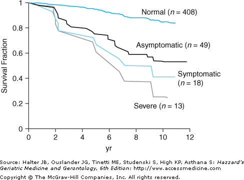
PAD is not a static disease. Progression from intermittent claudication to rest pain or gangrene can occur in anywhere from 2% to 7% of patients per year. Furthermore, PAD is associated with increased risk for mortality, and this risk becomes greater as the severity of PAD increases (Figure 80-2).
Patients with PAD are at increased risk of coronary heart disease, coronary vascular disease, and all-cause mortality. This risk is independent of traditional risk factors, including age, sex, smoking, systolic blood pressure, plasma lipids, fasting glucose, body mass index, and preexisting clinical cardiovascular disease. Risk for mortality as a consequence of coronary heart disease and cardiovascular disease is three to six times higher in subjects with PAD than in subjects without PAD, even after accounting for traditional risk factors.
Pathophysiology
The atherosclerotic lesions that lead to PAD tend to form in a few well-defined locations in the vascular tree, and patients often will have lesions in more than one location. Thirty percent of the patients have lesions in the abdominal aorta, 80% to 90% have lesions in the femoral and popliteal arteries, and 40% to 50% have lesions in the tibial and peroneal arteries. The location of the arterial lesion will determine the clinical presentation. For example, aortoiliac disease (Leriche syndrome) leads to buttock, hip, and thigh pain along with erectile dysfunction; the more common femoral–popliteal disease leads to symptoms in the calf. The atherosclerotic process is similar to that in other major arteriostenoses and is discussed in Chapter 75.
Presentation
Clinical PAD was recognized as early as 1831, when Jean-Francois Bouley Jeune observed a horse that, when pulling a cabriolet, limped with the hind legs. At autopsy, the horse was noted to have a partially thrombosed aneurysm of the abdominal aorta and occlusion of both femoral arteries.
Two schemes, both based on symptoms and clinical measures, are commonly used to classify the severity of PAD (Tables 80-2 and 80-3). In the early stages of PAD, the reduction in blood flow and ABI does not result in any noticeable symptoms (asymptomatic PAD) and is defined as stage I according to the Fontaine classification system and grade I, category 0 according to the more widely accepted Rutherford classification system. As PAD progresses, ischemic pain in the leg musculature occurs when patients walk (intermittent claudication) and is classified as either Fontaine stage II-a or II-b, or Rutherford grade I, category 1, 2, or 3, depending on the walking distance and the change in ABI following walking. In more advanced stages of disease, ABI is reduced to such an extent that pain is experienced even while at rest, classified as either Fontaine stage III or Rutherford grade II, category 4. Further progression of the disease leads to ischemic ulcerations on the lower extremities, gangrene, and tissue loss, classified as either Fontaine stage IV or Rutherford grade II, category 5, or grade III, category 6. Patients in these categories have critical limb-threatening ischemia in which the ischemia endangers part or all of the lower extremity. These patients are candidates for aggressive limb salvage interventions such as percutaneous transluminal angioplasty or bypass surgery.
STAGE | SYMPTOMS |
|---|---|
I | Asymptomatic |
II | Intermittent claudication |
IIa | Pain free, claudication walking >200 m |
IIb | Pain free, claudication walking <200 m |
III | Rest/nocturnal pain |
IV | Necrosis/gangrene |
GRADE | CATEGORY | CLINICAL DESCRIPTION |
|---|---|---|
I | 0 | Asymptomatic; not hemodynamically correct |
1 | Mild claudication | |
2 | Moderate claudication | |
3 | Severe claudication | |
II | 4 | Ischemic rest pain |
5 | Minor tissue loss; nonhealing ulcer, focal gangrene with diffuse pedal ischemia | |
III | 6 | Major tissue loss extending above transmetatarsal level; foot no longer salvageable |
The presentation of PAD varies widely among patients. PAD can be present without any clinical signs, in which case diagnosis of PAD can only be made with laboratory tests. With mild disease, the peripheral pulses can be decreased. With more advanced disease, there may be an audible bruit or the distal pulses may be absent. The extremity may be pale or cyanotic at rest, upon raising the leg, or with exercise. The skin of the extremity can be cool, smooth and shiny with hair loss, and thickened nails. Dangling the legs following elevating the extremity can lead to delayed return of color to the skin (usual time approximately 10 seconds), delayed filling of the veins of the feet and ankles (normal approximately 15 seconds), and the development of a dusky rubor in the legs. Patients with the most severe disease will present with ulcers on their extremity or frank gangrene.
Evaluation
A number of noninvasive tests have been used to screen for and to evaluate the extent of a patient’s PAD. The most basic clinical test is palpation and auscultation of the peripheral pulses. Little is known about the sensitivity of pulse palpation for the diagnosis of PAD. In situations in which direct examination of subjects is not feasible, such as epidemiological studies, the San Diego Claudication Questionnaire, which is a version of the Rose Questionnaire, is often used to screen subjects for PAD. Using these questionnaires, PAD is defined as leg pain associated with walking that goes away with rest. The sensitivity of the Rose Questionnaire has been reported as being anywhere from 10% to 50%.
Stay updated, free articles. Join our Telegram channel

Full access? Get Clinical Tree


