The classification applies to carcinomas. Papilloma is excluded. There should be histological or cytological confirmation of the disease. The regional lymph nodes are the hilar, abdominal para‐aortic, and paracaval nodes and, for ureter, intrapelvic nodes. Laterality does not affect the N classification. The pT and pN categories correspond to the T and N categories. Note pM0 and pMX are not valid categories.
RENAL PELVIS AND URETER (ICD‐O‐3 C65, C66)
Rules for Classification
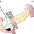
Anatomical Sites (Fig. 506)
Regional Lymph Nodes (Fig. 507)
TNM Clinical Classification
T – Primary Tumour
TX
Primary tumour cannot be assessed
T0
No evidence of primary tumour
Ta
Noninvasive papillary carcinoma (Fig. 516)
Tis
Carcinoma in situ
T1
Tumour invades subepithelial connective tissue (Fig. 516)
T2
Tumour invades muscularis (Fig. 516)
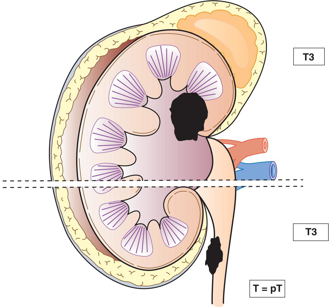
T3
(Renal pelvis) Tumour invades beyond muscularis into peripelvic fat or renal parenchyma (Fig. 517)
(Ureter) Tumour invades beyond muscularis into periureteric fat (Fig. 517)
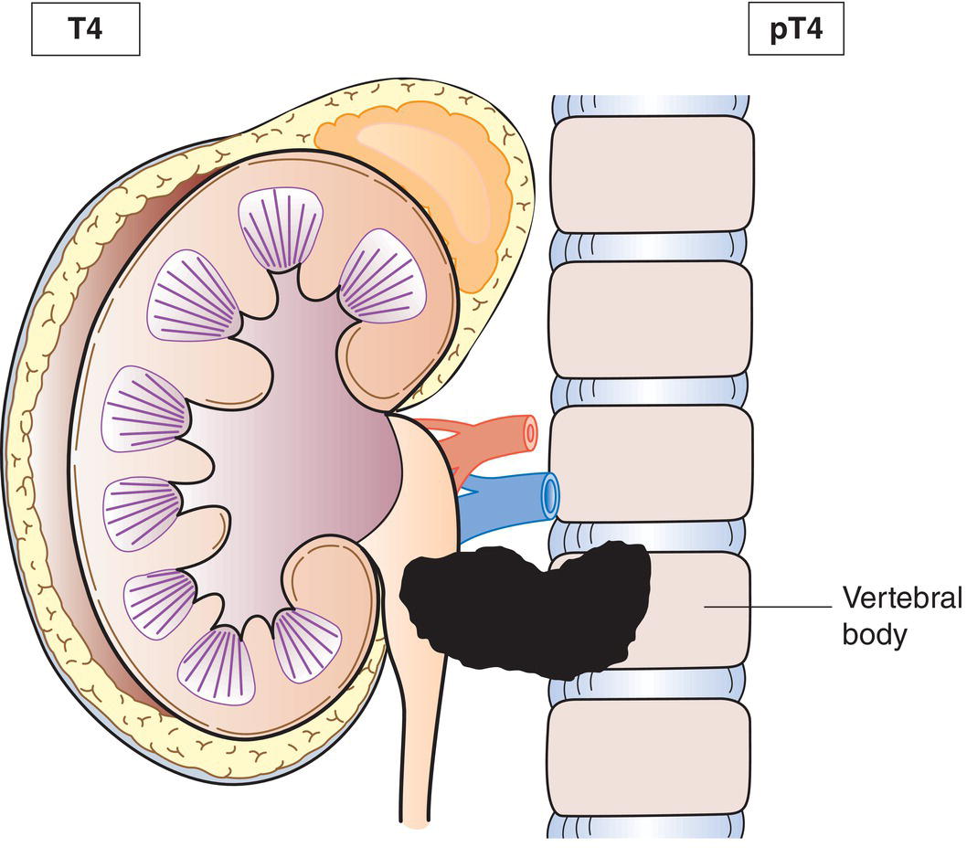
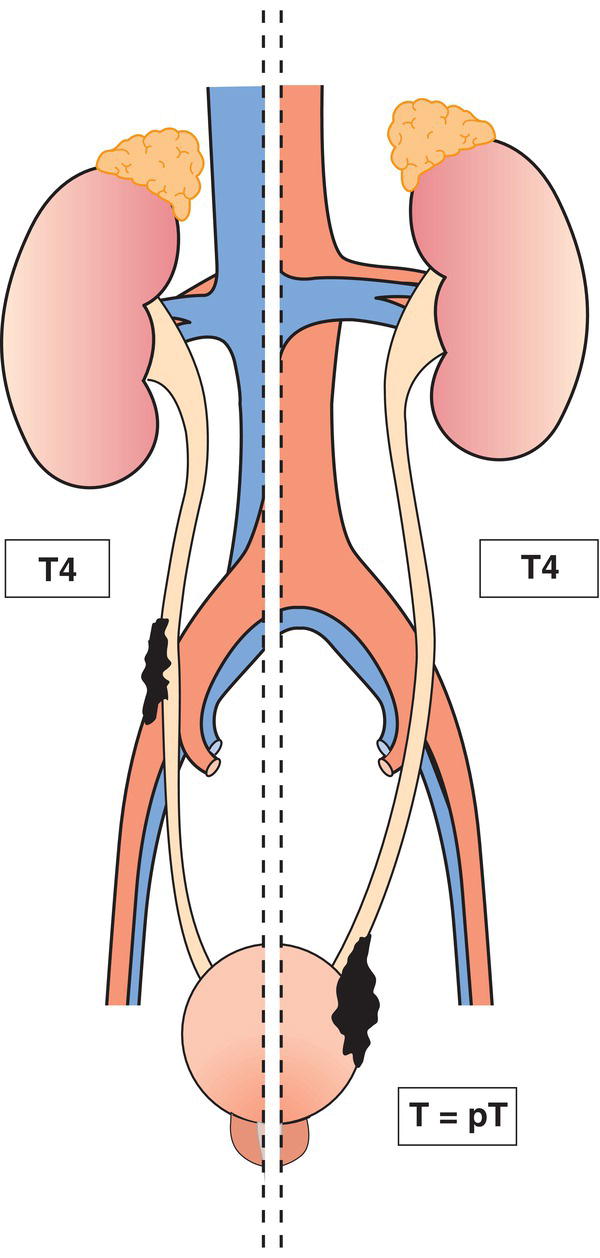
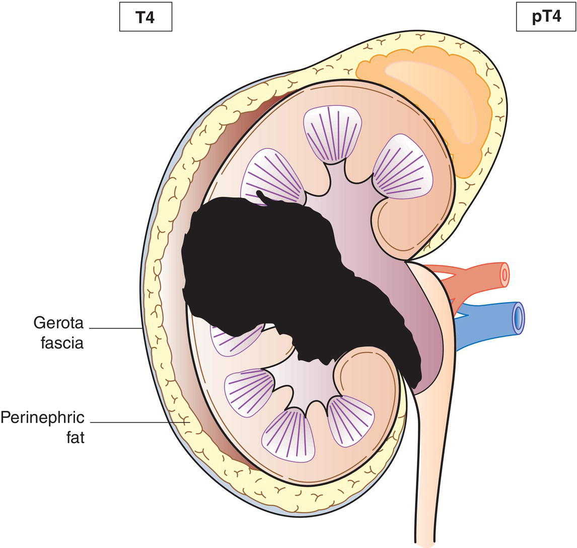
T4
Tumour invades adjacent organs (Figs. 518, 519) or through the kidney into perinephric fat (Fig. 520) 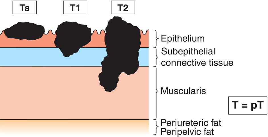
N – Regional Lymph Nodes
NX
Regional lymph nodes cannot be assessed
N0
No regional lymph node metastasis
N1
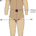
Metastasis in a single lymph node 2 cm or less in greatest dimension (Fig. 521)
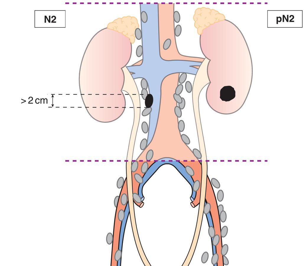
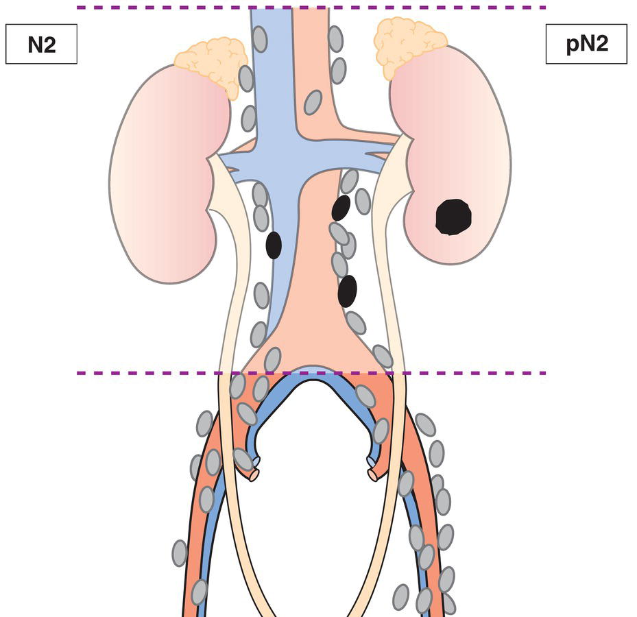
N2
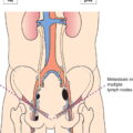
Metastasis in a single lymph node more than 2 cm or multiple lymph nodes (Figs. 522, 523) 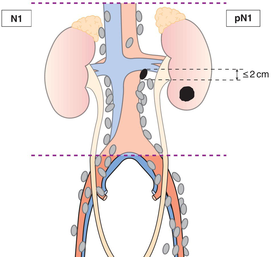
M – Distant Metastasis
M0
No distant metastasis
M1
Distant metastasis
pTNM Pathological Classification
pM1
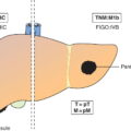
Distant metastasis microscopically confirmed
Summary
Stay updated, free articles. Join our Telegram channel

Full access? Get Clinical Tree


