Pathology and Molecular Diagnosis of Leukemias and Lymphomas


Pathology and Molecular Diagnosis of Leukemias and Lymphomas


Amy Heerema-McKenney

Michael L. Cleary

Daniel A. Arber


INTRODUCTION

This chapter focuses on the pathologic diagnosis of pediatric hematopoietic neoplasms and their classification using the World Health Organization’s WHO Classification of Tumours of Haematopoietic and Lymphoid Tissues,1 published in 2008. An update to the WHO classification is currently underway, but details of that revision were still pending at the time of the publication of this text. Pathologic diagnosis and classification requires proper specimen collection and communication between the treating physician and the pathologist or hematologist examining the diagnostic material. Accurate diagnosis also requires correlation with appropriate immunophenotyping and molecular genetic studies, which are now integral components of the classification of these diseases.


SPECIMEN PROCESSING


Bone Marrow and Peripheral Blood

Leukemia, as well as involvement of the marrow by lymphoma or histiocytic tumors, may be diagnosed by the examination of peripheral blood and bone marrow. Bone marrow aspirates are ideally prepared at the bedside and may be stained in the laboratory with a variety of Romanowsky stains prior to examination. Smears can also be prepared in the laboratory if the bone marrow aspirate is mixed with ethylenediaminetetraacetic acid (EDTA) for transport, but morphologic preservation may be limited. Care should be taken not to crush the marrow cells when preparing slides. Slides must be adequately air-dried before staining with quality-controlled reagents, using a well-validated and reproducible procedure. Touch preparations from the biopsy specimen should be considered in all cases; they are especially useful when the aspirate smears contain insufficient material for evaluation. Peripheral blood and bone marrow aspirates are ideal for flow cytometry immunophenotyping, karyotype, and molecular studies. An additional sample should be saved in EDTA or heparin for such studies when a hematopoietic malignancy is suspected.


Tissue Specimens, Including Lymph Nodes

Fresh tissue specimens, such as lymph node biopsies, must be triaged properly for adequate diagnosis.2 While lymph node excision provides the best material for diagnosis, smaller biopsies or fine-needle aspirations may suffice in some cases. Proper communication between the oncologist, surgeon, and pathologist is essential. If a neoplasm with only rare tumor cells, such as Hodgkin lymphoma, is suspected, then a larger portion of tissue is required. If the initial attempt at diagnosis of a hematopoietic tumor is by fine-needle aspiration, a separate sample for flow cytometry is strongly recommended. It should be confirmed in advance that the laboratory is equipped to triage such specimens. When larger tissue portions are removed, they should be submitted fresh to the pathologist for immediate processing. Depending on the laboratory, samples will be fixed in formalin or other fixatives, and a portion may be submitted fresh for immunophenotyping, cytogenetic studies, or molecular studies, or may be frozen and saved for ancillary studies. While many immunophenotypic markers and molecular aberrations can now be assessed on paraffin-embedded formalin-fixed tissues, fresh or frozen tissue may be required.


USE OF ANCILLARY STUDIES

Ancillary studies are essential for the proper diagnosis and classification of most hematopoietic tumors and include immunophenotypic, cytogenetic, and molecular genetic studies. While all of these tests are not necessary for each specimen type, an understanding of the utility of the different methods is essential.


Immunophenotyping

Determining the immunophenotype of the neoplastic population is critical for the proper classification of hematopoietic neoplasms. Flow cytometry and immunohistochemistry are the two most common methods; more commonly used immunophenotypic markers are listed in Table 7.1. Flow cytometry is performed on liquid
specimens, such as blood, bone marrow, or body fluids, or cell suspensions prepared from solid tissue specimens.3 Multicolor flow cytometry evaluates multiple antigens per cell, facilitating rapid evaluation. Flow cytometry is the preferred method for characterizing leukemias.


TABLE 7.1 Selected Immunophenotyping Markers in Hematopoietic Tumors


General


B lineage


CD45


CD19


Myeloid


CD20


CD13


CD22


CD15


CD79a


CD33


PAX5


CD117


Kappa/lambda


Myeloperoxidase


T lineage


Myelomonocytic


CD2


CD14


CD3


CD64


CD5


CD163


CD7


Megakaryocyte


CD4/CD8


CD41


Immature lineage


CD61


TdT


CD34


Immunohistochemistry is performed on formalin-fixed, paraffin-embedded tissue, facilitating correlation between cellular morphology and immunophenotype. It usually evaluates only one marker at a time (in contrast to the multiple antibodies that can be simultaneously used in flow cytometry). Immunohistochemistry is appropriate for tissue that has already been fixed, tumors in which the cells are fragile and may not survive processing for flow cytometry, and tumors with small numbers of neoplastic cells, such as Hodgkin lymphoma.


Cytogenetic Studies and Array-Based Technologies


Karyotype

Performing a karyotype requires fresh cells to grow in culture. Cytogenetic studies are of critical importance for many hematopoietic tumors. The results are often requisite for classification, and provide essential prognostic information.1 These studies are most successful in proliferating tumors, particularly in acute and chronic leukemias, myelodysplastic syndromes (MDSs), and some high-grade lymphomas.


FISH

Fluorescence in situ hybridization (FISH) is a molecular-based cytogenetic test utilizing fluorescently labeled DNA probes to detect translocations, additions, or deletions of chromosomal regions. It does not require cell culture and may be performed on preparations of fresh cells, frozen cells, or, in some cases, paraffin-embedded tissue. FISH is routinely used to find diagnostic translocations in pediatric acute myeloid leukemia (AML), acute lymphoblastic leukemia (ALL), and some lymphomas. Derivations of this methodology that are less commonly available include spectral karyotyping or SKY analysis, which uses chromosome-specific painting probes that allow for the identification of cryptic chromosomal rearrangements, and the combined use of immunohistochemistry and FISH, which allows the identification of specific genetic lesions within defined cellular populations.

Molecular Karyotype

Newer methodologies allow a closer inspection of the genome, detecting small changes below the resolution of routine cytogenetics, including copy number variations, and loss of heterozygosity. Array-based competitive genomic hybridization (array CGH, or aCGH) does not require cells to grow in culture. It is most commonly performed on fresh or frozen cells, but can be performed on paraffin-embedded tissue in some laboratories. Briefly, denatured DNA from the test sample and a reference sample labeled with different fluorochromes are allowed to hybridize to a normal metaphase spread of chromosomes. With the aid of a computer, the relative amounts of each fluorescent signal are measured, indicating either an excess or deficiency of genomic regions in the test sample. aCGH only detects changes in gene copy number; balanced translocations where the total gene copy number is unchanged are not found. Single nucleotide polymorphism (SNP) array is another array-based technology utilizing fluorescently labeled oligonucleotide probes that facilitates detection of copy number variation, as well as loss of heterozygosity for a given locus (uniparental disomy).4 SNP array is performed on DNA isolated from fresh or frozen cells. DNA degradation in paraffin-embedded tissue limits utility for some molecular studies.


Molecular Genetic Studies


Targeted Sequencing

As resolution of genetic abnormalities in hematologic malignancy becomes increasingly refined, we now know of single gene mutations associated with prognosis for some pediatric cancers. Specific genes can be amplified by PCR and Sanger sequenced from fresh cells, frozen cells, or even paraffin-embedded tissue. Some of the most common gene mutations relevant to prognosis in pediatric hematopathology include FLT3, NPM1, and CEBPA in AML, and IKZF1 in B-ALL. Platforms are being developed in laboratories to facilitate targeted sequencing of relevant genes for a general diagnosis such as AML or ALL.


Next-Generation Sequencing

Whole genome sequencing, exome sequencing, and transcriptome sequencing are methodologies that are rapidly becoming part of clinical care and they are intense areas of research in pediatric oncology, with collaborations active in leukemia research especially. Next-generation sequencing methods, however, can be used to assess multiple genes in panels rather than performing targeted sequencing in multiple assays, and these methods are quickly moving into diagnostic laboratories.


ACUTE LYMPHOBLASTIC LEUKEMIA/LYMPHOMA (PRECURSOR LYMPHOID NEOPLASMS)

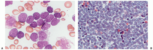
Acute lymphoblastic leukemia is the most common hematopoietic tumor of childhood, and is defined as a proliferation of immature lymphoid cells or lymphoblasts. A minimal threshold for defining leukemia has not been established by the WHO for ALL, in contrast to the 20% blasts used for AML. Protocols may require lymphoblasts to comprise 25% or more of bone marrow cells. The diagnosis is rarely made in the setting of less than 20% blasts. ALL is biologically similar to lymphoblastic lymphoma, distinguished by evidence of extramedullary disease and less than 25% bone marrow lymphoblasts in B-lymphoblastic lymphoma. The most clinically and biologically relevant classification schemes for ALL incorporate molecular genetic or cytogenetic features, in conjunction with immunophenotype. Lymphoblast morphology is not helpful in subclassifying ALL into clinically important prognostic groups. Burkitt leukemia/lymphoma, the older French-American-British (FAB) L3 type, is classified as a mature B-cell non-Hodgkin lymphoma and should not be termed B-ALL.

Lymphoblasts have a high nuclear-to-cytoplasmic ratio, round-to-convoluted nuclei, and lightly basophilic, agranular cytoplasm (Fig. 7.1). Morphology can vary; cytoplasmic granules may be seen in approximately 5% of cases (Fig. 7.2 and 7.3). Lymphoid lineage is established immunophenotypically. Neither morphology nor cytochemistry can reliably identify a blast to be lymphoid.

Detection of B-cell immunoglobulin or T-cell receptor gene rearrangements is not lineage-specific in the precursor lymphoid neoplasms as both types of rearrangements may occur in the same cell type. B-ALL characteristically expresses the B-cell markers CD19 and CD79a, as well as the immature lymphoid marker TdT. Most cases are CD10 positive, but there is variation in the expression of CD45, CD20, or CD34. Because these are neoplasms of immature B-lineage cells, they do not usually express surface immunoglobulin heavy or light chains; however, the presence of restricted immunoglobulin light or heavy chains in the presence of an otherwise immature cellular immunophenotype (TdT-positive) does not preclude a diagnosis of precursor B-cell ALL. Immunophenotyping discriminates B- and T lymphoblasts from minimally differentiated AML. Immunophenotyping is also useful for distinguishing residual or recurrent B-ALL from the presence of hematogones (benign precursor B cells) in the bone marrow. Minimal
residual disease monitoring may use multidimensional flow cytometry, patient-specific molecular assays for the immunoglobulin gene rearrangement of the leukemic clone, or the presence of a characteristic gene fusion or mutation.


Figure 7.1 Acute lymphoblastic leukemia and lymphoma. A: ALL shows a monotonous population of bone marrow cells displaying an immature lymphoid appearance with scant cytoplasm, fine chromatin, and small nucleoli. B: Lymphoblastic lymphoma displays a similar cell composition in tissue sections with monotonous small, irregular cells and a high mitotic rate.

The majority of B-lymphoblastic neoplasms present as leukemia, fewer cases present with extramedullary disease, and less than 25% present as blasts in the bone marrow (B-lymphoblastic lymphoma). The recurrent genetic abnormalities common to most cases of B-ALL are less often associated with B-lymphoblastic lymphoma. These cases have not been studied in molecular detail, but it appears that additional copies of regions of chromosome 21, particularly the RUNX1 region at 21q22, may be pathogenic.


B-Lymphoblastic Leukemia

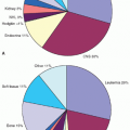
The 2008 WHO recognizes seven recurrent genetic aberrations (Table 7.2).5 The incidence of each subtype varies by age. These recurrent genetic factors are an important component of risk stratification, together with factors such as patient age, presenting white blood cell (WBC) count, and measures of early treatment response.6 Array-based molecular karyotyping identifies multiple submicroscopic alterations in many of these groups that may further influence disease risk and treatment in the future.7


Figure 7.2 Acute lymphoblastic leukemia with granules. Approximately 5% of pediatric ALLs contain cells with cytoplasmic granules, which may give a false impression of myeloid lineage.

The Philadelphia chromosome resulting from the (9;22) (q34;q11.2) chromosomal translocation is found in chronic myelogenous leukemia (CML), adult ALL, and 4% of pediatric B-ALL. The BCR/ABL1 fusion protein of pediatric ALL (p190) is smaller than that of CML (p210). Five to ten percent of cases display normal karyotypes, with a BCR/ABL1 fusion detectable by molecular methods. ALLs with t(9;22) are of precursor B-cell lineage (CD10, CD19, and TdT positive) and may show aberrant expression of the myeloid-associated antigens CD13 and CD33, as well as CD38. The 2008 WHO classification proposes strict criteria to distinguish B-ALL with t(9;22) from mixed phenotype acute leukemia (MPAL) with t(9;22), mainly based on evidence of myeloperoxidase (MPO) in the blasts of MPAL.8 Deletions of IKZF1, resulting in oncogenic Ikaros isoforms, are common in BCR-ABL1-positive B-ALL.
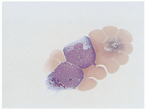
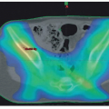
9

B-lymphoblastic leukemia with MLL (now known as KMT2A) abnormalities, particularly t(4;11)(q21;q23), is the most common ALL type in infants, and constitutes approximately 5% of ALLs in older children or adults (Fig. 7.4).10 The blast immunophenotype is typically CD10 negative with aberrant expression of myeloid-associated antigens CD15 or CD65. Other myeloid antigens are not usually expressed (specifically, no more than one marker of monocytic differentiation may be present, such as NSE, CD11c, CD14, CD64, or lysozyme), and the blast cells are routinely MPO negative. These features are necessary to distinguish B-ALL from the diagnosis of MPAL with t(v;11q23)—MLL rearranged. High-level expression of the FLT3 receptor tyrosine kinase within the leukemic blasts is a feature of this genetic subtype of ALL; however, FLT3-activating mutations are found in only a small subset of cases.11

B-ALL with t(12;21) (p13;q22) ETV6-RUNX1 is the most common genetic subtype of ALL in children, particularly in the 2- to 10-year age group, and constitutes at least 20% to 30% of childhood ALL.5 The t(12;21) fuses the ETV6 gene (aka TEL) on chromosome 12 with the RUNX1 (aka AML1) gene on chromosome 21. This aberration is not detectable by routine karyotype analysis and requires either RT-PCR or FISH evaluation for detection. The leukemic blasts are of precursor B-cell lineage (CD10, CD19, and TdT positive), lack expression of CD9 and CD20, and may show aberrant expression of CD13.

Blast clones with an abnormal hyperdiploid karyotype of more than 50 chromosomes (high hyperdiploid) are seen in 25% of childhood ALL cases. The identification of this subgroup can be made either by routine cytogenetics or by assessing DNA ploidy using flow cytometry. Nearly all hyperdiploid ALLs have trisomy of chromosomes 6, tetrasomy of chromosome 21, and duplication of an X chromosome. Other chromosomes that are frequently trisomic include 4, 10, 14, 17, and 18.12 No specific immunophenotypic
features are recognized. FLT3-activating mutations have been identified in 10% to 20% of cases.11


Figure 7.3 FAB Cooperative Group types of ALL. A: L1 blast cells have fine nuclear chromatin, small, indistinct nucleoli, and generally uniformly sized nuclei with scant cytoplasm. B: L2 blasts display more variation in nuclear size and often have more prominent nucleoli and more abundant cytoplasm. C: L3 blasts have more mature nuclear chromatin with chromatin clumping, multiple, distinct nucleoli, and darkly basophilic and vacuolated cytoplasm. Such cases correlate with Burkitt lymphoma and are no longer considered ALL.

Hypodiploid ALL is defined differently by different authors. The WHO assigns this diagnosis in B-ALL when the chromosome number is less than 46.5 However, stricter definitions of less than 44 chromosomes may be more clinically significant.13,14 Leukemias with less than 44 chromosomes are a rare subset comprising less than 1% of B-ALL, with a particularly poor prognosis. Near-haploid (23 to 29 chromosomes) or low-hypodiploid B-ALL can be missed by standard karyotyping if the hypodiploid clone has undergone endoreduplication, doubling the number of chromosomes. This discrepancy can be resolved by FISH. Molecular genetic studies of hypodiploid ALL identify unique differences from other ALL subtypes. Most near-haploid cases harbor activating mutations of Ras signaling (NFI, NRAS, KRAS, and PTPN11) and inactivating deletions and mutations of the IKZF3 (AIOLOS).
Nearly all low-hypodiploid cases have mutations of TP53 as well as inactivating mutations of IZKF2 (HELIOS). The TP53 mutations of low-hypodiploid ALL were also found in nontumor cells, suggesting possible Li-Fraumeni syndrome in many cases.7


TABLE 7.2 B-ALL with Recurrent Genetic Abnormalities


Diagnosis


Frequency, %


General Age, ya


Immunophenotype


Involved Genes


B-ALL with t(9;22) (q34;q11.2)


4


10


CD19+, CD10+, TdT+, CD13+, CD33+, CD38+


BCR/ABL1


B-ALL with t(v;11q23), MLL rearranged


5


<1


CD19+, CD10, TdT+, CD15+, CD65+


MLL, most commonly fused with AFF1 (AF4) on chromosome 4q21


B-ALL with t(12;21) (p13;q22)


25


2-10


CD19+, CD10+, TdT+, CD9, CD20, CD13+


ETV6/RUNX1 (TEL/AML1)


B-ALL with hyperdiploidy


25


2-10


CD19+, CD10+, TdT+, CD34+, CD45


>50 chromosomes, commonly with trisomies of 4, 10, and 17


B-ALL with hypodiploidy


1-5


All pediatric


CD19+, CD10+, no known distinctive immunophenotypic features


<46 chromosomes, some risk stratification schema restrict definition to <44 chromosomes


B-ALL with t(5;14) (q31;q32)


<1


All pediatric


CD19+, CD10+, with eosinophilia


IL3/IGH


B-ALL with t(1;19) (q23;p13.3)


5


˜5


CD19+, CD10+, cytoplasmic mu+, TdT+, CD20, CD34


TCF3/PBX1 (E2A/PBX1)


a Ages given are for most common age groups, but these disorders may occur less commonly at any age.


Figure 7.4 Infant acute lymphoblastic leukemia with t(4;11). These blasts often have irregular nuclei and nucleoli, which may suggest myeloid lineage. Immunophenotyping is necessary for determination of precursor B-cell lineage.

B-ALL with t(5;14)(q31;q32)—IL3-IGH is a rare subtype of B-ALL occurring in both children and in adults.5 Patients may present with ALL or asymptomatic eosinophilia. In the setting of eosinophilia, even small numbers of B lymphoblasts should prompt consideration of this entity and cytogenetic study to confirm the diagnosis. These cases should be distinguished from the rare lymphoblastic leukemias with mutations of PDGFRA or FGFR1 commonly associated with eosinophilia.

B-ALL with a balanced or unbalanced t(1;19)(q23;p13.3) TCF3-PBX1 chromosomal translocation occurs in approximately 6% of pediatric ALL, with an average age of 5 years. A rare variant chromosomal translocation t(17;19) fuses TCF3 and HLF. Up to 25% of cases with TCF3/PBX1 fusions detected by molecular genetic studies may have normal routine karyotypes.7 The blasts are of precursor B-cell lineage (CD10, CD19, and TdT positive), express cytoplasmic mu and CD9, and usually lack CD20 and CD34.

B-ALL lacking one of the aforementioned recurrent genetic abnormalities is categorized as B-lymphoblastic leukemia/lymphoma, not otherwise specified (NOS) by the 2008 WHO classification.15 Additional subtypes have emerged from molecular genetic studies. Some have prognostic significance that may be useful in risk-based stratification of therapy. These include the following: (1) B-ALL with intrachromosomal amplification of chromosome 21 (iAMP21), characterized by gain of at least three copies of a region of chromosome 21 that always includes RUNX1; this may be seen most frequently in B-ALL, NOS, but also occurs in cases with ETV6-RUNX1 and BCR-ABL1 rearranged; (2) ERG-altered B-ALL, occurring exclusively in B-ALL; NOS; (3) CRLF2 rearranged B-ALL, with rearrangement of the cytokine receptor gene CRLF2 at Xp22.3/Yp11.3 occurring in 50% of Down syndrome (DS)-associated ALL; (4) BCR-ABL1 like B-ALL, characterized by a similar gene expression profile as B-ALL with BCR-ABL1 but lacking the translocation, with numerous associated genomic alterations.7


T-Lymphoblastic Leukemia/Lymphoma

T-lymphoblastic tumors constitute approximately 15% of pediatric lymphoblastic leukemia and 85% to 90% of cases that present as lymphoblastic lymphoma.16 Cases with 25% or more bone marrow involvement are considered to be ALL. Approximately half of T-ALL patients have a mediastinal mass at the time of presentation, and other organ sites may be involved. Immunophenotyping is required to differentiate precursor T lymphoblasts from precursor B lymphoblasts. T-lymphoblastic neoplasms are TdT positive and usually express CD2, cytoplasmic CD3, CD5, and CD7, although loss of one or more of these T-cell antigens is common. Some cases also express CD1a, CD4, CD8, dual CD4 and CD8, or surface CD3. Surface and/or cytoplasmic CD3 expression is considered the most specific marker for the T lineage. T lymphoblasts also occasionally express CD79a (a B-lineage marker) or aberrantly express myeloid antigens such as CD13, CD33, or CD117.

The 2008 WHO classification does not further classify T-ALL by recurring cytogenetic abnormalities. A total of 50% to 70% of T-ALL have an abnormal karyotype, most commonly involving the T-cell receptor loci. Detection of these aberrations at diagnosis may be useful for disease monitoring, but distinct prognostic disease groups defined by recurring cytogenetic abnormalities are not yet part of any major classification scheme for risk group stratification.17 Mutations of PHF6, NOTCH1, and FBXW7 have been found in several cohorts of T-ALL, but a consistent role for these mutations in risk stratification has not been identified to date.7 One subtype of T-ALL, early T-cell precursor (ETP) ALL has been characterized immunophenotypically by expression of cytoplasmic or surface CD3, dim CD5, absence of CD8 or CD1a, and coexpression of CD34, CD117, HLA-DR, CD13, or CD117. ETP-ALL appears to have a poor prognosis, the karyotype may be complex, and numerous diverse genetic alterations are found. The transcriptional profile resembles that of hematopoietic stem cells and primitive AML more than that of human precursor T cells.7,18


ACUTE MYELOID LEUKEMIA

Major changes have occurred in the approach to the classification of AML in recent years. Cytogenetics, and recently gene mutation status, are more prognostically informative than the older morphologic descriptions of the FAB classification.19,20,21 The 2008 WHO AML classification includes seven recurrent chromosomal translocations and two provisional entities characterized by gene mutations: AML with mutated NPM1 and AML with mutated CEBPA.22 AML with myelodysplasia-related changes includes cases with a history of prior MDS, MDS-associated cytogenetic abnormalities, or severe multilineage dysplasia. FAB-like terminology is used for cases of AML, not otherwise specified (AML, NOS). These are cases of de novo AML not falling into one of the above groups. Therapy-related AML (t-AML) is a distinct category for any case arising after cytotoxic therapy. Myeloid proliferations of DS are also separately classified (Table 7.3). Myeloid sarcoma is a category of AML, but every effort should be made to recognize one of the aforementioned subtypes if possible for optimal classification. The threshold for AML is 20% (or more) blasts in most AML subtypes, with exception of the two core-binding factor (CBF) AMLs (AML with t(8;21) and AML with inv(16)) and acute promyelocytic leukemia (APL) with t(15;17).1

The WHO AML classification diagnoses require correlation with cytogenetics and/or FISH for the characteristic genomic changes. Some have characteristic morphologic and immunophenotypic features useful for initial evaluation.23 Key features of the more common AML types are summarized in Table 7.4.


Acute Myeloid Leukemia with Recurrent Genetic Abnormalities


Acute Myeloid Leukemia with t(8;21) (q22;q22)—RUNX1-RUNX1T1

The (8;21)(q22;q22) chromosomal translocation is one of the most common recurring cytogenetic abnormalities in AML and is present in 8% to 13% of pediatric AMLs.24,25 The RUNX1/RUNX1T1
chimeric fusion protein disrupts normal functioning of the CBF transcription factor complex regulating normal hematopoiesis. Blasts of AML with t(8;21) have fine pink granules in slightly basophilic cytoplasm. They usually show perinuclear clearing, or hofs, and a subset of the blasts have large, irregular pink cytoplasmic granules (Fig. 7.5). Thin Auer rods may also be present. Eosinophils may be increased in the marrow, and maturing granulocytes may show dysplastic changes. All cases with this translocation are considered AML, regardless of blast count.


TABLE 7.3 2008 World Health Organization Classification Categories of AML


AML with recurrent genetic abnormalities


AML with t(8;21)(q22;q22); (RUNX1-RUNX1T1)


AML with inv(16)(p13.1q22) or t((16;16)(p13.1;q22); (CBFB-MYH11)


APL with t(15;17)(q24.1;q21.1); (PML-RARA)


AML with t(9;11)(p22;q23); (MLLT3-MLL)


AML with t(6;9)(p23;q34); (DEK-NUP214)


AML with inv(3)(q21q26.2) or t(3;3)(q21;q26.2); (RPN1-EVI1)


AML (megakaryoblastic) with t(1;22)(p13;q13); (RBM15-MKL1)


Provisional entity: AML with mutated NPM1


Provisional entity: AML with mutated CEBPA


AML with myelodysplasia-related changes


Therapy-related myeloid neoplasms


AML, NOS


AML with minimal differentiation


AML without maturation


AML with maturation


Acute myelomonocytic leukemia


Acute monoblastic/monocytic leukemia


Acute erythroid leukemia


Pure erythroid leukemia


Erythroleukemia, erythroid/myeloid


Acute megakaryoblastic leukemia


Acute basophilic leukemia


Acute panmyelosis with myelofibrosis


Myeloid sarcoma


Myeloid proliferations related to DS


Transient abnormal myelopoiesis


AML associated with DS


APL, acute promyelocytic leukemia.


The blast cells express CD13, CD33, and CD34 with aberrant weak expression of the B-cell-associated antigen CD19.26,27 A subset of cases may also express CD56, which is associated with a worse prognosis in some studies. FLT3 mutations are uncommon in CBF AML, especially the FLT3 internal tandem duplication (FLT3-ITD) mutations.28 Mutations in KIT are present in 20% to 45% of adult AML with t(8;21) and roughly 20% to 30% of pediatric AML with t(8;21). In adults, exon 8 or 17 mutations are associated with a poor prognosis; in children, the presence of a KIT mutation does not seem to significantly affect clinical outcome.29


TABLE 7.4 Comparative Features of Common Pediatric AML Types with Recurrent Genetic Abnormalities


Diagnosis


Morphologic Features


Immunophenotype


Involved Genes


AML with t(8;21)


Perinuclear clearing of blast cytoplasm, large pink granules, dysplastic mature neutrophils


CD13+, CD33+, MPO+, CD34+, CD19+ (weak)


RUNX1/RUNX1T1


AML with inv(16)


Myelomonocytic blasts, abnormal eosinophils with large basophilic granules


CD13+, CD33+, CD14+/−, CD64+/−, CD2+ (subset)


CBFB/MYH11


Acute promyelocytic leukemia


Folded blast nuclei, cytoplasmic granules, abundant Auer rods


CD13+, CD33+, MPO+++, CD34, HLA-DR, CD2+ (subset)


PML/RARA


AML with t(9;11)


Monocytic or myelomonocytic blasts


CD13+, CD33+, MPO, CD14+, CD64+, CD34, CD56+/−


MLLT3/MLL


Acute megakaryoblastic leukemia with t(1;22)


Blasts with cytoplasmic blebs, basophilic cytoplasm, and fine granules


CD13+, CD33+, MPO, CD41+, CD61+


RBM15/MKL1


Acute Myeloid Leukemia with inv(16)(p13.1q22) or t(16;16)(p13.1;q22)—CBFB/MYH11

AML with inv(16)(p13.1q22) or t(16;16)(p13.1;q22) is also a relatively common type of AML (5%-10% of all childhood cases) and tends to occur in children or young adults. Both cytogenetic abnormalities result in a fusion of the CBFB and MYH11 genes on chromosome 16, which, similar to the t(8;21), disrupts the CBF transcription factor complex.30

The bone marrow shows a proliferation of myelomonocytic cells with a population of abnormal eosinophils. Some cases lack monocytic differentiation either by morphology or by cytochemistry. The maturing eosinophils in the bone marrow show large, coarse basophilic granules in a background of cytoplasmic eosinophilic granules (Fig. 7.6). Even if blasts are fewer than 20%, all cases with this cytogenetic abnormality are considered AML. The detection of abnormal eosinophils is highly predictive of a chromosome 16 abnormality, which may be subtle on routine karyotype. Additional studies, such as RT-PCR or FISH should be performed on cases with abnormal eosinophils and an apparently normal karyotype.

AML with inv(16) or t(16;16) shows expression of the expected myeloid antigens (CD13 and CD33), and often monocyte-associated markers, such as CD4, CD14, and CD64. A subset shows aberrant expression of the T-cell-associated marker CD2, but CD2 is not specific for this disease. As in AML with t(8;21), mutations in KIT are relatively common but do not have a demonstrated prognostic importance in children. FLT3-ITD mutations are uncommon.29


Acute Promyelocytic Leukemia/Acute Myeloid Leukemia with t(15;17)(q22;q12)—PML/RARA

APL represents 4% to 10% of pediatric AMLs and is frequently associated with disseminated intravascular coagulopathy (DIC). The risk for fatal intracerebral hemorrhage makes prompt recognition of the morphologic and immunophenotypic features imperative. All molecular subtypes contain mutations of the retinoic acid receptor α (RARA) gene on chromosome 17.31 The t(15;17) (q22;q12) is the most common genetic aberration in APL and results in fusion of RARA with the PML gene on chromosome 15. APL is subdivided into hypergranular and hypogranular (or microgranular) disease (Fig. 7.7). The classic hypergranular APL has “blast” cells that resemble promyelocytes with abundant cytoplasmic granules. Numerous Auer rods are present in individual blasts (so-called faggot cells). In contrast to the blasts of AML with t(8;21) or reactive promyelocyte proliferations, APL cells do not show differing stages of maturation or perinuclear clearing. Hypogranular
APL is recognized by characteristic folded or bilobed nuclei with variably visible fine cytoplasmic granules. Both types of APL show strong MPO positivity in virtually all blast cells—helpful in differentiating hypogranular APL from myelomonoblasts or monoblasts. FLT3 mutations are common in APL, present in approximately 40% of patients, with the majority being internal tandem duplication mutations. FLT3-ITD in APL is strongly associated with the hypogranular subtype, high WBC counts in peripheral blood, and the bcr-3 breakpoint in PML.32

Only gold members can continue reading. Log In or Register to continue

Stay updated, free articles. Join our Telegram channel

Aug 25, 2016 | Posted by in ONCOLOGY | Comments Off on Pathology and Molecular Diagnosis of Leukemias and Lymphomas

Full access? Get Clinical Tree

Get Clinical Tree app for offline access