PATHOLOGY
Part of “CHAPTER 43 – ENDOCRINE OPHTHALMOPATHY“
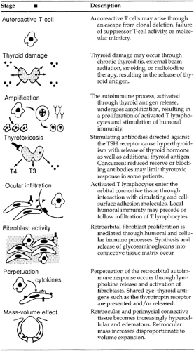
The major pathologic changes that occur in Graves disease are inflammatory infiltration by lymphocytes and chronic inflammatory cells in all orbital tissues with proliferation of the connective tissue (Table 43-1). Because autopsy findings are rare in this condition, precise clinicopathologic correlation has not been made for each of the physical signs that are described. Most of the material examined by pathologists is obtained at
the time of surgery for orbital decompression or correction of extraocular muscle difficulties. Thus, the earliest changes in the orbital tissues are rarely studied, whereas abundant reports exist of some of the pathologic findings in the more severe forms of ophthalmopathy.
the time of surgery for orbital decompression or correction of extraocular muscle difficulties. Thus, the earliest changes in the orbital tissues are rarely studied, whereas abundant reports exist of some of the pathologic findings in the more severe forms of ophthalmopathy.
 |
All orbital structures, including lacrimal glands, are affected. Massive swelling of the extraocular muscles is commonly seen; occasionally, they may become 8 to 10 times their normal volume (Fig. 43-2). Histologically, marked interstitial inflammatory edema and focal lymphocytic infiltration of the muscles is noted. The myofibrillar structure remains normal, but loss of striations and disorganization may occur later. With thyrotoxicosis, a diffuse increase in orbital fat content may be present, and a deposition of adipose tissue cells lying in strands between the muscle fibers may be noted. In the absence of thyrotoxicosis, the fat content is decreased and is replaced by connective tissue. The mucopolysaccharide and water content of the tissues increases, and large numbers of mast cells are seen in the perivascular areas.
Related posts:


Stay updated, free articles. Join our Telegram channel

Full access? Get Clinical Tree


