PARATHYROID ADENOMA
The parathyroid adenoma is more frequently located among the lower glands than among the upper ones. Grossly, a parathyroid adenoma is oval, reddish-brown, and smooth, circumscribed, or encapsulated (Fig. 48-3). It may show areas of hemorrhage and, if large, cystic degeneration. In small adenomas, a grossly visible rim of yellow-brown normal parathyroid tissue occasionally may be seen. Weights vary from 300 mg to several grams. The overall size ranges from smaller than 1 cm to larger than 3 cm.4
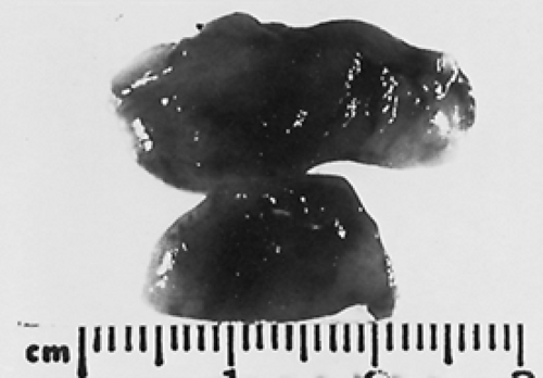 FIGURE 48-3. Transected parathyroid adenoma is shown. It weighed 1.1 g and had a brown, homogeneous surface. No rim is visible. |
Histologically, parathyroid adenomas are composed of sheets of parathyroid chief cells interspersed with a delicate capillary network. If not very large, most adenomas—consonant with their gross appearance—reveal a rim of normal or atrophic parathyroid tissue beyond the adenoma capsule1,4,12,13 (Fig. 48-4). The extraadenomatous, normal cells tend to be smaller and more uniform than those in the adenoma. Stromal and cytoplasmic fat is frequently abundant here but absent in the adenoma.13,14 The absence of a rim of normal tissue does not preclude the diagnosis of an adenoma, because large tumors may have overgrown the preexisting normal gland; alternatively, the rim may be lost when the tissue is sectioned.15 Zones of fibrosis with hemorrhage, cholesterol clefts, and calcification may be found in larger tumors.3,5
Related posts:


Stay updated, free articles. Join our Telegram channel

Full access? Get Clinical Tree


