Introduction
Sir James Paget described the disease that bears his name as osteitis deformans in a series of monographs published in the latter half of the nineteenth century.1 At this time, we understand much more about the disease, including how to treat it, but its aetiology remains uncertain, perhaps even mysterious.
Paget’s disease occurs in monostotic or polyostotic forms. The monostotic form occurs in a single bone, most frequently in a small quarter to half-dollar size lesion in the pelvis or lumbar spine. The pathologic description given below describes both monostotic and polyostotic Paget’s disease. Monostotic Paget’s disease is usually asymptomatic, although unusual placement of monostotic disease in the higher vertebrae, for example, with pathologic fracture can become symptomatic. Polyostotic Paget’s disease is generally described as involving more than one bone. It is worth noting that monostotic disease of a long bone, the femur for example, behaves more like polyostotic disease than monostotic disease, even if only one bone is involved. The rest of the presentation will describe polyostotic Paget’s disease unless otherwise noted.
Pathology of Paget’s Disease
Microscopic
Pathology at the cellular level is frequently divided into three types that correspond to early, middle and late stages of Paget’s disease (Table 89.1). These may all be found in a single bone, proceeding in an orderly manner from early to late.2 The early descriptions of the stages were generally called osteoclastic (early), osteoblastic (mid) and mixed (late). One could occasionally run across a very late stage of the mixed phase that was described as ‘burned out’. More modern descriptions combine the osteoblastic and mixed phases to one and include the ‘burned-out’ phase as the third. The early stage corresponds to increased osteoclastic activity without compensatory increase in osteoblastic activity. Examination of such areas of pagetic bone demonstrate large osteoclasts, frequently several times bigger than normal osteoclasts. All osteoclasts appear to have multiple nuclei, perhaps in the range of four to eight, but pagetic osteoclasts frequently have 20 to 40 nuclei. In the second phase (Table 89.1) osteoblastic activity in the area appears to be dramatically increased, but severely disorganized. Routine findings include disorganized matrix, woven bone and occasional areas of malacic or non-mineralizing osteoid. The characteristic order and architecture of cortical bone is lost and the cortical-trabecular boundary is lost. In this phase, both osteoclastic and osteoblastic activity is increased. Finally, in the ‘burned-out’ phase, activity of the pagetic bone appears to have returned to about normal, but the abnormal architecture remains. Most monostotic Paget’s is found in either phase two (mixed) or in the ‘burned-out’ form. In the third phase the abnormal architecture remains despite the relative normalization of cellular activity. Normal bone in the same individual, from another skeletal site or from an uninvolved portion of the pagetic bone, appears perfectly normal. It has appropriately sized osteoclasts with normal activity as well as normal osteoblasts and osteoblastic activity.3 This appears to be true even for areas that might be expected at some later date to become pagetic.
Table 89.1 Phases of Paget’s disease.
| Phase | Bone morphology | Activity |
| Osteoclastic | Normal, but increased osteoclastic size and activity | Osteoclast |
| Mixed | Changes of osteoclastic phase plus great increases in osteoblastic activity, poorly mineralized or osteomalacic bone, woven bone, large seams of osteoid, loss of cortical trabecular interface | Osteoclast and Osteoblast |
| Burned out | Activity returning to normal but may still appear somewhat increased | Osteoclast and Osteoblast |
Macroscopic
Pagetic bone is larger than normal bone, but significantly weaker. The bone appears ‘coarse’ rather than with the smooth surface of normal bone. Although the origin of the bone remains recognizable, it is frequently misshapen or bent, virtually always in weight-bearing long bones. The bone is hypervascular and may have multiple arteriovenous shunts.4 Paget’s disease is one reported cause of high output congestive heart failure.
Pagetic involvement of bones occurs idiosyncratically within very specific parameters. Large bones are much more likely to be involved than small bones. Anyone who has treated multiple cases of Paget’s will have seen some unusual small bone involvement in wrist, hand or foot, for example. A list of bones frequently involved is shown in Table 89.2. Involvement in long bones generally starts at one end and proceeds toward the other end over a period of years. The disease does not cross joint spaces for long bones. Similarly, involvement in the pelvis or skull begins at one location and gradually extends across the entire bone. Unlike long bones, Paget’s disease does frequently seem to spread across sutures in the bones of the skull or pelvis. Involvement of one long bone, the left femur for example, does not determine the involvement of the other, in this example the right femur. Usually, it is not involved, but occasionally it is.
Table 89.2 Bones commonly affected by Paget.
|
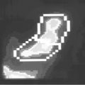
The sequelae of Paget’s disease may generally be inferred from the observations of micro- and macroscopic changes in bone. Changes in measures of osteoclastic activity either in serum or urine occur first and remain elevated. Urinary hydroxyproline was the measurement first used, but required specific dietary restrictions to avoid gelatine and gelatine-containing foods to be used accurately. Serum acid phosphatase, a potential serum marker of osteoclast activity, was occasionally used in some studies, but suffered from a series of difficulties. It was only elevated in about 20% of cases of Paget’s and was also a marker for prostatic disease, a fairly common finding in many male patients with Paget’s. More recent measures of bone-specific collagen breakdown markers in serum or urine have obviated these difficulties. These markers include N-telopeptide and pyridinium cross-links and provide much better estimates of osteoclastic activity. Urine studies should always be accompanied by a measurement of urinary creatinine to ensure comparability of one specimen to the next. These measures are routinely elevated in all phases of Paget’s disease.5 Measures of osteoblastic activity, primarily in serum, are also routinely elevated in Paget’s disease. Older measures of this activity include serum alkaline phosphatase and the heat-stable fraction of the serum alkaline phosphatase. Although the first measure is routinely available in chemistry panels, it is not terribly specific. The heat-stable fraction is more specific, but is relatively difficult to do. It is still not entirely specific for bone alkaline phosphatase as opposed to the enzyme from other sites. Additional tests assays of osteoblastic function including bone-specific alkaline phosphatase and other markers like osteocalcin are now available They are not reported to be as sensitive a marker for Paget’s disease as the older assays despite the fact they are generally considered to be more specific.6 Radiological changes can be seen from the earliest (osteoclastic) stages of the disease on both plain films and radionuclide scans. On plain films, the early lesions appear as lucencies. Depending on the site they frequently have specific names. The most common of these include ‘osteoporosis circumscripta’. This describes an early lesion in the skull that appears as a circular lucency on plain film of the skull. Similarly, the ‘blade of grass’ lesion describes a chevron-shaped lucency in a long bone. Over time (years), this lesion may be observed to march down the length of the bone at an approximate rate of one centimetre per year followed by signs of the osteoblastic/mixed stages of the disease.7 These signs include enlargement of the bone, irregular calcification and loss of the cortical trabecular demarcation in bone. Radionuclide scans of bone will also show significant changes. Affected areas are hot. In long bones or other large bones, clear demarcation can be seen between affected and unaffected. All of these radiological findings are generally considered pathognomonic for Paget’s disease except that ‘osteoporosis circumscripta’ needs to be clearly delineated from the ‘punched out’ lesions observed in the skull with multiple myeloma. Further, very occasionally, lesions of the lower lumbar vertebra may be impossible to differentiate from metastatic (prostate) carcinoma without biopsy. Magnetic resonance imaging (MRI) has been a major boon in this regard, however.8
Sequelae of Paget’s Disease
Most of the sequelae of Paget’s disease can be inferred from the knowledge of its effect on bone, that is the bone becomes larger, but weaker. The major exception to this is the most feared complication of Paget’s disease, osteosarcoma. This tumour is a rare but deadly outcome arising from pagetic bone, usually decades after it was first affected. Estimates of its frequency are quite low perhaps 1–3% of individuals with Paget’s disease.9 Mortality in affected individuals is quite high, in some series 100%, even after the development of successful protocols for the treatment of childhood osteosarcomas. The reasons for the abysmal outcomes in osteosarcomas related to Paget’s are at least twofold. First, the central location of Paget’s disease prevents the common first-step treatment, excision/amputation. Secondly, the symptoms of osteosarcoma are very non-specific and therefore frequently missed. The major presenting complaint is a significant worsening of Paget’s symptoms, particularly bone pain. Bone pain waxes and wanes spontaneously throughout the course of Paget’s disease. After many years or decades of such spontaneous changes, patients frequently do not complain of them to their physicians until it has been present for several months. In this setting, the osteosarcoma has frequently progressed too far for successful treatment upon its initial discovery.
A second feared complication of Paget’s is probably more unusual, platybasia, or basilar invagination. Individuals with severe Paget’s disease of the skull will be noted to have a ‘sharpening’ or ‘lipping’ of the occiput on lateral skull films. Instead of the normal curve of the occiput noted on lateral film, the bone begins to protrude downwards or ‘lip’. This is a radiological sign of a falling down of the skull around the spinal column. Essentially, the skull is too weak to hold its own weight and over a period of time it will collapse while the falxes hold the brain in place. This circumstance will cause hydrocephalus and eventual herniation of brain with death. This process extends over years. Neurosurgical intervention for the hydrocephalus can maintain normal pressure.10
Congestive heart failure is a reported complication of Paget’s disease. Arteriovenous shunting of blood through affected bone is reported to produce high output congestive heart failure in individuals with more than 15% of their skeleton involved by the disease. Even in very active polyostotic Paget’s disease, this complication is very rare in my experience and is only included for completeness and because the causes of high output failure are so limited. Urgent treatment to limit the activity of the pagetic bone is recommended. This reduces the arteriovenous shunting and decreases the need for the high output state. Altered blood supply (‘steal syndrome’) is suspected or reported in individuals with Paget’s disease of the skull who complain of somnolence. Similarly, this ‘steal syndrome’ has been implicated in an individual with Paget’s of a vertebra and paralysis below that level.4, 11
Other complications are more common, but generally less severe. Fracture through weaker pagetic bone is relatively common. Considerable excess blood loss may occur because of the vastly increased vascularity of pagetic bone. Longitudinal fractures or fissures are described. Bowing of affected weight-bearing long bones is an integral part of the disease and is associated with gait abnormalities and/or unsteadiness, osteoarthritis in nearby or adjacent joints and in painful microfractures that occur in severely bowed lower extremities. Some of these microfractures extend through the bone and become full-blown displaced fractures. Such microfractures occur when weight placed on the bowed extremity increases the curvature (circumference) of the bone, stretching the bone beyond its endurance. Treatment of this type of fracture requires either bracing or osteotomy to straighten the bone in order to heal and prevent further fractures in that bone. In the case of osteotomy, patients should be treated prior to surgery to reduce the vascularity of the affected bone.
Any place a nerve passes through a bony foramen or through bone, the potential exists for Paget’s disease to narrow the channel and impinge upon the nerve, causing pain and/or eventually loss of the nerve function.12 The most common of these is probably seen in Paget’s of the skull where approximately 70% of patients are reported to have mixed sensineural hearing loss. Other nerves pass through foramen in the skull. Blindness is an unusual but reported complication also. Facial nerve palsies can also be observed. Hypercementosis in teeth, an idiopathic condition of increased accretion of bone on one or more roots, causing tooth pain has also been reported in individuals with Paget’s of the skull.13
Similar nerve palsies may be observed in effected peripheral locations. Certainly the most severe of these may be related to weakness/fracture, as is the case in (partial) spinal cord transaction related to thoracic or cervical vertebra collapse due to pagetic involvement. Spinal stenosis, related to involvement of one or more vertebra with Paget’s resulting in critical narrowing of the spinal canal has also been reported, but is thought to be less common than the steal syndrome.10, 12
Stay updated, free articles. Join our Telegram channel

Full access? Get Clinical Tree


