PAGET DISEASE OF BONE
Ethel S. Siris
Paget disease of bone is a localized disorder of skeletal remodeling in which abnormally increased rates of both bone resorption and new bone formation change bone architecture. Typically, pagetic bone shows a characteristic radiographic appearance (Fig. 65-1). The affected bone is often larger, generally less compact, more vascular, and more susceptible to deformity and fracture than normal bone. Paget disease occurs predominantly in persons older than 40 years, affecting men slightly more often than women. In the United States, up to 2% to 3% of the population in their 50s and 60s have this bone disease, and the frequency rises in the ensuing decades.1a Most patients are asymptomatic, although a significant minority do experience signs and symptoms related to the area of involved bone, the degree of increased bone turnover, and the structural changes that result at affected sites.
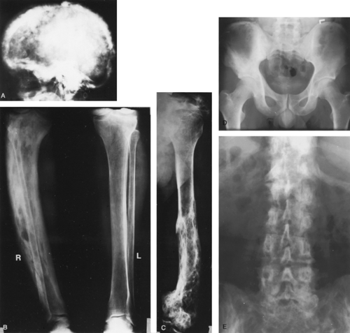 FIGURE 65-1. Radiographic findings in pagetic bone. A, Typical “cotton wool” appearance of the skull. Areas of extensive blastic change are present, and the cortex of the bone is markedly thickened. B, A normal left tibia and fibula contrasted with a pagetic, bowed right tibia. The affected tibia is larger, has areas of increased density as well as localized hyperlucent foci, and is bowed. C, Evidence of the localized nature of Paget disease, showing a normal proximal half of the humerus with pagetic involvement in the entire distal half. A sharp line of demarcation exists between normal and involved bone at the midshaft point. This radiograph also demonstrates the poor architectural quality of the pagetic bone. D, Relatively early changes of Paget disease in the right hemipelvis include thickening of the ilioischial and ilio-pectineal lines as well as sclerosis in the ilium and ischium, in contrast to the normal findings in the left hemipelvis. E, Extensive Paget disease involving the entire lumbosacral spine (and visible pelvis), with marked coarsening of trabecular markings. Secondary degenerative changes with loss of disc spaces are seen as well. (R, right; L, left.) (A through C from Siris ES, Canfield RE, Jacobs TP. Paget’s disease of bone. Bull N Y Acad Med 1980; 56:285.)
Related posts:Stay updated, free articles. Join our Telegram channel
Full access? Get Clinical Tree
 Get Clinical Tree app for offline access

|