Introduction and Definitions
As life expectancy increases beyond the eighth decade worldwide, particularly in developed countries, an increasing proportion of the female population is postmenopausal. With the average age of menopause being 51 years, more than one-third of a woman’s life is now spent after menopause. Here symptoms and signs of estrogen deficiency merge with issues encountered with natural aging. As the world population increases and a larger proportion of this population is made up of individuals over 50, medical care specifically directed at postmenopausal women becomes an important aspect of modern medicine.
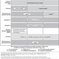
In an attempt to define the stages of reproductive aging and its clinical and biochemical markers, the Stages of Reproductive Aging Workshop was held in 2001 to develop a useful staging system and to revise the nomenclature. This system provides useful clinical definitions of the menopausal transition, perimenopause, menopause and postmenopause as follows:
- Menopausal transition. The menopausal transition begins with variation in menstrual cycle length and an elevated serum follicle-stimulating hormone (FSH) concentration and ends with the final menstrual period (not recognized until after 12 months of amenorrhoea). Stage—2 (early) is characterized by variable cycle length (>7 days different from normal menstrual cycle length, which is 21–35 days). Stage—1 (late) is characterized by two or more skipped cycles and an interval of amenorrhoea of ≥60 days; women at this stage often also have hot flushes.
- Perimenopause. Perimenopause means ‘around the menopause’, and begins in stage—2 of the menopausal transition and ends 12 months after the last menstrual period.
- Menopause. Menopause is defined by 12 months of amenorrhoea after the final menstrual period. It reflects complete, or near-complete, ovarian follicular depletion and absence of ovarian estrogen secretion.
- Postmenopause. Stage +1 (early) is defined as the first 5 years after the final menstrual period. It is characterized by further and complete damping of ovarian function. The majority of women in this stage have symptoms. Stage +2 (late) begins 5 years after the final menstrual period and encompasses the ageing process until death.
Epidemiology
Menopause occurs secondary to a genetically programmed loss of ovarian follicles.
Although the average age at menopause is approximately 51 years, for 5% of women it occurs after age 55 years (late menopause) and for another 5% between ages 40 and 45 years (early menopause). Menopause occurring prior to age 40 years is considered to be premature ovarian failure. Unlike the average age of menarche, which has been affected over time by trends in nutritional status and general health, the average age of menopause has not changed much over time. A number of factors are thought to play a role in determining an individual woman’s age of menopause, including genetics, ethnicity, smoking and reproductive history.
- Genetics. Based on family studies, heritability for age of menopause averaged 0.87, suggesting that genetics explain up to 87% of the variance in menopausal age. Other than gene mutations that cause premature ovarian failure (explained later in this chapter), no specific genes have been discovered to date that account for this genetic influence. However, several genes are likely involved, including genes coding telomerase, which is involved in ageing.
- Ethnicity. Ethnicity and race may also affect the age of menopause. Natural menopause occurs earlier among Hispanic women and later in Japanese-American women, compared with Caucasian women.
- Smoking. The age of menopause is reduced by about 2 years in women who smoke.
- Reproductive history. There is a tendency for women who never had children or who had shorter cycle length during adolescence (a predictor of high basal FSH) to have earlier menopause.
Endocrinology and Neuroendocrinology of Menopause
Menopause is associated with a marked decline in oocyte number that is attributable to progressive atresia of the original complement of oocytes. However, the evidence for absolute oocyte depletion is limited. Residual oocytes and differentiating follicles have been identified in the ovaries of some postmenopausal women, although the follicles are frequently atretic.
Late Reproductive Stage
A decline in fertility begins in the third to fourth decade of life but accelerates rapidly after the age of 35 years in association with a well-documented decrease in the pool of ovarian follicles. This age-related decrease in follicle number and fertility is marked by a rise in FSH in the early follicular phase before increases in luteinizing hormone (LH) or decreases in estradiol. The presence of regular menstrual cycles and an increase in follicular-phase FSH define the late reproductive stage. In older ovulatory women, the early follicular phase peak in FSH occurs earlier with reference to the onset of menses than in their younger counterparts and the day 3 FSH value is widely used as an indicator of reproductive ageing in clinical settings.
Studies of reproductive ageing have provided important insights into the physiology of inhibin in humans and the role of the inhibins in the selective negative feedback regulation of FSH. Inhibin A and B are secreted differentially during the normal menstrual cycle (for a review, see the key references at the end of this chapter). The pattern of inhibin B secretion suggests that it is primarily secreted from small antral follicles; inhibin B increases across the luteal–follicular transition, reaching a peak in the mid-follicular phase, and is not correlated with the size of the dominant follicle. Inhibin B expression in the ovary is confined to the granulosa cells and is absent from the theca. In the quiescent ovary, inhibin B levels in serum increase in response to FSH administration, preceding secretion of estradiol and inhibin A. Secretion of inhibin B from human granulosa cells is not stimulated directly by FSH or cyclic adenosine monophosphate (AMP), however, but rather its secretion is constitutive.
Thus, the increase in inhibin B in response to endogenous or exogenous FSH is secondary to the recruitment of a cohort of follicles into the growing pool accompanied by a dramatic increase in granulosa cell number. During folliculogenesis, peak levels of inhibin A occur in the late follicular phase and inhibin A is correlated with the size of the dominant follicle in normal cycles, as is estradiol. Inhibin A is produced by granulosa cells in response to stimulation by FSH in small follicles and both LH and FSH at later stages of follicle development. Inhibin A is also secreted from the corpus luteum, its secretion paralleling that of progesterone.
The decrease in developing follicles is reflected in a parallel decrease in the serum concentration of inhibin B, which is probably the earliest easily measurable marker of follicular decline. The rise in the serum concentration of FSH in early menopause is also closely related to the fall in inhibin B; this suggests that inhibin B plays an important role in the normal control of FSH secretion. Serum concentrations of Müllerian-inhibiting substance (MIS) [also known as anti-Müllerian hormone (AMH)] may be a useful marker reflecting reproductive ageing. Low serum AMH concentrations were predictive of a poor ovarian response to exogenous gonadotropin stimulation and may mark a critical juncture in the timing of the menopausal transition.
Menopause Transition
The onset of irregular cycles defines the transition from the late reproductive years to the menopausal transition. Variable cycle length is defined as cycle lengths that are more than 7 days different from an individual’s normal cycle length and may therefore include cycles with both abnormally long and abnormally short intermenstrual intervals.
The menopausal transition is characterized by a dynamic period of markedly changing hypothalamic–pituitary feedback from the ageing ovary. There is a progressive decrease in menstrual cycle regularity and dramatic swings in estradiol from undetectable to levels that are several times higher than those observed in the ovulatory cycles of younger women. Levels of inhibin A and B are further decreased and FSH levels are generally much higher than during the regular cycles of the late reproductive years. FSH levels may occasionally decrease to near the normal range in association with prolonged increases in estradiol, however. The majority of longitudinal studies indicate that the menopausal transition is not a low-estrogen state but is characterized by widely fluctuating levels of estrogen that are often increased in comparison with earlier stages of reproductive life. These fluctuations in estrogen levels in particular are likely to account for many of the symptoms of the transition to menopause.
In addition to the decline in follicular number, there may be a decrease in hypothalamic–pituitary sensitivity to estrogen-positive feedback during perimenopause.
Postmenopause
Later, with the final menstrual period, levels of inhibin A and estradiol decrease dramatically and there is a further increase in FSH and LH. In particular, the loss of estradiol feedback on LH and FSH and inhibin feedback on FSH associated with menopause results in a 15-fold increase in FSH levels and a 10-fold increase in LH levels in comparison with the early follicular phase in healthy women in whom estradiol levels are at their nadir in the menstrual cycle. The loss of ovarian function at menopause is also associated with marked changes in hypothalamic and pituitary function. There is now evidence, however, that age-related neuroendocrine changes occur that are independent of those caused by the loss of ovarian feedback on the hypothalamic and pituitary components of the reproductive axis. There is an increase in the overall amount of gonadotropin-releasing hormone (GnRH) secreted despite a decrease in GnRH pulse frequency with ageing.
In addition, following menopause, gonadotropin levels decrease progressively with age. Studies in postmenopausal women indicate that between the ages of 45–55 and 70–80 years, there is a decrease of approximately 30% in mean levels of FSH from 148.6 ± 8.4 IU l−1 in younger postmenopausal women to 107.0 ± 5.4 IU l−1 in older women. There is a similar degree of change in LH from 95.8 ± 7.3 to 60.4 ± 3.9 IU l−1 in younger and older postmenopausal women, respectively. Whether the decline in gonadotropin secretion with age is caused by hypothalamic or pituitary effects has been an area of active recent investigation.
Ovarian Ageing and Sex Steroids Changes
Hormonal integration of the reproductive system is dramatically affected by reproductive ageing. At the menopause, the final menstrual cycle, a dramatic decline in plasma estradiol level occurs and the postmenopausal ovary will cease to contribute to estradiol levels in blood. Instead, peripheral conversion of androstenedione into estrone becomes prominent. Only 5% of the thus formed estrone is converted to estradiol through the action of 17-hydroxysteroid dehydrogenase. The activity of this enzyme is in a reversible reaction converting estrone to estradiol and back, depending on the oxido-reductive state that prevails in the cell. Further, the amount of estrone generated and the associated conversion to estradiol continue to decline during the first year after menopause and stabilizes thereafter. The amount of estrone generated is a function of the abundance of androstenedione and age. The corpus luteum synthesizes progesterone and in the absence of ovulation only basal levels derived from the adrenal glands are detected. In postmenopausal women, administration of adrenocorticotrophic hormone (ACTH) dramatically increases whereas human chorionic gonadotrophin has no effect on progesterone levels, attesting to the negligible role of postmenopausal ovaries in progesterone production.
Dehydroepiandrosterone (DHEA) is produced in both the ovaries and the adrenal glands under the influence of LH and ACTH, respectively. DHEA sulfate (DHEAS) is exclusively produced by the adrenal glands and is converted to DHEA by steroid sulfatase. Their declining plasma levels are due to age-related reduced steroid synthesizing capacity of the zona reticularis and due to ovarian ageing. During the reproductive years, both the adrenal glands and the ovaries share equally in androstenedione production. Bilateral oophorectomy in premenopausal women results in a 50% reduction in serum androstenedione levels while postmenopausal ovaries contribute only 20% of its total circulating levels. Since the metabolic clearance of androstenedione is not affected by ovarian function or age, the 30% drop represents the effect of ovarian senescence.
In premenopausal women, 50% of circulating testosterone is derived from peripheral conversion of androstenedione, while the remaining testosterone production is shared between the ovaries and the adrenal glands. In postmenopausal women, testosterone levels decrease compared with young women, although ovarian synthesis after the menopause appears to contribute a higher proportion of circulating testosterone. This may be due to higher LH levels and their effect on ovarian stromal steroidogenesis. In a recent cross-sectional study, a different aspect of ovarian ageing was reported. The circulating levels of DHEA, androstenedione and total and free testosterone were found to be highest during the third decade of life and to decline afterwards in the remaining reproductive years. Around the age of 50 years, free and total testosterone levels decrease by about 50%. Testosterone exists in circulation as free testosterone (1–2% of the total), loosely bound to albumin (31%) and tightly bound to sex hormone binding globulin (SHBG) (66%). It is the free and albumin-bound testosterone that is available to cells. Many clinicians and clinical investigators use the ratio of total testosterone to SHBG to derive the free testosterone index.
SHBG is a protein synthesized in the liver. Estrogen stimulates its synthesis whereas all androgens suppress its hepatic synthesis. Obesity, particularly with upper abdominal distribution, also suppresses SHBG levels. In the postmenopausal period, SHBG levels decline and that may account for the higher bioavailability of testosterone. A decrease in bioavailable testosterone level may result from impaired testosterone production or from increased SHBG levels in the presence of normal testosterone production. It is therefore necessary to consider SHBG levels in the assessment of bioavailable testosterone in women.
Surgical Menopause
As can be expected, the removal of both ovaries in a premenopausal woman results in an abrupt decline in estrogen to undetectable levels, a 50% reduction in androstenedione and about a 70% drop in DHEA and testosterone levels. These women experience a sudden onset of the menopausal transition. In at least 30–50% of cases, symptoms of androgen deficiency are experienced despite ‘adequate’ estrogen replacement.
Specific Healthcare Problems in Relation to The Menopause
As many as 80% of women experience one or more physical or psychological symptoms of estrogen deficiency as ovarian function declines during the menopause, with almost one half of sufferers finding their symptoms distressing.
Short-Term Consequences of Estrogen Deficiency
The change in hormone levels that occurs during the climacterium, particularly the decline of estrogen, can cause acute menopausal symptoms.
Brain Symptoms
Symptoms include the following:
- vasomotor symptoms (hot flushes and night sweats)
- sleep problems
- mood changes (depression and anxiety)
- sexual dysfunction
- impaired concentration and memory.
Sex steroids play pivotal neuroactive and brain region-specific roles on the central nervous system through genomic and non-genomic mechanisms. Therefore, their protective effects are multifaceted and brain region dependent. They encompass systems that range from chemical to biochemical and genomic mechanisms, protecting against a wide range of neurotoxic insults. Consequently, gonadal steroid withdrawal, during the reproductive senescence, dramatically impacts brain function, negatively affecting mood, anxiety behaviour and cognitive vitality.
The hallmark feature of declining estrogen status in the brain is the hot flush, which is more generically referred to as a vasomotor episode. Hot flushes occurs in up to 75% of women in some cultures. Hot flushes are most common in the late menopausal transition and early postmenopausal periods. They are self-limited, usually resolving without treatment within 1–5 years, although some women will continue to have hot flushes until after age 70 years. Hot flushes typically begin with a sudden sensation of heat centred on the upper chest and face that rapidly becomes generalized. The sensation of heat lasts from 2 to 4 min, is often associated with profuse perspiration and occasionally palpitations and is often followed by chills and shivering and sometimes a feeling of anxiety. Physiological studies have determined that hot flushes represent thermoregulatory dysfunction; there is inappropriate peripheral vasodilatation with increased digital and cutaneous blood flow and perspiration, resulting in rapid heat loss and a decrease in core body temperature below normal. Hot flushes usually occur several times per day, although the range may be from only one or two each day to as many as one per hour during the day and night. Hot flushes are particularly common at night. The fall in estrogen levels precipitates the vasomotor symptoms. Although the proximate cause of the flush remains elusive, the episodes result from a hypothalamic response (probably mediated by catecholamines) to the change in estrogen status. A speculative mechanism for the initiation of hot flushes is endogenous opioid peptide withdrawal. Estrogen increases central opioid peptide activity and estrogen deficiency may be associated with decreased or absent endogenous central opioid activity.
A distressing feature of hot flushes is that they are often associated with arousal from sleep. This association has been well documented by EEG studies, although, primary sleep disorders are common in this population, even in the absence of hot flushes. This disturbed sleep often leads to fatigue and irritability during the day and deficit of memory is often reported.
Psychological symptoms have been associated with the menopause, including depressed mood, anxiety, irritability, lethargy and lack of energy. Women with a longer menopausal transition and/or with more intense climacteric symptoms, surgical menopause, a history of depression, menstrual cycle-related and postpartum mood changes (premenstrual syndrome, postpartum depression), thyroid disease and unfavourable socio-environmental conditions are at greater risk of developing depression and mood disorder. Observational studies have demonstrated that, in women with a history of depression, the menopausal transition is accompanied by a significant risk of relapse.
Stay updated, free articles. Join our Telegram channel

Full access? Get Clinical Tree


