There should be histological confirmation of the disease and division of cases by histological type – for example, basal cell, squamous cell, sebaceous carcinoma. Melanoma of the eyelid is classified with malignant melanoma of skin. The regional lymph nodes are the preauricular, submandibular and cervical lymph nodes. The pT and pN categories correspond to the T and N categories. Note
CARCINOMA OF THE SKIN OF THE EYELID (ICD‐O‐3 C44.1)
Rules of Classification
Regional Lymph Nodes
TNM Clinical Classification
T – Primary Tumour
T0
No evidence of primary tumour
Tis
Carcinoma in situ
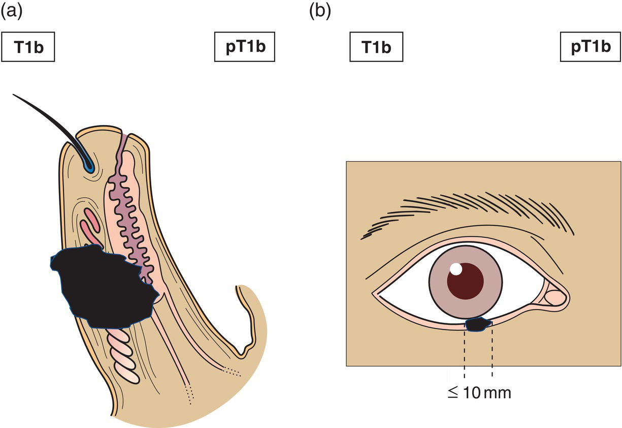
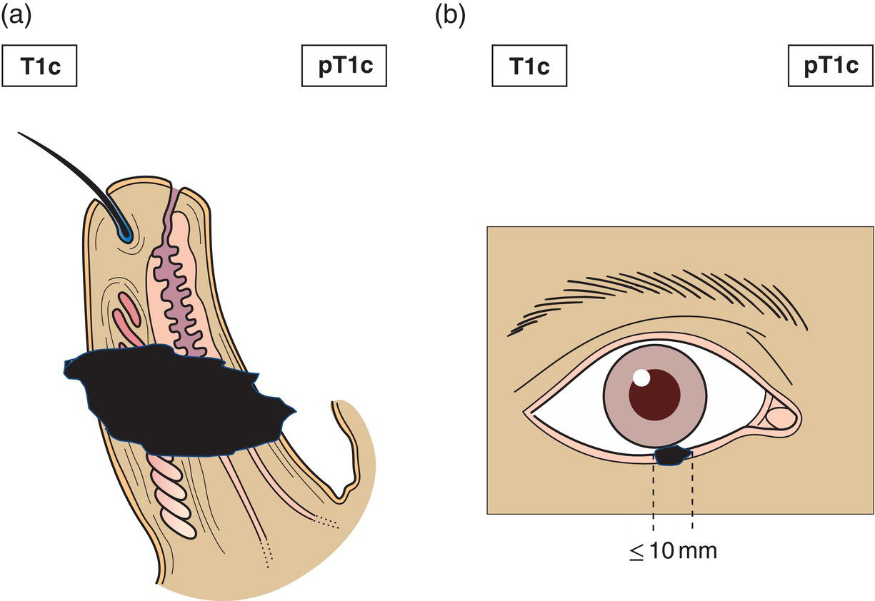
T1
Tumour 10 mm or less in greatest dimension
T1a
Not invading the tarsal plate or eyelid margin (Fig. 346)
T1b
Invades tarsal plate or eyelid margin (Fig. 347)
T1c
Involves full thickness of eyelid (Fig. 348)
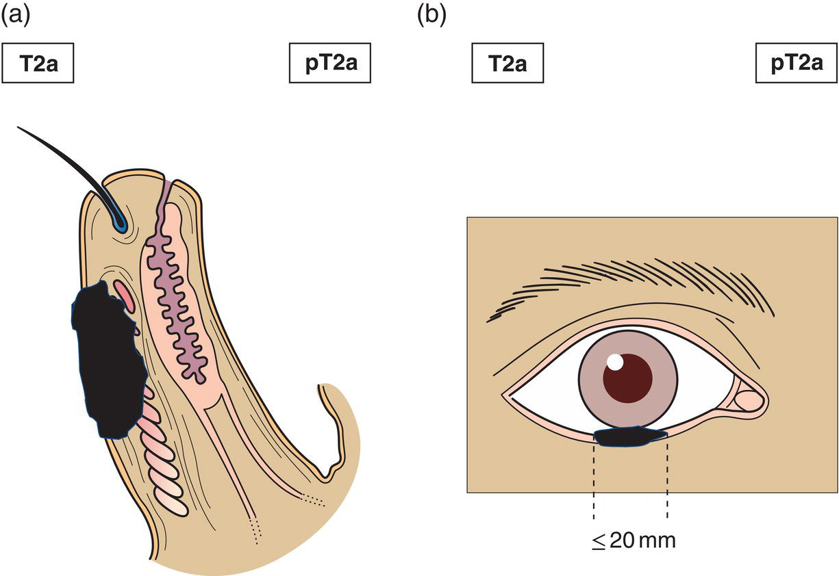
T2
Tumour > 10 mm, but 20 mm or less in greatest dimension
T2a
Not invading the tarsal plate or eyelid margin (Fig. 349)
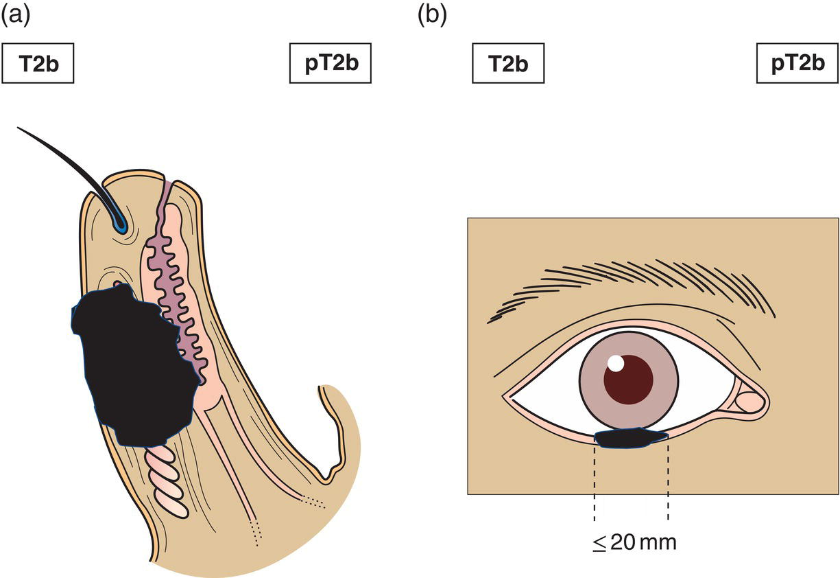
T2b
Invades the tarsal plate or eyelid margin (Fig. 350)
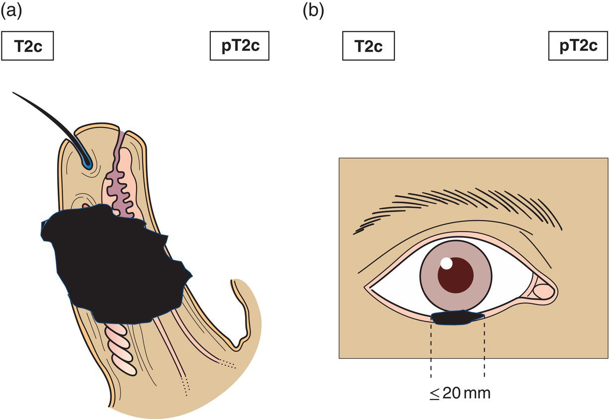
T2c
Involves full thickness of eyelid (Fig. 351)
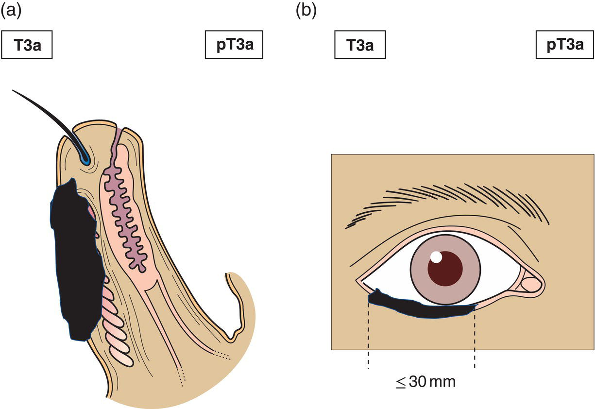
T3
Tumour > 20 mm, but more than 30 mm in greatest dimension
T3a
Not invading the tarsal plate or eyelid margin (Fig. 352)
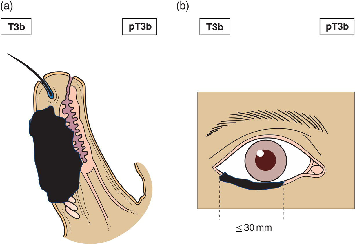
T3b
Invades tarsal plate or eyelid margin (Fig. 353)
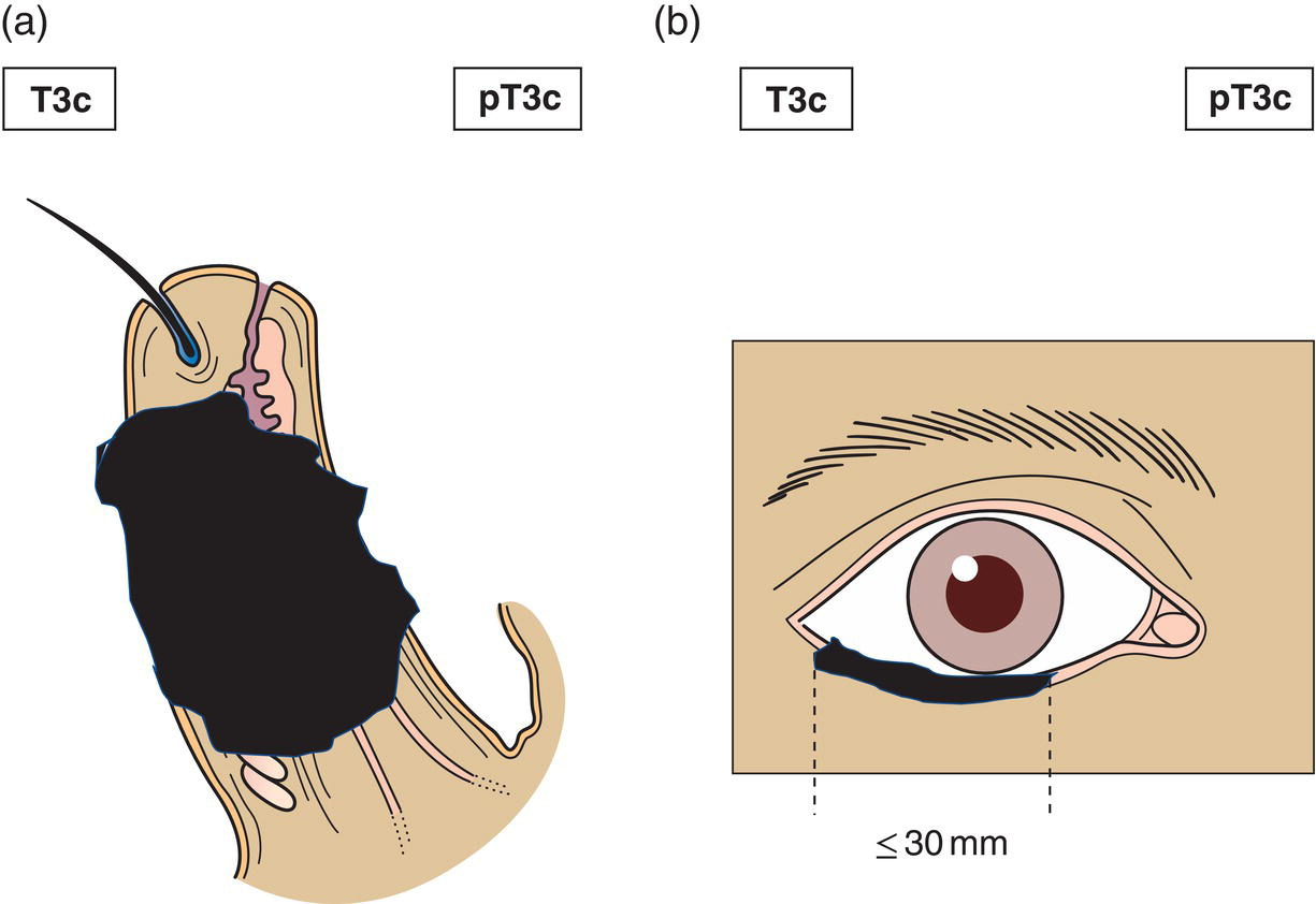
T3c
Involves full thickness of eyelid (Fig. 354)
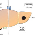
T4
Any eyelid tumour that invades adjacent ocular, orbital or facial structures
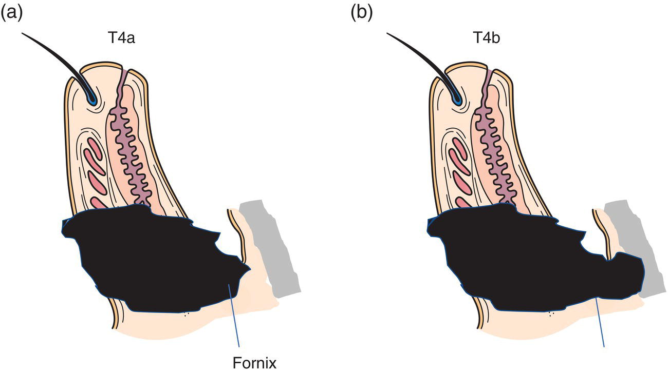
T4a
Tumour invades ocular or intraorbital structures (Fig. 355a)
T4b
Tumour invades (or erodes through) the bony walls of the orbit or extends to paranasal sinuses or invades the lacrimal sac/nasolacrimal duct or brain (Fig. 355b) 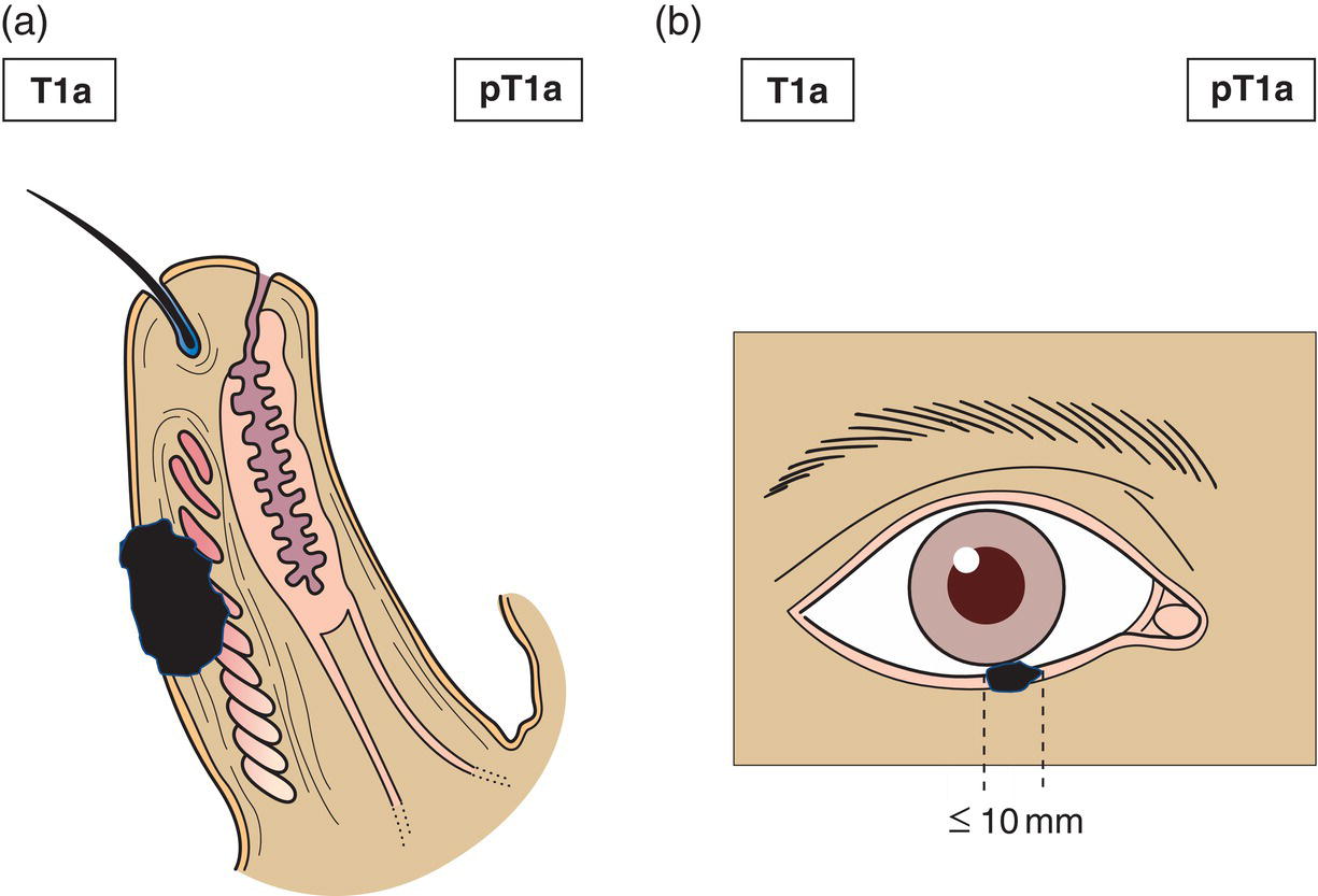
N – Regional Lymph Nodes
NX
Regional lymph nodes cannot be assessed
N0
No evidence of lymph node involvement
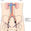
N1
Metastasis in a single ipsilateral regional lymph node, 3 cm or less in greatest dimension (Fig. 356a)
N2
Metastasis in a single ipsilateral lymph node more than 3 cm in greatest dimension (Fig. 356b), or in bilateral or contralateral lymph nodes 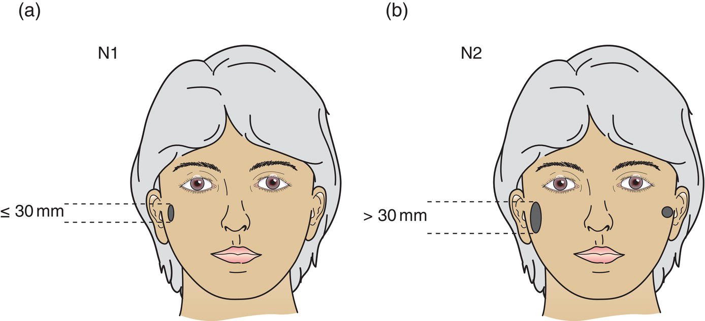
M – Distant Metastasis
M0
No distant metastasis
M1
Distant metastasis
pTNM Pathological Classification
pM1
Distant metastasis microscopically confirmed
pM0 and pMX are not valid categories.
Summary
Related posts:


Stay updated, free articles. Join our Telegram channel

Full access? Get Clinical Tree


