Introduction
Thyroid eye disease (TED), also known as thyroid orbitopathy and Graves’ ophthalmopathy, is an autoimmune-driven process that results in pathologic changes of the extraocular muscles, orbital fat, and surrounding tissues. Most patients with TED have a self-limited course, not requiring medical or surgical intervention. Mild ophthalmic complaints include ocular surface irritation, excessive tearing, or pressure sensation. More moderate involvement includes diplopia, eyelid retraction, and proptosis. Vision-threatening disease includes compressive optic neuropathy, corneal ulceration and perforation, globe subluxation, and choroidal folds. The diagnosis and management of TED can be particularly challenging. The goal of this chapter is to help providers recognize emergency ophthalmic conditions in TED that warrant referral to ophthalmologists or oculoplastic specialists and their current treatment strategies.
Demographics
Thyroid eye disease more commonly affects women, with a bimodal distribution that peaks in incidence between 50 and 70 years of age. Annually, 16 women and 3 men per 100,000 people are newly diagnosed. 1 Among patients with TED, the majority have Graves’ disease (90%), while a small subset are primarily hypothyroid (1%), have Hashimoto’s thyroiditis (3%), or are euthyroid (6%). Overall, among patients with Graves’ disease, approximately one in four will develop TED. Risk factors for developing TED include smoking, older age, extreme physical or psychological stress, prior treatment with radioactive iodine, and increased titers of antithyroid stimulating hormone receptor antibodies. ,
The exact pathophysiology of TED is incompletely understood. However, it is believed to be an autoimmune inflammatory condition. A combination of genetic, environmental, and epigenetic factors that result in the production of autoreactive T-cells, B-cells, and antibodies stimulates the proliferation of orbital fibroblasts and adipocytes and upregulates a cascade of inflammatory mediators. The cytokine-mediated activation results in tissue remodeling and production of fibrosis and adipogenesis.
The natural course of the disease consists of an initial inflammatory phase followed by a durable quiescent phase. The inflammatory (active) phase typically lasts between 6 to 18 months in a nonsmoker and 24 to 36 months in an active smoker. The quiescent/cicatricial phase follows after the inflammation subsides and features varying degrees of fibrosis and fat expansion in the orbit and eyelid. Differentiating between the active inflammatory phase and the quiescent phase is critical in guiding treatment of TED. The timing of surgical management of TED depends on the patient’s overall clinical stability.
Clinical Evaluation
A systematic approach is critical when evaluating a patient with TED, as the clinical presentation may be highly variable. A comprehensive medical history of thyroid condition should be documented including timing and clinical presentation of diagnosis, thyroid status at the time of diagnosis, and prior treatment history (antithyroidal medication, surgical thyroidectomy, radioactive iodine). One must establish the stability of the thyroid status and recent changes in thyroid medications. Family history of thyroid and autoimmune conditions should be noted. If the patient is an active smoker, smoking cessation counseling should be actively pursued.
A careful history and duration of the eye symptoms should be documented to help determine where in the timeline of thyroid eye disease activity the patient resides. The International Thyroid Eye Disease Society’s VISA (Vision, Inflammation, Strabismus, Appearance) classification can be a useful framework to help monitor activity and severity. It includes both subjective and objective measures of disease. Subjective assessment of vision loss includes blurriness and decreased color perception. Inflammatory symptoms include retrobulbar ache at rest or with gaze, and lid swelling. Symptoms of diplopia may occur at rest, intermittently, or constantly and may produce a compensatory head tilt. Appearance changes include the presence of lid stare, tearing, irritation, or light sensitivity.
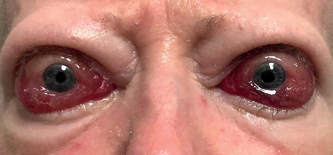
Objective measures of vision and optic nerve function are assessed with tests of visual acuity, color perception, pupillary response, and visual field. Additional pertinent clinical findings include chemosis, conjunctival injection, periocular redness or edema, extraocular motility restriction, upper or lower eyelid retraction, lagophthalmos, proptosis, corneal abnormalities, and elevation of intraocular pressure ( Fig. 3.1 ). An exophthalmometer is used to quantify the distance from the orbital rim to the corneal surface of each eye as a measure of proptosis. Photographs taken prior to the development of TED help determine the magnitude of change in the eyes from baseline.

Laboratory investigations should include T3, free T4, thyroid-stimulating hormone (TSH) levels, thyroid peroxidase, TSH receptor antibody, and thyroid stimulating immunoglobulins. Neuroimaging with computed tomography or magnetic resonance imaging aid in determining the degree of orbital fat and extraocular muscle expansion. , Fusiform enlargement of the extraocular muscles, which spares the tendons helps differentiate TED from other etiologic causes of muscle enlargement, including idiopathic orbital inflammation and lymphoma.
External Pathology
PERIORBITAL EDEMA
Periorbital edema involving the upper and lower eyelids is an early sign of orbital congestion. Over time, the eyelids can become thickened in chronic cases. Severe periorbital congestion may be seen in cases featuring compressive optic neuropathy. However, it should be noted that among octogenarians, compressive optic neuropathy often occurs in the absence of periorbital edema or erythema. High-salt-content foods should be avoided to mitigate the swelling.
UPPER AND LOWER EYELID RETRACTION
The most common ocular manifestation of TED is eyelid retraction, which is seen in 90% of patients. Eyelid retraction may manifest prior to symptoms of hyperthyroidism or serologic evidence of hyperthyroidism by as many as 6 to 12 months. The internist and general ophthalmologist should remain vigilant in a patient who presents with asymptomatic unilateral eyelid retraction and maintain a low threshold for serial serologic thyroid evaluations. Once the patient is within the quiescent phase of TED, upper eyelid retraction can be corrected surgically by procedures including graded blepharotomy. , Lower eyelid retraction similarly may be corrected by placement of an autologous or xenograft.
PROPTOSIS
Thyroid eye disease is the most common cause of unilateral and bilateral proptosis. In adults, exophthalmos is seen in almost 60% of cases. Proptosis or exophthalmos may produce eyelid malposition, corneal exposure, and ocular dysmotility. The degree of proptosis is quantified using an exophthalmometer and helps differentiate real from pseudoproptosis produced by eyelid retraction. The range of normal exophthalmos is established in the literature; however, normative averages provide little guidance for the individual patient where baseline measurements are unknown. A review of old photos is therefore helpful to understand the magnitude of change consequent to the TED.
After establishing that the TED has been stable for a period of 6 months, decompression may be considered. Depending upon the phenotype of the proptosis (fat or muscle predominant expansion) and the degree of proptosis reduction desired, bony decompression and/or fat decompression can be employed to reverse proptosis. Typically, decompression should be avoided in the active phase of TED, unless medically urgent relief of proptosis is required.
SPONTANEOUS SUBLUXATION OF THE GLOBE
Globe subluxation occurs when the eyelids slip behind the equator of the globe resulting in acute stretch of the optic nerve and exquisite pain. Approximately 0.1% of patients with TED develop globe subluxation. Subluxation is more typically seen when proptosis is severe, and results from expansion of the orbital fat and when there is upper lid retraction or preexisting laxity of the eyelids.
Spontaneous subluxation is a vision-threatening condition that can produce stretch optic neuropathy. Although reversable if the globe can be repositioned, if there is prolonged stretch or repeated insults, optic nerve damage may be irreversible. The severe pain results from both the stretching of the orbital tissues and the frequent association with corneal abrasion. The pain and shock of the unexpected event make home management of subluxation challenging. The globe can be repositioned in the emergency room by first applying topical anesthetic to relieve corneal pain. The patient is asked to maintain a downward gaze. Then, using one hand to pull on the upper eyelid skin and the other hand on the globe to place posterior pressure, the upper lid is returned to its normal position. If this fails, the use of Desmarres retractors or a paper clip bent as a shoehorn can help reposition upper lid over the globe. Intravenous corticosteroids may be used to limit acute inflammatory swelling and potentially provide neuro-protection. A temporary lateral tarsorrhaphy may be used to reduce the acute risk of recurrent subluxation. Definitive treatment options include orbital decompression to reduce proptosis, lid retraction repair, and permanent tarsorrhaphy.
DIPLOPIA
Diplopia is a debilitating symptom of TED and may manifest in the horizontal, vertical, or oblique planes. Up to 42% of adult TED patients will demonstrate progressive extraocular motility changes. Extraocular muscle involvement can be assessed by measuring the amount of ocular rotation of the eye in degrees from 0 to 45 in each field of gaze. Ocular misalignment resulting in symptomatic diplopia is measured with neutralizing prisms and often with the assistance of a certified orthoptist.
Diplopia is the most disabling feature of TED. Treatment varies based on the phase of the orbitopathy. If only present in eccentric gaze, aggressive immunosuppression may be helpful in preventing progression of diplopia into primary gaze. If the diplopia is in primary or reading gaze, temporary relief can be obtained by occluding vision from the nondominant eye with frosted adhesive tape on the lens of eyeglasses. Alternatively, an orthoptist can help provide temporary (Fresnel) prisms applied to either or both distance or reading glasses to eliminate diplopia and maintain binocular depth perception. Orthoptics measurements should be repeated quarterly with adjustments in the prisms as required. When orthoptics measurements remain unchanged for 6 months, TED is considered to have entered the stable phase and rehabilitative surgery may be considered. Strabismus surgery is an effective measure to restore single binocular vision in primary and reading gaze. ,
Anterior Segment Pathology
CORNEAL KERATOPATHY
Assessment of the cornea in patients with TED is critical, as vision loss from corneal keratopathy may be confused with that caused by compressive optic neuropathy. Severe corneal keratopathy can result in permanent vision loss due to scarring or perforation. A slit lamp examination should evaluate for abnormal tear film, superior limbal keratitis, mild punctate epithelial erosions, and signs of chronic exposure with corneal scarring. The pathogenesis of corneal keratopathy is multifactorial including unstable tear film, eyelid retraction, proptosis, reduced motility, and poor Bell’s reflex.
Many patients in the active phase of TED complain of ocular irritation, light sensitivity, and tearing. For mild disease, treatment includes topical lubrication with preservative-free artificial tears and lubricating gel at night. Moisture chambers and Saran Wrap occlusion at bedtime should also be considered. If there are signs of corneal keratopathy with progressive corneal thinning, the cornea may be further protected by closure of the eyelids by placement of a temporary tarsorrhaphy or injection of botulinum toxin to induce a ptosis of the upper eyelids. A longer-term solution can be achieved with a permanent tarsorrhaphy. Amniotic membrane grafts to the cornea have been reported, especially if a corneal dellen (focal thinning of the cornea) develops. Emergency corneal gluing or corneal transplantation may be required if perforation occurs.
Corneal ulcers or infectious keratitis develops when the epithelial surface disruption becomes colonized with bacteria. This is a rare complication, as only 1.3% of TED patients develop microbial keratitis. The spectrum of the corneal involvement includes corneal infiltrates, corneal melt, severe corneal thinning, and, in rare cases, corneal perforation. A mixture of gram-negative and gram-positive flora has been isolated from corneal cultures.
The majority of the corneal keratopathy is related to upper and/or lower eyelid retraction and lagophthalmos. Once in the stable phase, eyelid retraction repair reduces the risk of corneal disease.
CONJUNCTIVAL INJECTION AND CHEMOSIS
Conjunctival injection or hyperemia and chemosis are common clinical features of active TED disease. Symptomatic medical management with topical lubrication is recommended. Over-the-counter topical redness-relief medications have active decongestants containing either selective or mixed alpha-1 and alpha-2 adrenergic receptor agonists. These agents include tetrahydrozoline, naphazoline, and brimonidine, which should be avoided due to their association with rebound redness and irritation. Conjunctival injection should be managed with lubricating artificial tear drops.
Posterior Segment Pathology
RETINAL PATHOLOGY—CHOROIDAL FOLDS
Chorioretinal folds are rarely seen in TED. The etiology of chorioretinal folds in TED is variably speculated to be secondary to vascular engorgement from apical crowding, traction on the optic nerve, and posterior pressure on the globe due to enlarged rectus muscles. The visual acuity in these patients can vary; however, many patients will report metamorphopsias. A dilated fundoscopic examination and use of optical coherence tomography can aid in diagnosis. Despite medical therapy or surgical decompression, chorioretinal folds may variably persist indefinitely. ,
COMPRESSIVE OPTIC NEUROPATHY
Compressive optic neuropathy occurs in 4% to 8% of TED patients. Male and older patients are more likely to develop compressive optic neuropathy. The etiology is multifactorial and thought to be secondary to the mass effect of the pathologically enlarged and inflamed extraocular muscles in the orbital apex, producing variable degrees of optic nerve compression, stretch, and inflammation. Optic neuropathy may be challenging to diagnose and relies on changes in visual acuity, pupillary reactions, color vision, and visual fields. Younger patients with compressive optic neuropathy tend to have better visual acuity and color vision, fewer visual field deficits, and greater exophthalmos as compared with older patients. A mathematical formula to aid in the diagnosis of compressive optic neuropathy has been found to have good specificity and sensitivity, when employing multiple clinical parameters.
Oral corticosteroids have been a primary treatment for moderate to severe active TED for the past 50 years. Intravenous corticosteroids may be more effective and have a lower rate of therapeutic morbidity. The most widely utilized treatment protocol, developed by EUGOGO (European Union Graves Ophthalmopathy Group) infuses 500 mg IV methylprednisolone weekly for 6 weeks, followed by 250 mg weekly for 6 weeks to suppress active TED. Although the therapeutic response is favorable, the relapse rate is between 20% and 40% after completion of the infusion protocol. The use of high-dose corticosteroids has been shown to be effective in reversing vision loss from compressive optic neuropathy in active-phase TED, but it does not shorten the overall duration of the active phase. Conversely, compressive optic neuropathy identified in the stable phase is not responsive to medical therapy. In these cases, and those in which active-phase compressive optic neuropathy is unresponsive to corticosteroid therapy, surgical decompression is required to restore optic nerve function.
The first report of surgical decompression performed for severe TED with compressive optic neuropathy in 1911 described removal of the lateral orbital wall to relieve the orbital congestion. Subsequently, surgical innovation has expanded and refined both the decompressive techniques and surgical planning. Surgical decompression in the acute phase of TED should be reserved for patients who do not respond to medical therapy or have contraindications to medical treatment. In the majority of patients surgical decompression should be postponed until the stable phase is achieved and the outcome of surgery can be more reliably predicted.
Complications associated with bony decompressions should not be taken lightly as they include ptosis, worsening strabismus, trigeminal nerve paresthesia, orbital cellulitis, cerebral spinal fluid leak, hypoglobus, change in voice quality, and permanent vision loss. Surgical decompressions during the active phase have increased risk of orbital hemorrhage, enophthalmos, hypoglobus, and diplopia. ,
Orbital radiotherapy was first used to treat TED in 1936. The standard radiotherapy protocol is 2000 cGy delivered in 10 treatments to the orbital tissue over a 2-week course. It is most commonly used in the acute phase to supplement corticosteroid treatment of compressive optic neuropathy, rapidly progressive orbitopathy, and steroid-dependent orbitopathy. In a study of patients with compressive optic neuropathy responsive to corticosteroids who received adjuvant orbital radiation therapy, only 6% of patients required an urgent surgical decompression as a result of persistent optic neuropathy.
Conclusion
Management of TED poses many challenges. The clinical presentation can range from mild to severe ocular. Recognizing the signs and symptoms of TED and making the appropriate referral for treatment is vital to the preservation of both form and function. A multidisciplinary approach including endocrinology and ophthalmology is required to optimize management.
References
Stay updated, free articles. Join our Telegram channel

Full access? Get Clinical Tree


