Nuclear Medicine

Chapter 10 Nuclear Medicine


Positron Emission Tomography


Despite the exquisite resolution of computed tomography (CT) and magnetic resonance imaging (MRI), some disease processes may go undetected by these anatomically based modalities. The anatomic modalities also have shortcomings in assessing the response to treatment and in distinguishing responders from nonresponders.


Over the past half-century, a variety of nuclear medicine probes have been used to evaluate disease processes at the cellular level. Nuclear medicine is the only clinical discipline using intracellular contrast agents in imaging, and it is therefore more sensitive than anatomic modalities in detecting certain disease processes. Some of these probes, such as radioiodine, have been very successful and are heavily used. Nevertheless, most nuclear medicine imaging techniques have suffered from low specificity and low spatial resolution; the latter problem is associated with the physics of single-photon-emitting radiotracers. Synthesis of biologically important radiopharmaceuticals with single-photon-emitting radiotracers has been a major challenge.


Positron emission tomography (PET) is a nuclear medicine modality that uses positron emitters such as fluorine-18, oxygen-15, nitrogen-13, and carbon-11. The fact that these nuclides are components of common biologic molecules makes PET particularly suitable for visually capturing various biologic pathways. Although PET has been used for several decades in the research setting, its clinical use has grown substantially in the past decade. PET with 18F-fluorodeoxyglucose (FDG), or “radioactive sugar,” as its workhorse has substantially influenced clinical oncology practice by refining the processes of diagnosis, staging, and restaging. Although many of the strengths and limitations of FDG-PET in diagnosing cancer are well known, its role in staging and restaging is evolving. The data indicate that FDG-PET will likely play an important role in monitoring therapy and predicting the course of the disease.


Basic Physics of Positron Emission Tomography


The radioisotope portion of the molecule used in PET imaging emits a positron (i.e., positively charged electron), which travels a distance of a few millimeters in tissue before it collides with a negatively charged electron. This collision annihilates the entire mass of the positron and electron, generating two photons with energy of 511 KeV each. These two photons travel at the speed of light in exactly opposite directions (i.e., 180 degrees apart). Coincident detection of these two photons by two oppositely positioned detectors in the PET scanner results in images with a much higher resolution compared with the conventional, single-photon nuclear medicine studies and produces the possibility of quantitative measurement of the tracer uptake in a volume of interest.


PET/CT allows the fusion of the metabolic information from PET with the anatomic information from CT and thus increases the diagnostic accuracy compared with stand-alone PET. In PET/CT, the patient undergoes a CT scan, followed by a PET scan, without changing the patient’s position. PET scans for most oncologic indications (whole-body PET scans) are acquired from the base of the skull through the upper thighs. In some instances, as in patients with melanoma, PET scans are acquired from the vertex of the skull through the toes. The CT portion (i.e., transmission scan) of PET/CT is acquired within seconds to minutes, whereas the PET acquisition time for each bed position (about 15 cm) is several minutes; the total PET acquisition time in newer machines is 15 to 25 minutes.


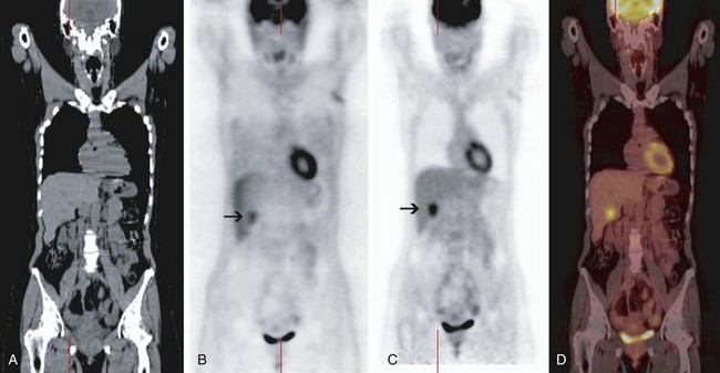
In addition to delivering anatomic information, the CT portion of PET/CT is used to measure the attenuation of the x-ray photons traveling through the patient to produce the so-called attenuation map and correct the PET data for tissue attenuation. During PET acquisition, photons from structures deep in the abdomen or pelvis are more strongly attenuated than those from superficial structures and the chest. The intensity of uptake in deeper structures is underestimated on non–attenuation-corrected PET images; the intensity of uptake in the deeper structures is normalized to the intensity of uptake in the superficial structures on the attenuation-corrected PET images (Fig. 10-1). Although correction of the PET data for tissue attenuation is indispensable, misalignment between PET and CT can cause mislocalization of lesions on the fused PET/CT images. This may be caused by the changed position of a body part (e.g., neck, legs) or physiologic changes in the position of an organ (e.g., respiratory movement) between the transmission scan (CT) and the emission scan (non–attenuation-corrected PET). Because the degree of misalignment and resulting mislocalization can be significant, the physician must be cautious when interpreting the attenuation-corrected PET/CT images or using PET/CT images for radiation therapy planning. The magnitude of this misalignment can be assessed by fusing the non–attenuation-corrected PET images with the CT images; this can be performed on any PET review station. In case of significant misalignment, the non–attenuation-corrected PET images should be reviewed without fusion with the CT images, and the metabolic findings of PET should be correlated side by side with the anatomic findings of CT (Fig. 10-2).


At its introduction, the CT portion of the PET/CT was acquired without contrast and used solely for attenuation correction and anatomic localization of the PET findings. However, with the development of multislice CT machines and their introduction to PET/CT scanners, an increasing number of institutions are performing the CT portion of the PET/CT studies with diagnostic-quality CT, using intravenous and oral contrast media as needed. However, because the density of intravenous and oral contrast media used in CT can produce artifacts, with overestimation of FDG uptake in areas of high-contrast density, the CT scanning protocol has to be modified with the general approach of dilution of the contrast medium.


General Aspects of Tumor Visualization on 18F-Fluorodeoxyglucose Positron Emission Tomography


FDG (2-[18F] fluoro-2-deoxy-D-glucose) is the most commonly used radiotracer in clinical PET imaging. Tumor imaging with FDG is based on the principle of increased glucose metabolism of cancer cells. Like glucose, FDG is taken up by the cancer cells through facilitative glucose transporters (GLUTs). GLUTs are glycoproteins; 12 isoforms have been identified. Once in the cell, glucose or FDG is phosphorylated by hexokinase to glucose-6-phospate or FDG-6-phosphate, respectively. Expression of GLUTs and hexokinase, as well as the affinity of hexokinase for phosphorylation of glucose or FDG, is generally higher in cancer cells than in normal cells. Glucose-6-phosphate travels farther down the glycolytic or oxidative pathways to be metabolized, in contrast to FDG-6-phosphate, which cannot be metabolized. In normal cells, glucose-6-phosphate or FDG-6-phosphate can be dephosphorylated and exit the cells. In cancer cells, however, expression of glucose-6-phosphatase is usually significantly decreased, and glucose-6-phosphate or FDG-6-phosphate therefore can become only minimally dephosphorylated and remains in large part within the cell. Because FDG-6-phosphate cannot be metabolized, it is trapped in the cancer cell as a polar metabolite, and it constitutes the basis for tumor visualization on PET.


The intensity of a malignant tumor on PET correlates with the number of malignant cells in the tumor mass. Although most malignant tumors are markedly intense on PET, tumors such as bronchoalveolar lung cancer may be only moderately or mildly intense or may not be visualized on PET at all. Likewise, the intensity of a tumor decreases as soon as only one cycle of effective chemotherapy has been completed, possibly because of a decreased number of viable cells or the decreasing metabolic activity of those cells. Although this constitutes the basis for monitoring therapy by PET, the effect can also cause false-negative PET findings.


The intensity of malignant lesions on PET also depends on their location in the body. For example, the FDG avidity of a lung or liver lesion may be underestimated, or the lesion may even go undetected on PET; because of the repetitive craniocaudal movement of the lung and liver lesions during PET acquisition, the effective acquisition time is decreased. Detectability of lung and liver lesions can be improved by respiratory gating, in which only emission data collected in certain parts of the respiratory cycle are used for image reconstruction, resulting in better visualization of small lesions with the disadvantage of longer acquisition time. A practical approach in systems not equipped with respiratory gating is to increase the PET acquisition time and view the non–attenuation-corrected images; the latter eliminate any underestimation of uptake due to misalignment between PET and CT.


18F-fluorodeoxyglucose is also taken up in benign processes such as infection and inflammation because white blood cells and fibroblasts are highly avid for FDG. This is probably the most common reason for false-positive findings on PET scans obtained for oncologic indications. A false-positive reading on PET can partly be avoided by obtaining the patient’s medical history and determining the pretest likelihood of an infectious or inflammatory process. There are no published data on the time interval after an invasive procedure or radiation therapy in which a PET scan can be falsely positive, and the inflammatory cell reaction after such a procedure varies among organs. For example, there is evidence that only minimal reactive cell accumulation occurs in the liver within the first 3 days after radiofrequency ablation and that the reactive cells do not cause false-positive PET findings at least in the first 7 days after radiofrequency ablation.1


To minimize FDG uptake in the muscle while maximizing its uptake in tumor, patients are instructed to fast for at least 4 hours and avoid excessive physical activity for 24 hours before the PET appointment. Glucose-containing drinks and intravenous glucose must be avoided at least 4 hours before FDG injection. The fasting state lowers the serum level of glucose so that FDG has less competition for uptake by the tumor, whereas muscle uptake is minimized by fasting (by lowering the serum insulin level) and by avoiding excessive physical activity; low FDG uptake in the muscles improves the tumor-to-background ratio and the image quality.


High glucose levels in patients with diabetes can decrease the image quality. Although a normal glucose level in a patient with diabetes is desirable before FDG injection, it often cannot be achieved. Most institutions perform PET for patients with diabetes after one or two attempts to reduce the serum glucose level below an empirically set level of 200 to 250 mg/dL. Although the positive predictive value of the findings on such a scan remains high, the negative predictive value may be reduced. In patients with diabetes, the image quality (i.e., muscle and soft tissue uptake) should be assessed before interpretation and mentioned in the report.


18F-fluorodeoxyglucose uptake in the brown fat in the neck and supraclavicular regions may obscure pathologic findings in these areas. FDG uptake in brown fat is even more extensive in pediatric patients, and it can be seen in the mediastinum, paraspinal region, and upper abdomen. Diazepam administration can reduce the FDG uptake in brown fat.


The standardized uptake value (SUV) is a semiquantitative measure of the tracer uptake in a region of interest that normalizes the lesion activity to the injected dose and body weight; SUV does not have a unit. Despite initial enthusiasm, it is generally accepted that SUV should not be used to differentiate malignant from benign processes, and that the visual interpretation of PET studies by an experienced reader provides the highest accuracy. There are many factors influencing the calculation of SUV, such as the body weight and composition, the time between tracer injection and image acquisition, the spatial resolution of the PET scanner, and the image reconstruction algorithm. Nonetheless, SUV may be useful as a measure to follow the metabolic activity of a tumor over time within the same patient and to compare different subjects within a research study under defined conditions. For example, the SUV of an individual tumor can be measured before and at different time points after therapy, and any change can be used as an index of therapeutic response. However, it is unclear whether this measure can be used for clinical decision making. Some indicate that the intensity of FDG uptake by itself can assess the aggressiveness of a tumor and therefore correlate with prognosis, regardless of the treatment modality. However, there is no firm evidence that patient management should be modified on the basis of intensity of uptake.2


Most Common Indications for 18F-Fluorodeoxyglucose Positron Emission Tomography in Oncology


Lung Cancer


Positron emission tomography has an overall sensitivity of more than 90% and a specificity of about 85% for diagnosing malignancy in primary and metastatic lung lesions; the sensitivity and specificity of PET for small cell lung cancer are similar. The sensitivity of PET for bronchoalveolar lung cancer and carcinoid of the lung is about 60%, and the specificity of PET for lung cancer is lower in areas with a high prevalence of granulomatous lung disease. PET is particularly useful in patients with a low (5% to 20%) or intermediate (20% to 70%) risk of lung cancer, as determined by an evaluation of symptoms, risk factors, and radiographic appearance. In these cases, PET is helpful in moving the patient to the very-low-risk (<5%) or high-risk (>70%) category.3 It is expected that the use of PET for diagnosing malignancy in indeterminate lung nodules will continue to grow as more patients are diagnosed with nodules on CT performed for other indications or as a screening test. Most current PET scanners are capable of detecting lung lesions as small as 6 mm, and the resolution of PET is likely to improve. Most of the literature in this regard is based on data from the 1990s, when the resolution of PET was above 1 cm; studies are being conducted to systematically assess the sensitivity and specificity of PET for detecting malignancy in subcentimeter lung lesions.


In mediastinal staging of non–small cell lung cancer (NSCLC), patients with clinical stage I and II disease have by definition a radiographically negative mediastinum. However, in patients with central tumors, adenocarcinoma, or N1 lymph node enlargement, the false-negative rate of CT for mediastinal involvement is 20% to 25%. It is unclear whether PET should be used instead of mediastinoscopy in staging the disease of these patients. In mediastinal staging of clinical stage III tumors, positive results of PET need to be confirmed by tissue diagnosis because of a relatively high false-positive rate (15% to 20%). The false-negative rate of PET and mediastinoscopy in assessing enlarged mediastinal lymph nodes is 5% to 10%, and some authorities therefore do not pursue biopsy in the case of a negative PET result for disease in the mediastinum, whereas others argue that mediastinoscopy can detect microscopic metastases, and they are not comfortable accepting a negative PET result.4 Practically, in larger centers, patients with stage III tumors undergo both PET to assess for distant metastases and mediastinoscopy, but a strong argument for staging of the mediastinum with PET can be made in communities without an experienced mediastinoscopy service.


For patients with clinical stage I peripheral tumors, most authorities do not request mediastinoscopy before surgery, because the rate of mediastinal or systemic involvement is very low (about 5%); however, this detection rate is based on investigations before implementation of PET, and PET may increase the detection rate. Among patients with clinical stage II tumors, the rate of metastatic disease is higher, and PET may be warranted in these patients to assess for systemic disease. For stage III tumors, the false-negative rate of clinical evaluation for systemic disease is 15% to 30%, and PET is justified instead of a battery of other tests (e.g., bone scan, CT, MRI) to assess for distant metastases.4 PET is more sensitive (90% vs. 80%) and more specific (90% vs. 70%) than bone scan in detecting bone metastases from NSCLC; PET has a sensitivity and specificity of greater than 90% in detecting adrenal metastases from NSCLC. Brain CT or MRI is still needed because PET cannot reliably detect brain metastases because of physiologically intense brain uptake of FDG. For patients with stage IV tumors, PET may be able to indicate the most accessible site for biopsy.


PET is also useful in restaging NSCLC. In particular, PET appears to be more sensitive than CT in differentiating postirradiation change from local recurrence, although differentiating these two entities remains a challenge. The postirradiation change in the chest can remain intense on PET for up to several years. In differentiating local recurrence from postirradiation change, the intensity of uptake and its shape should be taken into account (Fig. 10-3).

Jun 13, 2016 | Posted by in ONCOLOGY | Comments Off on Nuclear Medicine

Full access? Get Clinical Tree

Get Clinical Tree app for offline access