VII.A.001 Lymph Node, Normal
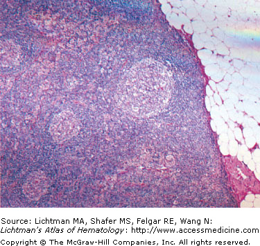
VII.A.002 Lymph Node, Normal
VII.A.003 Lymph Node, Normal
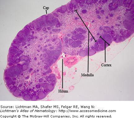
VII.A.003
Normal human lymph node. Low power. Capsule (Cap) is a thin connective tissue covering. Below the capsule is the subcapsular sinus. Lymphatics penetrate the capsule and enter the subcapsular sinus. The cortex is composed of adjacent lymphatic nodules, usually with fine connective tissue trabeculae extending from the capsule separating the nodules. The nodule has a germinal center that stains lighter than the outer mantle zone because of the proliferating medium-sized and large lymphocytes with less dense staining properties. The medulla is composed of interconnecting medullary cords composed of lymphocytes and interspersed light staining channels, the medullary sinuses. Lymph flows from the subcapsular sinus down the trabecular sinuses and into the medullary sinuses and exits the node via efferent lymphatics at the hilum.
VII.A.004 Lymph Node, Normal
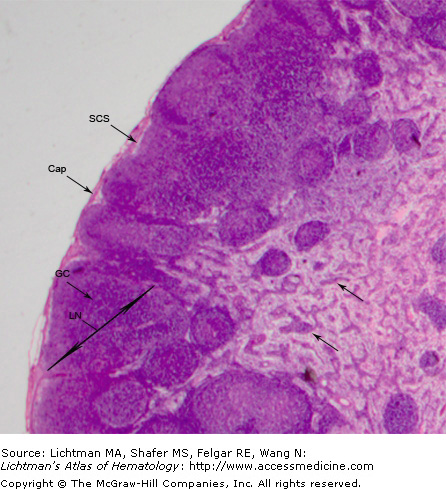
VII.A.004
Normal human lymph node. Higher power. Capsule (Cap) is a thin connective tissue covering. Below the capsule is the subcapsular sinus (SCS). Lymphatics penetrate the capsule and enter the subcapsular sinus. The cortex is composed of adjacent lymphatic nodules (LN), usually with fine connective tissue trabecula extending from the capsule separating the nodules. The nodule has a germinal center (GC) that stains lighter than the outer mantle zone because of the proliferating medium-sized and large lymphocytes with less dense staining properties. The medulla is composed of interconnecting medullary cords (arrows) composed of lymphocytes and interspersed lighter staining channels, the medullary sinuses. Lymph flows from the subcapsular sinus down the trabecular sinuses and into the medullary sinuses and exits the node via efferent lymphatics at the hilum.
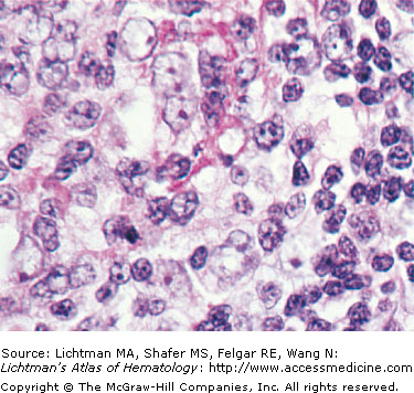
VII.A.005 Lymph Node, Germinal Center
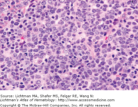
VII.A.005
Lymph node, germinal center. Biopsy section. Typical cellular content of a normal germinal center, showing mostly small lymphocytes, apoptotic lymphocytes and tingible-body macrophages. During normal lymphocyte development and gene rearrangement of immunoglobulin gene, non-functional B-cells undergo an apoptotic, programmed cell death. The resultant nuclear debris is phagocytized by macrophages, which show stainable (tingible) bodies in the cytoplasm that represent nuclear fragments.
VII.A.006 Lymph Node. CD20, CD3, BCL-6, Ki-67 Immunostains
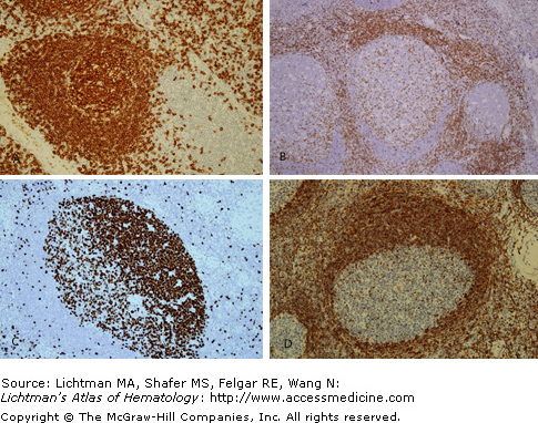
VII.A.006
Related posts:

Stay updated, free articles. Join our Telegram channel

Full access? Get Clinical Tree


