Normal & Abnormal Monocytes & Macrophages


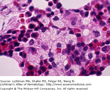
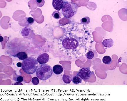
V.H.001 Gaucher Cells


V.H.001


Gaucher cells in Gaucher disease. The two macrophages are engorged with glucocerebroside as a result of the inherited absence of glucocerebrosidase, required for glycolipid degradation.


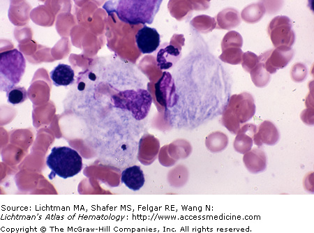
V.H.002 Gaucher Cell


V.H.002


Gaucher cell. Marrow film. Large macrophage with gray-blue stained, striated cytoplasm. The lack of the enzyme glucocerebrosidase results in an inability to metabolize glycolipids normally with a resultant accumulation of glucocerebroside in macrophages.


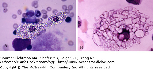
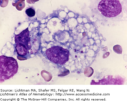
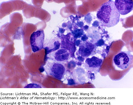
V.H.003 Gaucher Cells. Spleen Imprint
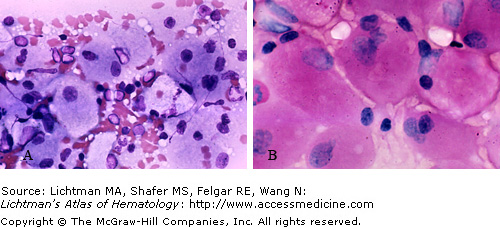


V.H.003


Gaucher cells. Spleen Imprint. (A) Numerous macrophages with gray-blue stained, striated cytoplasm. The lack of the enzyme glucocerebrosidase results in an inability to metabolize glycolipids normally with a resultant accumulation of glucocerebroside in macrophages. Splenic enlargement as a result is common. (B) Periodic acid Schiff stain imparts characteristic reddish-pink coloration to the cytoplasm of Gaucher cells.


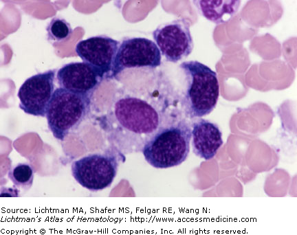
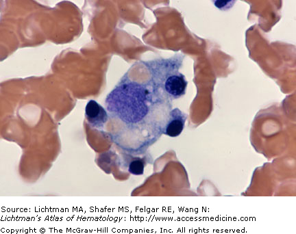
V.H.004 Macrophage, Cystine Crystals


V.H.004


Macrophage with cystine crystals. Marrow film. Macrophage engorged with cystine crystals in a case of cystinosis.


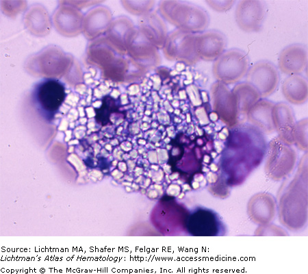
V.H.005 Macrophages with Cystine Crystals


V.H.005


Macrophages with cystine crystals. Marrow section. Case of cystinosis with macrophages engorged with cystine crystals.


V.H.006 Macrophages with Cystine Crystals


V.H.006


Macrophages with Cystine Crystals. Marrow films. (A, B) Case of cystinosis with macrophages engorged with cystine crystals. (A) Lower magnification of two crystal engorged macrophages. (B) Higher magnification of macrophage engorged with cystine crystals.


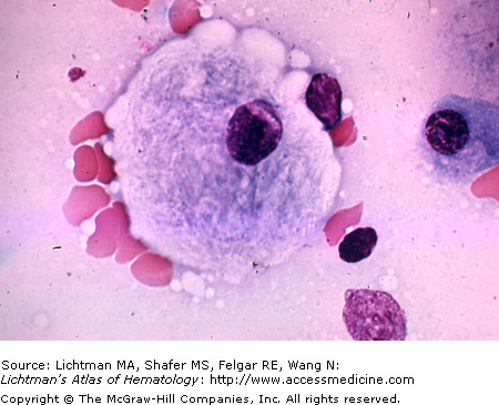
V.H.007 Macrophage, Erythroid Islet


V.H.007


Macrophage, erythroid islet. Marrow film. Macrophage with ingested erythrocytes and surrounding erythroblasts.


V.H.008 Macrophage, Erythrophagocytosis


V.H.008


Macrophage, erythrophagocytosis. Marrow film. Erythrophagocytosis. Note pale erythrocyte and erythrocyte ghost (hemolyzed) in macrophage cytoplasm (arrows).


V.H.009 Macrophage, Erythrophagocytosis


V.H.009


Macrophage, erythrophagocytosis. Marrow film. Erythrophagocytosis. Note pale erythrocyte and numerous shrunken erythrocyte ghosts (hemolyzed) in macrophage cytoplasm. Cytoplasm intensely vacuolated and several apparent nuclear vacuoles.


V.H.010 Macrophage, Erythrophagocytosis


V.H.010


Macrophage, erythrophagocytosis. Marrow film. Two macrophages. One containing numerous presumptive iron particles. The other macrophage has ingested several erythrocytes and has a heavy load of iron particles.


V.H.011 Macrophage, Erythrophagocytosis


V.H.011


Macrophage, erythrophagocytosis. Marrow film. Macrophage with ingested erythrocytes and four erythroblasts apparently attached to macrophage periphery.


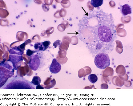
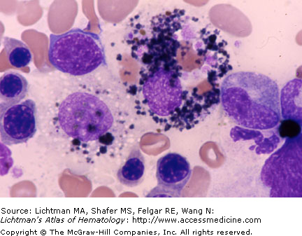
V.H.012 Macrophage, Hemophagocytic


V.H.012


Macrophage, hemophagocytic. Marrow film. Macrophage with ingested platelets and cellular residue and large vacuoles with cellular residue in various phases of disintegration.


V.H.013 Macrophage, Hemophagocytic


V.H.013


Macrophage, hemophagocytic. Marrow film. Macrophage engorged with neutrophils and red cell nuclei in various phases of disintegration.


Jun 12, 2016 | Posted by in HEMATOLOGY | Comments Off on Normal & Abnormal Monocytes & Macrophages

Full access? Get Clinical Tree

Get Clinical Tree app for offline access