Introduction
Any attempt to describe the neurological signs of ageing immediately prompts a number of challenging questions. Perhaps the most pertinent of these is how to ascribe neurological signs observed in later life to normal ageing per se, rather than concurrent (and possibly subclinical) age-related neurological conditions such as cerebrovascular disease, Alzheimer’s disease (AD), Parkinson’s disease or combinations thereof. Differing quantitatively or qualitatively from normal ageing, such diseases fall within the purview of geriatric neurology. From the clinical perspective, this is not merely a question of dry academic interest, since disease-related changes might be amenable to disease-specific therapeutic interventions, whereas age-related change might require acceptance as part of the human condition, perhaps abetted by sympathetic attempts at neurorehabilitation.
To define the neurological signs of normal ageing, one requires a definition of ‘normal’, which will determine the optimal requirements for a study which aims to describe such signs. The one absolute of studies of ageing is substantial heterogeneity, in both control and age-related disease groups. Hence it must be decided if individuals selected for study should be free of all age-related disease and medication use. Such individuals may be described as having undergone ‘successful’ ageing or ‘optimal’ ageing. By contrast, the category of ‘typical’ ageing accepts common age-associated disease (and medication use) as physiologically typical of the ageing process. Such definitions may be informed by biological models of ageing, such as senescence (age as a disease) or lifespan (age as development) models. Considerations of this nature will also determine the locus of study populations (community based, hospital based, nursing home based) and whether an attempt should be made to include all relevant subjects or only those who volunteer for study.
‘Successful’ ageing or ‘optimal’ ageing occurs in ‘super-normals,’ individuals whose function or performance clusters at the upper end of any normal distribution. Genetically, these individuals may differ from those undergoing ‘typical’ or ‘normal’ ageing. Relatively greater or lesser vulnerability of ageing tissues to disease processes and relative preservation or loss of neural regenerative capacities with ageing1 may also be relevant.
Study logistics also need careful consideration. Cross-sectional studies, in which, for example, 20-year-olds are compared with 50- and 80 year-olds, with assessment occurring at one time point, may overestimate age-related changes because of cohort effects such as differences in education or nutrition. Moreover, with lack of follow-up, it may be that ‘normals’ in the older age groups were in fact in the subclinical phase of age-related disease. Longitudinal studies of cohorts which might address such difficulties, by following individuals for many years with repeated examinations at successive time points, risk underestimating change due to loss of subjects to follow-up. Such studies are also expensive, sometimes prohibitively so.
The definition of signs to be examined and standardization of testing procedures are also fundamental requirements. Disagreement between experienced examiners in the interpretation of neurological signs is well recognized.2, 3 Hence prespecified operationalization of neurological examination and agreement on scoring or quantitation of signs are required for robust results.4 Without specifying these parameters, it is difficult to realize a quantitative measure of the sensitivity and specificity of neurological signs with respect to ageing.
Similar arguments apply to age-related changes in investigation findings, such as neuroimaging and electrodiagnostic studies, and likewise to the definition of the neuroanatomical substrates of change in neurological signs with ageing. For example, cerebral atrophy on structural brain imaging or in postmortem tissue is not an uncommon finding in cognitively normal older individuals and is not in itself an inevitable signature of AD.5, 6
Neurological Signs of Ageing
An early and comprehensive review of the neurology of old age was given by Critchley in his three Goulstonian Lectures delivered to the Royal College of Physicians of London in March 1931.7 Since then, many reviews have appeared, some indicating the need to revise the designation of ‘senile’ for certain signs in favour of aetiological explanations which may carry therapeutic implications.
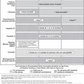
Some neurological signs particularly associated with ageing are briefly described (summarized in Table 53.1), along with details of their investigational correlates and neuroanatomical substrates where these are known. Techniques for eliciting neurological signs and their semiological value are not covered here.8 The description follows the traditional, and somewhat arbitrary, sequence of the neurological examination.
Table 53.1 Topographical overview of age-related neurological signs.
Cognitive function:
Cranial nerves:
Motor system:
Sensory system:
|
Cognitive Function
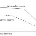
Ageing has both structural and functional effects on the brain.9 Serial registered magnetic resonance imaging scans show that there is a decrease in global and regional (temporal lobe, hippocampus) brain volumes, the rate of which may increase after the age of 70 years.5 Hence brain atrophy per se is not specific for the diagnosis of pathological change, an assumption which may lead to clinical misdiagnosis of AD if undue weight is placed on structural neuroimaging findings.6 The neuroanatomical correlates of this volume loss are uncertain, possibly including neuronal loss or shrinkage, dendritic pruning and synaptic loss and white matter change. There is also evidence for plasticity in the ageing brain, with dendritic sprouting which may help to maintain synaptic numbers, although such compensatory abilities may decline with age. Vascular change becomes more frequent in the ageing brain, as manifested by leukoaraiosis (small vessel ischaemic change in white matter) and silent infarcts, possibly related to rises in blood pressure. These changes may not only reflect brain ageing but may also contribute to pathological disorders, both AD and vascular cognitive impairment. Interaction of AD-type pathology with vascular changes may lower the threshold for the clinical appearance of cognitive decline.10
Neuropathological studies of ageing brain have focused on both positive and negative phenomena. Of the former, neurofibrillary pathology (neurofibrillary tangles, neuropil threads) and senile neuritic plaques, hallmarks of the AD brain, may be seen in cognitively normal older individuals. The development of neurofibrillary pathology follows a relatively stereotyped hierarchical pattern with age, appearing first in the transentorhinal cortex.11 Spread to hippocampal and association cortex is associated with progressive appearance of cognitive decline. Senile plaques have a broader and more variable distribution; a significant burden may be associated with normal cognition. Negative phenomena include neuronal and synaptic loss. There is relative preservation of cortical and hippocampal neuronal populations with ageing, although subcortical structures such as the basal forebrain, locus ceruleus and substantia nigra do show neuronal losses.
What are the functional consequences of these changes? Typical cognitive ageing involves losses in processing speed, cognitive flexibility and the efficiency of working memory (or sustained attention). In other words, it may take more time and/or more trials to learn new information. Other cognitive domains, such as access to remotely learned information including semantic networks and retention of well-encoded new information, are spared with typical ageing. Hence impairments in these latter domains may be sensitive indicators of pathology rather than physiology.12 Memory decline in healthy ageing may be secondary to a decline in processing speed and efficiency: controlling for processing speed may attenuate or eliminate age-related differences in memory performance, unlike the situation with memory impairment in dementia. Longitudinal studies of neuropsychological function, such as the Mayo’s Older Americans Normative Studies, indicate that there is considerable variability in normal older adults across different skills and consistency across different domains may not necessarily be observed.12 Clearly this needs to be taken into account when assessing whether perceived cognitive decline is pathological or normal, that is, in defining neuropsychological norms for ageing. Likewise, norms may need to be age weighted rather than age corrected to detect cognitive impairment related to AD, the prevalence of which increases exponentially with increasing age. Many other situational influences may also impact on testing of cognitive skills, such as fatigue, emotional status, medication use and stress. These also need to be taken into account when considering the results of cognitive testing, as may factors such as educational and background experience. Many norms are also culturally-weighted.
Age-related cognitive decline may exist in individuals who do not fulfil validated criteria for the diagnosis of dementia or AD.13 Various terms have been used over the years to describe this state, dating back to Krol’s ‘benign senescent forgetfulness’ and including age-associated memory impairment (AAMI), age-associated cognitive decline (AACD), cognitive impairment, no dementia (CIND) and mild cognitive impairment (MCI). Some consensus has developed around the concept of MCI, criteria for which are the presence of a memory complaint, preferably corroborated by an informant; evidence of objective memory impairment for age and level of education; largely normal general cognitive function; essentially intact activities of daily living; and failure to fulfil criteria for dementia.14 MCI is certainly a heterogeneous clinical entity, some examples of which certainly represent ‘prodromal AD’. The annual conversion rate of MCI to AD is ∼5–10%, such that most individuals with MCI do not progress to dementia even after 10 years of follow-up.15 Updated diagnostic criteria for AD16 seek to abolish the MCI category altogether in favour of an earlier, biologically based, diagnosis of AD, even without the clinical correlate of dementia which was insisted upon in earlier criteria.13 This is logical if the hope is for earlier diagnosis and intervention to prevent cognitive decline to dementia. However, as yet no disease-modifying intervention has been discovered in the clinical arena, cholinesterase inhibitors proving robustly negative in trials aimed at slowing MCI to AD conversion and other interventions (blockers of amyloid production such as secretase inhibitors, immunotherapies to prevent amyloid deposition, tau aggregation inhibitors) remain at the clinical trial stage.
Since ageing per se is a significant risk factor for dementia, the ageing of the world population has important clinical, social and economic implications.17 Primary and secondary prevention measures, perhaps facilitated by predicting risk of dementia in 20 years time based on factors such as age, education, blood pressure, cholesterol and obesity,18 may be a more appropriate public health strategy, emphasizing a life-long, lifestyle approach to cognitive well-being.
Cranial Nerves, Including Special Senses
Olfaction (Olfactory Nerve; Cranial Nerve I)
Stay updated, free articles. Join our Telegram channel

Full access? Get Clinical Tree


