4
Neoplastic Mimics of the Stomach
INTRODUCTION
Neoplastic mimics of the stomach predominantly comprise polyps, submucosal lesions, and ulcers (Table 4.1). Many of these lesions are not specific to the stomach, and are described in Chapter 2. Two lesions that are specific to stomach, fundic gland polyp and hyperplastic polyp, are exaggerations of one or both of the two normal compartments of the gastric mucosa, that is, the superficial foveolar and the deep glandular compartments. Hamartomatous polyps occur as part of polyposis syndromes and the syndromes are described at length in the intestinal section of this chapter. All of these benign polyps are considered neoplastic mimics to adenomatous polyp, adenocarcinoma, or carcinoid tumor of the stomach.
TABLE 4.1 Neoplastic Mimics of the Stomach
GROSS CONFIGURATION | NEOPLASTIC MIMICS | NEOPLASM |
Mucosal plaques or submucosal nodules | Xanthoma/xanthelasma | Diffuse/signet ring cell adenocarcinoma |
| Pancreatic heterotopia | Adenocarcinoma |
| Gastritis cystica profunda/polyposa | Adenocarcinoma |
Mucosal granularity | Gastric foveolar hyperplasia | Adenomatous polyp |
Polyps | Gastric fundic gland polyp | Adenomatous polyp |
| Gastric hyperplastic polyp | Adenomatous polyp, dysplasia |
| Inflammatory fibroid polyp | Gastrointestinal stromal tumor, leiomyoma |
| Hamartomatous polyp | Adenomatous polyp, dysplasia |
Mucosal thickening and/or ulceration | Gastric fundic gland hypertrophy (proton pump inhibitor effect) | Diffuse/signet ring cell adenocarcinoma |
| Reactive/chemical gastropathy | Mucosal dysplasia |
| Crohn’s disease | Adenocarcinoma |
FUNDIC GLAND POLYP
Fundic gland polyp is a benign polyp composed of proliferation and dilatation of gastric fundic glands. Fundic gland polyp was first described by Elster (281) and had been thought to be a form of hamartoma (282), hence in older literature referred as Elster’s polyp or cystic hamartomatous gastric polyp. These polyps occur exclusively in the body and fundus of the stomach; therefore, as a rule, multiple small polyps in the fundus are fundic gland polyps (283). They are asymptomatic, often found incidentally and almost always appear in healthy gastric mucosa or only in minimally chronic nonspecific gastritis in Helicobacter pylori-negative patients. When multiple, they may be associated with proton-pump inhibitor therapy (284,285) or with familial adenomatous polyposis (286). Fundic gland polyposis is, in fact, a far more common manifestation of familial adenomatous polyposis than gastric adenomas and occur earlier in life (287). The findings of APC gene and beta catenin mutation raises the possibility of a neoplastic nature of fundic gland polyps, at least those associated with familial adenomatous polyposis (288,289).
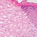
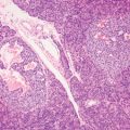
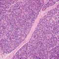
Fundic gland polyps are small, sessile polypoid nodules with smooth surfaces in the body and fundus of the stomach. They usually measure less than 1 cm in greatest diameter, but large examples occur. The bulk of the polyp is composed of disorganized hyperplastic fundic or oxyntic glands with focal cystic dilatation (Figures 4.1 and 4.2). The surface foveolar epithelium shows varying degree of hyperplasia and dilatation. The superficial lamina propria is occasionally edematous but is rarely inflamed. Very small polyps may have subtle microscopic changes with mild and focal fundic gland distortion and rare fundic gland cysts. Those associated with familial adenomatous polyposis may show changes resembling low grade dysplasia with crowding, hyperchromasia, and pseudostratification of the nuclei (Figure 4.3), but they rarely progress to high-grade dysplasia or carcinoma.
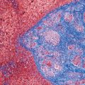
Although fundic gland polyps are often associated with proton pump inhibitor therapy, they have to be distinguished from diffuse fundic gland hypertrophy with cystic dilatation, apical snouts, and occasionally cytoplasmic vacuolization secondary to proton pump inhibitor therapy itself (Figures 4.4 and 4.5
Stay updated, free articles. Join our Telegram channel

Full access? Get Clinical Tree


