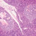
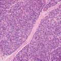
6 Neoplastic mimics of the serosa of the gastrointestinal tract are frequently encountered during laparoscopic or open abdominal surgery, including fat necrosis, foreign body giant cell reaction, splenosis, and endometriosis. These lesions may be multiple, scattered throughout the peritoneal surface or serosal surface of abdominal organs, resembling metastatic carcinoma or peritoneal carcinomatosis. TABLE 6.1 Neoplastic Mimics of the Serosa and Mesentery of the Gastrointestinal tract LOCATION NEOPLASTIC MIMICS NEOPLASM Mesentery Sclerosing mesenteritis Desmoid tumor, inflammatory myofibroblastic tumor, carcinoid tumor Serosa Fat necrosis Metastatic neoplasm Foreign body giant cell reaction Metastatic neoplasm Endometriosis Metastatic neoplasm Splenosis Metastatic adenocarcinoma, angiosarcoma, lymphoma Sclerosing or retractile mesenteritis is an idiopatic inflammatory fibrosing process of the mesenteric adipose tissue (380,381). Mesenteric panniculitis and mesenteric lipodystrophy are other terms that are often used to reflect the predominant component of the inflammation or fat necrosis in this fibroinflammatory process (382,383). Sclerosing mesenteritis occurs most often in middle-aged or older adults, predominantly males. It is associated with previous abdominal surgery and other inflammatory disorders. The association of sclerosing mesenteritis with IgG4-related diseases remains unclear as a small percentage of sclerosing mesenteritis may have significant IgG4-positive plasmacellular infiltrate (384,385). The most common site of involvement is the small bowel mesentery, but rare cases have been reported to involve the large bowel (386,387). The presenting symptoms are abdominal pain, weight loss, fever, and bowel obstruction (388,389). Protein-losing enteropathy as the result of disturbed lymphatic drainage caused by the sclerosed mesentery has been reported as the first manifestation of sclerosing mesenteritis (390). In addition, lymphatic blockage by the fibroinflammatory process can cause multiple mesenteric lymphatic cysts (391). Although the imaging features can be nonspecific, the “fat ring” sign and the presence of a tumoral pseudocapsule have been suggested for the diagnosis of the disease (392). Sclerosing mesenteritis can regress spontaneously but can also have a prolonged debilitating course with a fatal outcome. The process can be extensive involving multiple organs and major vessels and, therefore, unresectable. Symptomatic patients with unresectable lesion may benefit from steroid or immunosuppressive therapy (393). Sclerosing mesenteritis forms an ill-defined solid mesenteric tumor and retracts the overlying bowel loop. It is characterized by alternating areas of fat necrosis in lobules, separated by haphazard myofibroblastic proliferation and dense hyalinized fibrous tissue with eosinophilic collagen bands (Figures 6.1–6.3
Neoplastic Mimics of the Serosa and Mesentery of the Gastrointestinal Tract
INTRODUCTION
SCLEROSING MESENTERITIS
![]()
Stay updated, free articles. Join our Telegram channel

Full access? Get Clinical Tree


