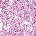
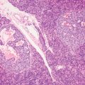
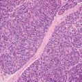
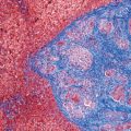
3 Neoplastic mimics of the esophagus take the form of white plaques such as seen in glycogenic acanthosis, polyp in squamous papilloma, and ulceration in gastroesophageal reflux disease and Crohn’s disease (Table 3.1). Endoscopically and microscopically, these lesions can mimic neoplasms of the esophagus, that is, squamous cell carcinoma and adenocarcinoma. Squamous cell carcinoma tends to occur in the proximal to middle third of the esophagus, whereas adenocarcinoma occurs in the distal third of the esophagus in association with Barrett’s esophagus. TABLE 3.1 Neoplastic Mimics of the Esophagus GROSS CONFIGURATION NEOPLASTIC MIMICS NEOPLASM Mucosal plaque Glycogenic acanthosis Squamous cell dyplasia, carcinoma Gastric heterotopia “inlet patch” Adenocarcinoma Polyps Squamous papilloma Squamous cell carcinoma Fibrovascular polyp Squamous cell carcinoma Inflammatory fibroid polyp Gastrointestinal stromal tumor, leiomyoma Ulcerating lesion Crohn’s disease Squamous cell carcinoma, adenocarcinoma Gastroeosphageal reflux ulcer Adenocarcinoma
Neoplastic Mimics of the Esophagus
INTRODUCTION
![]()
Stay updated, free articles. Join our Telegram channel

Full access? Get Clinical Tree


Oncohema Key
Fastest Oncology & Hematology Insight Engine


