Natural Killer Cells


Natural Killer Cells


Wayne M. Yokoyama


INTRODUCTION

Natural killer (NK) cells were initially described because they spontaneously kill certain tumor targets.1,2,3,4 However, they are now recognized to play important roles in early innate immune responses, especially to viral infections. They interact with other innate immune components and modulate the subsequent adaptive immune response. These effects are due to NK cell responses to proinflammatory cytokines or susceptible targets, which stimulate NK cells to secrete other cytokines and/or kill targets. In this chapter, we will consider all of these issues in detail by describing how they differ from other lymphocytes, their functions, unique target recognition strategies, tolerance, development, and role in immune responses and human diseases. There will be an emphasis on their target recognition receptors because their discovery made it possible to understand NK cell biology more precisely.


GENERAL DESCRIPTION

Developmental studies have provided strong evidence that NK cells belong to the lymphocyte lineage (discussed in detail in the following). Morphologically, NK cells are typically large lymphocytes containing azurophilic granules.5 However, the large granular lymphocyte morphology is not invariably associated with NK cells because small, agranular lymphocytes may display natural killing,6 activated cytotoxic T-lymphocytes (CTLs) can display this morphology,7 and human large granular lymphocyte leukemias contain NK- and T-cell variants.8 Among lymphocytes, NK cells more closely resemble T cells than B cells. Thus, it is useful to compare and contrast these two lymphocyte populations as well as consider another enigmatic cell termed the “lymphokine-activated killer” (LAK) cell.


Natural Killer Cells versus T Cells

NK cells are most often confused with T cells because they may have similar morphologies, express several cell surface molecules in common,9,10 and share functional capabilities. While this confusion was frequent before the molecular description of the T-cell receptor (TCR)/cluster of differentiation (CD)3 complex, their similarities remain a potential source of uncertainty. However, mature NK cells are clearly not T cells by several criteria.11 Conventional NK cells do not require a thymus for development and are normal in athymic nude mice (though this is not the case for the newly described “thymic” NK cell subset, discussed subsequently). NK cells do not express the TCR on the cell surface, do not produce mature transcripts for TCR chains, and do not rearrange TCR genes.12,13 Mice with the scid mutation or deficiencies in Rag1 or Rag2 lack TCR gene rearrangements and mature T cells but possess NK cells with apparently normal function.14,15,16,17 Several CD3 components may be found in the cytoplasm of NK cells, particularly immature NK cells, but they are not displayed on the cell surface18 with the exception of CD3ζ. But CD3ζ is expressed in association with FcγRIII (CD16) and other NK cell activation receptors instead of the TCR/CD3 complex.19,20 Whereas mice lacking CD3ζ lack most T cells, NK cell number and function are minimally affected.21 On the other hand, NK cells are completely absent in mice with only partial defects in T-cell subsets, such as in mice lacking components of the IL-15R (see following discussion). NK cells do not require the presence of major histocompatibility complex (MHC) class I (MHC-I) molecules on their targets for lysis in an important functional distinction with CD8+ MHC-I-restricted T cells. Instead, NK cells kill more efficiently when their targets lack MHC-I expression. Thus, NK cells can be clearly distinguished from T cells, even from so-called CD3+ NKT cells that express NK cell markers (see following discussion).


Natural Killer Cells, Lymphokine-Activated Killer Cells, and Interleukin-15

Another area of overlap between NK and T cells concerns cytokine responses. When mouse splenocytes or human peripheral mononuclear cells are exposed to high concentrations of interleukin (IL)-2 (800 to 1000 U/mL), robust lymphocyte proliferation ensues (ie, LAK cells are generated).22,23,24,25 Although most are CD3- NK cells, TCR/CD3+ T cells are also produced. To distinguish NK cells within this population, they are sometimes called “CD3 LAK” cells or “IL-2-activated NK cells.”

In a related phenomenon, NK cells are activated when mice are injected with polyinosinic-polycytidylic acid (poly-lic) or other agents that trigger through Toll-like receptors (TLRs), often on plasmacytoid dendritic cells (DCs).26 NK cells can also be activated in vitro with interferon (IFN)α/β, IFNγ, or low concentrations of IL-2 that are insufficient to induce proliferation.3,4,27

NK cells activated in these various ways, with or without proliferation, display enhanced killing of typical NKsensitive targets. They also kill a broader panel of targets, including those that are generally resistant to freshly isolated NK cells, such as the murine P815 mastocytoma cells and
freshly explanted tumors. Many agents that enhance killing by NK cells may also activate T cells, such that even T-cell clones may display promiscuous killing of targets that is no longer MHC-restricted.28 This phenomenon was a frequent source of confusion between NK cells and T cells during the initial characterization of both cell types.

Why cytokine activation leads to enhanced killing is incompletely understood. Interestingly, mouse NK cells constitutively express messenger ribonucleic acid (mRNA) for cytotoxicity components, perforin, and granzymes, but no protein.29 Cytokine activation enhances expression of mRNA for perforin and granzymes,30 and translation into expressed proteins that contributes to more lytic capacity,29,31 but this effect may not explain the capacity of LAK cells to kill a broader panel of targets. Activated NK cells express additional receptors that may deliver stimulatory signals,32,33 but their contribution to the LAK phenomenon remains unclear. In mice, cytokine activation of NK cells results in expression of an alternatively spliced form of a receptor termed NKG2D (see subsequent discussion), which may play a role,34,35 and IL-15 contributes to LAK-like activity of CTLs, although how much this applies to conventional NK cells is not understood.36 Enhanced killing may also be due to effects on adhesion molecules.37,38 Nonetheless, it remains unclear how each of these factors contribute to the LAK phenomenon of enhanced and broader killing capacity as compared to resting NK cells.

It seems unlikely that high concentrations of IL-2 can be achieved, even locally, to stimulate NK cells in vivo. Furthermore, NK cells tend to be early responders in immune responses whereas the prime reservoir of large amounts of IL-2 is the activated T cell that produces it somewhat later. Although naïve T cells can make IL-2 soon after stimulation39,40 and DCs can produce IL-2 to enhance NK lytic activity,41 whether the resultant IL-2 concentrations are sufficient to generate LAK cells in vivo is unclear.

Interestingly, NK cells are apparently normal in mice with a targeted mutation in the IL-2 gene or the IL-2Rα chain,42,43 indicating that IL-2 itself is not required for normal NK cell development. Paradoxically, NK cells are deficient in mice with a mutation in either IL-2Rβ or IL-2Rγ.44,45,46 The discrepancy in NK cell dependence on IL-2 versus IL-2 receptor (IL-2R), as well as the LAK cell phenomenon, may be best understood by comparing the components of the IL-2 and IL-15 receptors.

In brief, the high affinity IL-2R is a heterotrimeric receptor complex comprised of α (p55), β (p75), and γ (p64) chains.47 Though individual components may bind IL-2 with low affinity, only the intermediate-affinity βγ receptor (Kd ˜1 nM) and the high-affinity αβγ receptor (Kd ˜10 pM) are capable of signaling. Resting NK cells constitutively express IL-2Rβγ47,48 and upon activation, may induce IL-2Rα and further upregulate IL-2Rγ chain expression.47 In contrast, resting T cells generally do not express any functional IL-2 receptors, and most naïve T cells do not respond to high concentrations of IL-2.49 The IL-2Rγ chain is also termed the common γ subunit (γc) because it is a required component of the multimeric receptor complexes for other cytokines, including IL-15,50 that is particularly relevant to NK cells. IL-15 does not bind to IL-2Rα but instead utilizes a unique IL-15Rα chain to form a high-affinity complex with IL-2Rβγ.51,52 The IL-15Rα chain does not directly signal. Its distribution is widespread on numerous cell and tissue lineages including NK cells.

IL-15 has a number of effects on NK cell biology. It is required for NK cell development; mice lacking IL-15 or any component of the trimeric IL-15R complex lack NK cells.44,45,46,53,54 Not surprisingly, mice deficient in other components of the IL-15R complex and its signaling pathway (IL-2Rβ, Jak3, and STAT5α/β) exhibit similar defects in NK cell development.44,55,56,57 Depending on its relative concentration, IL-15 has an antiapoptotic or proproliferative effect.58,59 When NK cells are transferred to NK cell-deficient mice, they undergo “homeostatic” proliferation,60,61 akin to T-cell homeostatic proliferation.62 Like memory CD8+ T-cell homeostasis, NKcell homeostasis is IL-15-dependent,60,61 to a more or less degree.63 Finally, LAK cells can be generated with IL-15.64 These studies strongly suggest that LAK cells are generated because high-dose IL-2 acts through the IL-2Rβγ that is normally expressed with IL-15Rα as components of the constitutively expressed trimeric IL-15R complex on resting NK cells. Thus, the LAK cell phenomenon is related to the role of IL-15 and its receptor in NK cell biology.

Interestingly, IL-15 is expressed at very low levels and is difficult to detect in vivo.65 The IL-15Rα chain can present IL-15 in trans to NK cells that can respond through IL-2/15Rβγ alone.66 For example, IL-15Rα-deficient NK cells develop in bone marrow (BM) chimeric mice in which IL-15Rα-deficient BM was used to reconstitute IL-15Rα-sufficient animals,67 indicating that IL-15Rα on another cell can allow IL-15Rα-deficient NK-cell development. In certain inflammatory situations in vivo, DC presentation of IL-15 in trans can enhance NK cell responses (also known as priming).31,68 In DCs expressing both IL-15 and IL-15Ra, the IL-15Ra chain provides a chaperone function to stabilize receptor-cytokine complexes on the cell surface.69 Taken together, trans presentation of IL-15 may be physiologically important to NK cell function.


SELECTIVE NATURAL KILLER CELL SURFACE MARKERS

The constitutive expression on NK cells of IL-15R complex with IL-2Rβ has practical usefulness because anti-IL-2Rβ (CD122) is sometimes used to identify naïve CD3- NK cells or deplete them in mice,70 but anti-CD122 is less useful during an ongoing immune response and CD122 is expressed on regulatory T cells. Anti-IL-15Rα antibodies have not been widely used. Other markers have proven to be more useful for analysis of NK cells.

In the mouse, the NK1.1 molecule is an especially important marker on NK cells in C57BL strains.11 NK1.1 is an activation receptor encoded by Nkrp1c (Klrb1c),71 a member of the Nkrp1 gene family (see following discussion). In FACS sorting experiments, the NK1.1+ fraction contained all of the natural killing activity in the spleen.72 In vivo administration of the anti-NK1.1 mAb PK13673 completely abrogated natural killing but did not affect adaptive immune responses74
(mAb PK136 is available from the American Type Culture Collection [ATCC], Manassas, VA [HB-191] and is an IgG2a isotype [ATCC, and data not shown], not IgG2b as originally described73). While mAb PK136 is very efficient at NK-cell depletion and is widely used for this purpose, unfortunately it recognizes an epitope on NK1.1 that is confined to C57BL/6, C57BL/10, and a few other strains.73 Moreover, in Swiss, NIH and SJL/J mice, mAb PK136 recognizes another NKRP1 family member, NKRP1B.75,76 However, there are now available NK1.1+ congenic strains, such as BALB.B6- Cmv1r (catalogued as C.B6-Klra8Cmv1-r /UwaJ, stock number 002936 at The Jackson Laboratory, Bar Harbor, ME) in which the C57BL/6 allele of NK1.1 has been genetically bred onto the BALB/c background that otherwise lacks the NK1.1 epitope.77 Similarly, the NK1.1 allele has been introgressed onto the nonobese diabetic (NOD) background.78

A subpopulation of T cells expresses NK1.1, described in detail in another chapter 18. These “natural killer T (NKT) cells” express the TCR/CD3 complex and typically are restricted by the nonclassical MHC-I molecule, CD1, which presents glycolipid antigens to NKT cells. NKT cells respond early during the course of an immune response and may potently activate conventional NK cells.79 Nonetheless, NKT cells can be distinguished from conventional NK cells by expression of the CD3 complex (ie, conventional NK cells are NK1.1+ CD3-).

The NKG2D (Klrk1) activation receptor is expressed on all NK cells in human and all strains of mice examined.80 In humans, NKG2D is also expressed on all γδTCR+ and CD8+ T cells, whereas in mice, NKG2D is expressed on most NKT and γδTCR+ T cells but not on resting CD8+ T cells.81,82,83 However, essentially all activated mouse CD8+ T cells express NKG2D. In both humans and mice, CD4+ T cells do not express NKG2D, but it is found on a subset CD4+CD28- T cells in patients with rheumatoid arthritis.84 Blocking anti-NKG2D mAbs and NKG2D-deficient mice have been described.80,85,86,87 Regardless, conventional NK cells are NKG2D+ CD3-.

The NKp46 (Ncr1) activation receptor appears to be expressed on all CD3- NK cells in humans and all strains of mice. However, recent reports indicate expression of NKp46 on immune cells in the gut that may be developmentally distinct from conventional NK cells.88,89,90,91,92 Moreover, depleting anti-NKp46 mAbs have not been described, limiting its usefulness for in vivo functional experiments. Nonetheless, recently developed mice may allow other approaches, such as a mouse where a green fluorescent protein (GFP) cassette was inserted into Nkp46 and two different transgenic (Tg) mice with a Nkp46 promoter contruct for expression of Cre or diptheria toxin receptor.93,94,95

The mAb DX5 recognizes a molecule that is coexpressed on most NK1.1+ CD3- cells and on small populations of splenocytes in NK1.1- strains, consistent with identification of NK cells in all strains. However, mAb DX5 recognizes the α2 integrin that is widely expressed on other leukocytes, not just NK cells,96,97 and its expression is regulated.98 Nevertheless, the DX5 epitope has been used to identify NK cells in mouse strains that do not express NK1.1, but it has been largely supplanted by other nonpolymorphic markers such as NKp46.

The glycolipid determinant asialo-GM1 is expressed by most if not all murine NK cells and a subpopulation of T cells.99,100,101 Although the functional significance of this molecule is unknown, polyclonal rabbit anti-asialo-GM1 (Wako Chemicals USA, Richmond, VA) has been used to effectively deplete NK cells. In more recent studies, the anti-NK1.1 mAb PK136 has become the reagent of choice for NK-cell depletion because of an available defined mAb and its more restricted reactivity with NK cells.11,72,73,74 However, anti-asialo-GM1 remains in use for NK-cell depletion when anti-NK1.1 cannot be employed.73

In addition to NKG2D and NKp46, human NK cells selectively express CD56. Although it is also found on neural tissues and some tumors, CD56 is generally not expressed by other hematopoietic cells or lymphocytes.102,103,104 This 140 kDa molecule is derived from alternative splicing of the gene encoding neural cell adhesion molecule (NCAM) involved in nervous system development and cell-cell interactions.105,106 CD56 may be involved in adhesion between NK cells and their targets,107 but this function is controversial. Curiously, mouse CD56 is not expressed on hematopoietic cells,108 indicating that its role on NK cells is not conserved. Nevertheless, CD56 is particularly useful as a pan-NK-cell marker in humans.

Human NK cells can be functionally divided according to the level of CD56 expressed.103,109 Most human peripheral blood NK cells are CD56dim, a phenotype associated with more cytotoxicity and less cytokine production than a smaller subset of NK cells that express CD56 at higher levels (CD56bright). These cells also tend to differentially express receptors involved in target recognition as well as CD16. The CD56bright cells may undergo a maturation process to become CD56dim cells110 and may be related to a subset of NK cells identified in mice, termed “thymic” NK cells.111

Other molecules selectively expressed on NK cells are better discussed below under the general topic of NK cell receptors because they are molecularly defined and their ligands are known.


A MOLECULAR DEFINITION OF NATURAL KILLER CELLS?

A precise molecular definition of NK cells has been elusive. There are no known molecules that are exclusively expressed on NK cells and are responsible for critical functions only displayed by NK cells. The NK cell is therefore still defined by function to the exclusion of other cells, a concept first articulated 25 years ago.11

The defining functional feature of NK cells remains their intrinsic ability to perform natural killing (ie, they spontaneously lyse certain tumor cells in a perforin-dependent manner). Unlike other lymphocytes, NK cells do not express surface immunoglobulin or the TCR/CD3 complex, and generally do not require MHC-I expression on targets for lysis. Therefore, a current working definition is that an NK cell is a sIg, TCR/CD3 lymphocyte that can mediate perforin-dependent natural killing against targets that may lack MHC-I expression.

It is noteworthy that T cells were historically defined by an awkward functional definition (thymus-derived, sIg lymphocytes responsible for cell mediated immunity).112
With the molecular definition of the TCR and coexpressed CD3 molecules, immunologists can now define a T cell as a cell expressing the TCR/CD3 complex.113 The availability of molecular probes and mAbs directed against this complex provides precise definition even in pathologic tissue sections, without the need for functional analysis (cell mediated immunity, thymus dependence). Similarly, a molecular definition should permit unequivocal identification of NK cells to define their role in normal immune responses and pathologic settings.

Presumably, such a definition will require further knowledge of the molecular basis for NK-cell function, such as the receptors involved in natural killing. On the other hand, one difficulty is that the function that is most attributed to NK cells, natural killing, can be displayed by other cells, such as cytokine-treated T cells. Moreover, NK cells can utilize more than one receptor for target recognition, and individual NK cells can simultaneously express several of these receptors. Thus, there is, as yet, no consensus on the elusive “NK-cell receptor” analogous to the TCR, and the general sentiment in the field is that there is unlikely to be such a receptor.

In the absence of a precise definition, most investigators currently consider the following phenotypes to be surrogate markers of bona fide NK cells. Mouse NK cells are typically NK1.1+ (in appropriate strains), FcγRIII+ (CD16), CD122+, and CD3-. Human NK cells are generally CD56+ and CD3-. In general, mouse and human NK cells also express NKG2D and NKp46, with caveats as elaborated previously.

Note that these markers are generally correlated with cells having natural killing capacity but the markers themselves are not required for natural killing. It should be emphasized that these phenotypes can lead to some confusion due to expression of other molecules on NK cells that are used to help define other immune cells. For example, NK cells express B220 (CD45R) that is often used as a B-cell-specific marker; CD19 is more reliable to distinguish B cells from NK cells.114 Similarly, NK cells express CD11b, first described as Mac-1 on macrophages.98 Moreover, NK cells express CD11c, a marker used to define certain DC populations, leading to publications describing a novel type of DC, termed killer DCs.115,116 However, detailed investigation suggests that these cells are developmentally unrelated to DCs and are actually activated NK cells.117,118,119 Therefore, markers associated with NK-cell function have been extremely useful in shaping our current concepts of NK cell biology and elaborate their effector functions, but caution may be necessary to avoid confusion with other immune cells.


EFFECTOR FUNCTIONS OF NATURAL KILLER CELLS


Cytotoxicity

A hallmark of NK-cell effector function is target killing, mediated by a process termed granule exocytosis that can be initiated by exposure to susceptible targets or cross-linking of specific activation receptors. Like CTLs, NK cells possess preformed cytoplasmic granules that resemble secretory lysosomes with properties of both secretory granules and lysosomes.120 Granule formation is affected by Lyst, the molecule defective in humans with Chediak-Higashi syndrome121,122 in which enlarged lysosomes are observed apparently due to decreased lysosome fission.123 Normal granules contain perforin and granzymes (granule enzymes). Perforin, a pore-forming protein, is rendered inactive by association with calreticulin and serglycin, and is activated by a cysteine protease.124 Granzymes are first produced as inactive proenzymes that are activated by N-terminal cleavage by dipeptidyl peptidase I, also known as cathepsin C. However, granzymes are rendered inactive by the acidic pH of the granules. Upon activation by a sensitive target, NK (and T) cells are triggered to rapidly polarize the granules and reposition the microtubule organizing center toward the target in a dynein-dependent manner.125 The granule membrane ultimately fuses with the plasma membrane, and externalizes, releasing granule contents. Calcium-dependent polymerization of perforin results in “perforation” of the target cell plasma membrane, and granzyme entry by an as yet incompletely understood process. A recent study suggests that perforin induces a plasma membrane repair process that results in endocytosis of perforin and granzymes into enlarged endosomes, called “gigantosomes.”126 Perforin pores in the gigantosome membrane then allow delivery of granzymes that mediate cleavage of caspases and Bid, ultimately leading to target cell apoptosis.127

Recently, many details of the granule exocytosis pathway have come from studies of the heterogeneous human disorder, hemophagocytotic lymphohistiocytosis (HLH).128,129 In particular, genetic studies of heritable HLH, termed familial HLH (FHL), led to identification of the first described FHL mutation in the perforin gene (PRF1), responsible for FHL2. Studies of patients with FHL without PRF1 mutations led to discovery of other genes whose products (MUNC13-4, syntaxin 11, MUNC18-2) affect granule exocytosis by cytotoxic lymphocytes. Fusion of the cytolytic granule with the plasma membrane requires vesicular RAB27a, a member of the small GTPase superfamily. Defects in RAB27a are associated with the human disorder Griscelli syndrome, type 2. Mice have been described with defects in granule exocytosis components including LYST (beige), perforin (Pfn1-/-), Unc13d (equivalent to MUNC13-4, also known as Jinx), and Rab27a (ashen). As highlighted by the names of the mutant mice, many mutations of molecules in the granule exocytosis pathway are associated with skin pigment changes because these molecules also affect melanosomes in melanocytes.130

Human T and NK cells also express another pore-forming molecule, granulysin, that is related to a family of saposin-like proteins.131 Based on crystallographic studies, these molecules appear to be active against bacteria, fungi, and tumor cells by charge association with target membranes and subsequent disruption, leading to target cell lysis.132 Granulysin is contained in cytolytic granules containing the other cytolytic proteins, such as granzymes.133 In a perforindependent manner, granulysin causes target apoptosis but is not expressed in mouse cytotoxic lymphocytes.134

NK-cell cytotoxcity can be demonstrated in several related ways. Natural killing refers to the process by which NK cells kill certain tumor targets without need for prior host sensitization with the target. Natural killing was first
assessed with a simple in vitro assay for target membrane integrity that is still used today, the standard 51Cr-release assay.135 The prototypical NK-sensitive tumor target for mouse NK cells is YAC-1 (TIB-160 from ATCC), a thymoma derived from Moloney virus-infected A strain mice, whereas the standard human target is K-562 (CCL-243 from ATCC), an erythroleukemic cell line derived from a human patient with chronic myelogenous leukemia in blast crisis.136 Maximal killing by enriched, IL-2-activated NK cells usually occurs with effector:target (E:T) ratios of <10:1 whereas unfractionated, freshly isolated peripheral blood or splenocyte preparations usually require E:T ratios of >100:1. Even at high E:T ratios, not all targets are killed, with percentagespecific cytotoxicity typically ranging from ˜10% with fresh NK cells to ˜80% with activated NK cells. Note that perforin-dependent leakage of 51Cr from the targets is mostly complete within about an hour; 4-hour assays are standard. Longer periods may reflect other apoptotic processes, such as Fas-induced apoptosis.

While 51Cr release is still the gold standard, there are also numerous nonradioactive tests for perforin-dependent killing, including release of intracellular enzymes or use of fluorochromes for target labeling.137,138,139 The release of granule components, including granzymes, into the supernatant can be determined by conversion of an appropriate substrate, such as granzyme A-mediated cleavage of alpha-N-benzyloxy-carbonyl-L-lysinethiobenzyl ester (also known as BLT-esterase activity).140

A particularly useful new flow cytometric assay exploits the orientation of lysosomal-associated membrane protein-1 (LAMP-1, CD107a) on the luminal side of cytotoxic granules in unactivated NK cells. During granule exocytosis, the granule fuses with the plasma membrane, resulting in externalization of the granule membrane and exposing CD107a on the external surface of the plasma membrane as an indicator of NK-cell activation.141,142,143 By contrast to other methods, the CD107a assay provides the opportunity for measuring NK-cell responses at the single cell level, isolating triggered NK cells,144 and possibly simultaneously assessing other NK-cell functions.

Activated NK cells and CTLs also induce perforin-independent target cell killing by expressing Fas ligand (tumor necrosis factor [TNF] superfamily 6) that binds Fas (TNF receptor superfamily 6, TNFRSF6) on the target, triggering apoptosis.145,146,147,148,149,150 Similarly, other TNF superfamily members, such as TNF-related apoptosis-inducing ligand (TRAIL TNFSF10), can be involved in related processes.151 However, mice deficient in TNF family members or their receptors may manifest significant alterations in lymphoid organogenesis and splenic architecture, and NK cell number and function,152,153,154 such that the relative contributions of these pathways to NK-cell function are incompletely understood. Moreover, NK cells from mice deficient in perforin, granzymes, or molecules involved in granule formation or exocytosis demonstrate profound defects in natural killing in vitro.155,156,157,158 Similar defects have been found with NK cells derived from patients with deficiencies in granule exocytosis.128 Thus, the available data strongly suggest that granule exocytosis is the predominant mechanism for natural killing.

In addition to natural killing, cytotoxicity by NK cells can be triggered by deliberate cross-linking of activation receptors (discussed in greater detail in the “Activation Receptors” section). Plant lectins can also trigger target killing.159 In general for all stimuli, cytotoxicity occurs via granule exocytosis and can be measured with the same assays for natural killing.


Cytokine Production

When exposed to NK-sensitive targets or cross-linking of receptors, NK cells also produce cytokines, including IFNγ, TNFα, and granulocyte-macrophage colony stimulating factor (GM-CSF).160,161,162 They can also be similarly triggered to produce chemokines, such as RANTES, lymphotactin, MIP-1α, and MIP-1β.163 Moreover, NK cells produce cytokines in response to other cytokines. For example, in response to IL-12, NK cells produce IFNγ.164 Similarly, NK cells respond to type I IFNs (IFNα/β) produced by DCs stimulated by in vivo administration of poly-I:C and other ligands for TLR and nucleic acid sensors.165 Cytokine-stimulated responses may obscure detection of specific activation by activation receptors in vivo.163,166

While cytokine production can be indirectly measured with RT-PCR for mRNA, it should be noted that resting NK cells typically already express abundant levels of cytokine mRNA even though the proteins are not synthesized,167 as described previously for granule components in mouse NK cells.29 Enzyme-linked immunosorbent assays (ELISA) of tissue culture supernatants are often used, but recent studies have utilized intracellular staining of cytokines, such as IFN, for analysis of individual NK-cell responses that may be more informative, akin to use of the CD107a degranulation assay.168

In immune responses, NK-cell production of cytokines should occur relatively early and may thereby influence the subsequent adaptive immune response. Moreover, their responses to cytokines are regulated by complex interacting pathways.169 A fuller description of NK-cell cytokine responses and production is provided in the following sections on NK cell responses during infections and interactions with DCs.


NATURAL KILLER CELL RECOGNITION OF TARGETS

Molecular dissection of NK-cell recognition of their targets opened new frontiers in NK-cell biology because it not only explained target recognition but it led to identification of receptors that are selectively expressed on NK cells. In addition to providing molecular tools for detailed studies of NK cell function, this analysis yielded several surprises. In contrast to CTL recognition: 1) NK cell receptors are germline encoded and are not strictly “clonotypic” as defined in terms of clonotypic TCRs (unique receptor only expressed by the rare effector clone and its progeny); 2) individual NK cells express both inhibitory and activation receptors for target recognition, and often simultaneously express several different receptors of each type; 3) the receptors are often promiscuous and may have overlapping ligand specificities; and 4) NK-cell receptors specifically bind MHC-I molecules but they are functionally and structurally distinct from other
receptors that bind MHC-I (ie, TCR and CD8). In the following sections, we will discuss NK-cell receptors involved in target recognition by first considering the relationship between target susceptibility to natural killing and expression of MHC-I.


Target Cell Major Histocompatibility Complex-I and Natural Killer Cells: The “Missing-Self” Hypothesis

Whereas initial studies suggested that natural killing was “non-MHC-restricted,”11 substantial progress in understanding NK-cell recognition began with ascertaining the role of MHC-I molecules in natural killing (Fig. 17.1). Kärre and colleagues discovered that MHC-I-deficient tumors remained susceptible to in vivo rejection, apparently by NK cells.170 Conversely, target cell expression of MHC-I molecules appeared to have a protective effect against NKcell -mediated lysis in vitro. A number of methods, such as IFNγ treatment, to upregulate MHC-I correlated with target protection but other effects could not be excluded.171 There was significant variability in capacity of specific MHC-I molecules to protect targets172,173; in vitro culture conditions could influence NK-cell specificities,174 and the specificities of individual human NK cell clones were not easily assignable to specific MHC-I alleles.175 Thus, the MHC-I effect on natural killing was controversial for some time.


FIG. 17.1. Major Histocompatibility Complex (MHC)-I Expression on Targets is Inversely Related to Natural Killing. Targets expressing MHC-I are more resistant to lysis by natural killer (NK) cells than targets lacking MHC-I expression. This is the exact opposite of the requirements for MHC-I-restricted cyotoxic T-lymphocytes that recognize foreign peptides presented by MHC-I. As depicted, T cells can recognize virus-infected cells, but some viruses may evade T cells by downregulating MHC-I. These infected cells then become more susceptible to NK cells, which generally tend not to discriminate between self- and viral-peptides, though there are some peptide contributions to NK recognition as described in the text.

Several groups, however, observed that MHC-I-expressing parental targets were resistant to natural killing, whereas mutants selected for absence of MHC-I expression became susceptible. The parental (resistant) phenotype could be restored by reconstitution of MHC-I expression by transfected expression of molecules to correct the defect, be it β2-microglobulin (β2m)176 or transporter associated with processing (TAP).177,178 Studies utilizing mice with a targeted mutation in the β2m gene added substantial support to the MHC-I protective effect, as normal expression of MHC-I heavy chains requires β2m.179,180 β2m-deficient lymphoblasts were susceptible to lysis by normal NK cells. Moreover, β2m-/- BM transplanted into otherwise syngeneic normal hosts was rejected by recipient NK cells.180,181 These results resembled hybrid resistance whereby NK cells in irradiated F1 hybrid mice reject parental BM transplants.182 Hybrid resistance is regulated by parental determinants that are genetically linked to the MHC-I region, H-2D.183 Thus, in several distinct NK-cell recognition systems, the target cell expression of certain MHC-I molecules correlated with resistance to
natural killing whereas absence of MHC-I was associated with susceptibility to NK cells.

NK cells, therefore, have a different relationship to target cell MHC-I molecules than MHC-I-restricted CTLs (see Fig. 17.1). Strictly speaking, NK cell lysis is “non-MHC-restricted,”11 at least as far as MHC restriction is precisely defined for T cells having a requirement for specific self-MHC molecules presenting a given peptide antigen.184 However, the term “non-MHC-restricted” (and its synonyms) is now somewhat outdated because it implies, when viewed in a broader sense, that MHC plays no role in NK-cell cytotoxicity. Avoidance of these terms will minimize confusion concerning the relationship of target cell MHC-I molecules with NK-cell specificity.

As initially observed and discussed by Kärre in the “missing-self” hypothesis, the relationship between target expression of MHC-I and resistance to natural killing highlights a fundamental distinction between NK and T cells185 (see Fig. 17.1). Whereas T cells are triggered by detection of “foreign” epitopes, Kärre proposed that NK cells are equipped to detect the absence of “self” epitopes. The [missing-self] hypothesis suggests that NK cells survey tissues for expression of MHC-I molecules that are normally ubiquitously expressed and that somehow prevent NK-cell activity. If MHC-I molecules are downregulated or mutated, NK cells can then lyse the target. The generally opposite requirements of NK and T cells for target cell MHC-I expression may be physiologically important. Several pathogens, including herpes viruses, possess mechanisms that prevent the normal expression of MHC-I molecules on infected cells, providing means to avoid MHC-I-restricted T cells.186 Moreover, tumorigenesis is frequently associated with alterations in MHC molecules, either mutation in structural genes or decreased expression, again leading to escape from T-cell surveillance.187,188,189 In either case, however, the MHC-I-deficient cells should become more susceptible to natural killing. The host, therefore, is endowed with two components (T and NK cells) with opposing requirements for self-MHC-I expression. This fail-safe system should eliminate pathologic processes that might otherwise evade immune responses by any alteration of MHC-I expression (either increased to avoid NK cells or decreased to avoid T cells). The missing-self hypothesis thus provided a tentative physiologic explanation for MHC-I-associated resistance, creating a framework for initial attempts to define NK-cell recognition of their targets.


FIG. 17.2. Current Principles of Target Recognition by Natural Killer (NK) Cells. Pictured are several scenarios of interactions between ligands on targets (top row) and receptors on NK cells (bottom row). Successful activation of NK cells is shown by the dashed upward line. A: Targets expressing majoe histcompatibility complex (MHC)-I are resistant to lysis by NK cells because of MHC-I-specific inhibitory receptors. B: The absence of MHC-I (or lack of receptors specific for target MHC-I, not depicted) does not automatically result in target killing. C: Activation receptor engagement is required to trigger target killing in absence of MHC-I. D: In the situation where both inhibitory and activation receptors are engaged, the inhibitory receptor effect often dominates and no killing occurs. E: In the induced-self model, induced expression of NKG2D ligands can overcome the inhibitory influence of MHC-I, resulting in NK cell activation. F: Normal epithelium masks ligands for NK-cell activation receptors at the tight junctions. G: Under pathologic situations, the epithelial architecture may be disrupted, leading to ligand exposure. (Not depicted are inhibitory ligands at the epithelial tight junctions that may inhibit NK cells when they transmigrate through epithelial barriers.) NK cells can also recognize pathogen encoded ligands on infected cells (not shown, but similar to C or E).


Current Principles of Target Recognition by Natural Killer Cells

The MHC-I-associated resistance to natural killing inspired a panoply of models and their variants to explain not only resistance but also natural killing.172 The target interference or masking model predicted that a single NK-cell receptor activates natural killing when it engages its putative target cell ligand.185 MHC-I molecules mask the putative target cell ligand and block its recognition by the NK-cell receptor. This hypothesis was initially favored because it was the simplest.190 Moreover, it made the most sense if one considered that NK cells should have only one defined receptor analogous to the TCR. The effector inhibition or inhibitory receptor model suggested that NK cells are inhibited from natural killing by an NK-cell receptor that binds MHC-I on the target and delivers negative signals overriding a default pathway of activation.185

Although the target interference model has not been refuted, it is now known that NK cells express inhibitory receptors that physically bind MHC-I in a manner that may be influenced by MHC-bound peptides191 (Fig. 17.2).
All known inhibitory receptors contain cytoplasmic immunoreceptor tyrosine-based inhibitory motifs (ITIMs) consisting of V/I/L/SxYxxL/V (single amino acid code where x is any amino acid).192 Ligand engagement leads to phosphorylation of the ITIM, which leads to inhibition, traditionally thought to be due to recruitment and activation of intracellular phosphatases, although recent evidence suggests more complexity.

MHC-I inhibitory receptors on NK cells have either of two general structures193: 1) C-type lectin-like receptors that are disulfide-linked dimers with type II transmembrane topology (extracellular carboxyl termini). These receptors are encoded in the NK gene complex (NKC) and were first described in mice. 2) Immunoglobulin (Ig)-superfamily receptors that have type I transmembrane orientation. These molecules are encoded in a different genetic region, termed the leukocyte receptor complex (LRC), and were first described in humans. Although ongoing studies indicate that both structural types of receptors are expressed on mouse and human NK cells, the lectin-like receptors (Ly49 receptors) are the major MHC-specific inhibitory receptors in mouse whereas the Ig-like receptors (killer Ig-like receptors [KIRs]) predominate in human.

The absence of MHC-I does not always result in killing, indicating that release from inhibition does not result in activation by default (Fig. 17.2B). Instead, it was suggested that NK cells express two functionally different receptors for target cell ligands.194,195 In this two receptor model, one receptor triggers activation upon ligand binding (Fig. 17.2C) whereas the MHC-I-specific receptor inhibits activation by negative signaling. In many circumstances, the inhibitory receptor effect dominates over the activation receptor (Fig. 17.2D), but the outcome usually reflects the integration of signals from both types of receptors, which can be affected by ligand expression or affinities (not shown).

Many, but not all, NK-cell activation receptors are encoded in the NKC and LRC, having similar structural properties as their inhibitory receptor counterparts except for absence of cytoplasmic ITIMs. The activation receptors typically do not have signaling motifs in their cytoplasmic domains but contain charged transmembrane residues that facilitate association with reciprocally charged residues in the transmembrane domains of signaling chains having immunoreceptor tyrosine-based activation motifs (ITAMs) analogous to ITAMs in TCR and B-cell receptor (BCR) complexes (D/ExxYxxL/Ix6-8YxxL/I). NK cells express three ITAM-containing signaling chains: CD3ζ, FcεRIγ, and DAP12 (DNAX associated protein of 12 kDa, also known as killer activating receptor-associated protein, KARAP; Ly83; tyrosine kinase binding protein, Tyrobp). NK cells also express DAP10 (hematopoietic cell signal transducer, Hcst) that lacks ITAMs and instead contains a motif for recruitment of phosphatidylinositol 3-kinase (PI3K) and Grb2. The signaling chains typically provide two major functions: facilitate cell surface expression of the associated activation receptor, and transduce signals.

To date, the ligands for activation receptors fall into several major groups. One group is encoded by the host and is expressed normally. Presumably, NK-cell attack against cells expressing these ligands is limited by inhibitory receptors (Fig. 17.2D). Another group of ligands is characterized by their relatively low expression on normal tissues and induced expression under “stress” conditions (Fig. 17.2E). Other ligands become exposed when tissue architecture is altered (Fig. 17.2F,G). Because the ligands are encoded in the normal host genome, they would be recognized by the NK cell as indicators of pathologic conditions, either as “induced-self” or “exposed-self,” respectively. Another group of ligands is found on infected cells and is encoded by the pathogen (not shown but similar to Fig. 17.2C or E).

Finally, many other NK-cell receptors have been discovered that do not fall neatly into the categories described here. Some appear to have similar inhibitory function as the MHC-specific inhibitory receptors but bind non-MHC ligands, strongly suggesting MHC-independent self-recognition. The function of these and other receptors remains under intense investigation.


NATURAL KILLER CELL RECEPTORS

In the following sections, we will describe the major receptors on NK cells in detail by first discussing the MHCspecific inhibitory receptors that helped elucidate NK recognition paradigms before delving into MHC-independent inhibitory receptors, activation receptors, and other receptors found on NK cells. Given the large number of receptors now identified (Table 17.1), this section will primarily discuss work on the receptors that have been studied most extensively. This summary will illustrate the experimental approaches that led to identification of these receptors, their features, and outline general principles applicable for study of other receptors that will not be discussed in detail due to space constraints. Nonetheless, description of the major activation receptors and their ligands help illustrate and provide molecular handles on the various functions of NK cells.


Inhibitory Natural Killer-Cell Receptors Specific for Major Histocompatibility Complex-I Molecules

The mouse and human MHC-specific inhibitory receptors are remarkably different in protein structure. Each will be discussed separately.


Mouse Ly49

The Ly49A receptor was the first inhibitory MHC-I-specific NK-cell receptor to be described in molecular terms.191,196 Ly49A was originally identified as a molecule of unknown function on a T-cell tumor.197,198 It is a disulfide-linked homodimer (44 kDa subunits) with type II membrane orientation, and C-type lectin superfamily homology.199,200 Previously termed Ly49, it is now appreciated that Ly49A (Klra1) belongs to a family of highly related molecules.
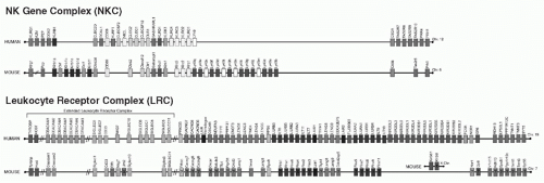
195,201,202,203 Indeed, genetic analysis revealed that the genes for Ly49A and NK1.1 are linked in the NKC (Fig. 17.3), leading to studies indicating that Ly49A is constitutively expressed on a distinct subpopulation (20%) of NK cells in C57BL/6 mice.203


TABLE 17.1 The Panoply of Receptors Expressed by Natural Killer Cellsa


Receptor


H


M


Inhibitory (I) or Activation (A)


Other Names


Ligand


Ly49A


X


I


Ly49, Ly-49, Klra1


H2Dd, Dk, Dp, alleles in H2r, and H2q


Ly49C


X


I


Klra3


H2Kb, numerous


Ly49E


X


I


Klra5


Urokinase plasminogen activator


Ly49G2


X


I


LGL-1, Klra7


H2Dd


Ly49I129


X


I


Klra9


m157


Ly49Q


X


I


Klra17


H2Kb


KIR2DL1


X


I


CD158a, p58.1, EB6


HLA-C2 (HLA-Cw2, Cw4, -Cw5, -Cw6)


KIR2DL2/KIR2DL3


X


I


CD158b, p58.2, GL183


HLA-C1 (HLA-Cw1, -Cw3, -Cw7, -Cw8)


KIR2DL4


X


I


CD158d


HLA-G


KIR3DL1


X


I


CD185e1, NKB1, p70, NKAT3


Bw4 (HLA-A and B)


KIR3DL2


X


I


CD158k, p140, NKAT4


HLA-A3, -A11


Lilrb4


X


I


gp49


αvβ3 integrin


CD94/NKG2A


X


I


Kp43


HLA-E


X


I


Qa-1


LILRB1


X


I


CD85j, ILT2, LIR1


Folded HLA, UL18


LILRB2


X


I


ILT4, LIR2


Folded, free HLA


LAIR-1


X


I


Collagen


Siglec-7


X


I


P75, AIRM1


Carbohydrates


Siglec-10


X


I


Carbohydrates?


Siglec-E


X


I


?


PILRa


X


I


CD99


PILRb


X


I


CD99


FcγRIII


X


X


A


CD16


Fc of IgG


Ly49D


X


A


Chinese hamster MHC-I, H2Dd


Ly49H


X


A


m157


Ly49PMA/My


X


A


M04 + H2Dk


KIR2DS1


X


A


CD158h, p50.1


HLA-Cw7


KIR2DS2


X


A


CD158j, NKAT5, p50.2, clone 49


KIR2DS3


X


A


NKAT7


KIR2DS4


X


A


CD158i, NKAT8, clone 39


HLA-Cw4


KIR2DS5


X


A


CD158g, NKAT9


KIR3DS1


X


A


CD158e2


CD94/NKG2C


X


A


HLA-E


CD94/NKG2C


X


A


Qa-1


NKG2D


X


A, costimulation


KLRK1


MICA, MICB, ULBP/RAET1


NKG2D


X


A, costimulation


Klrk1


H60, RAE1, MULT1


Nkrp1c


X


A


NK1.1


?


Nkrp1b(d)


X


I


Clrb


Nkrp1f


X


A?


Clrg


NKRP1A


X


I


LLT1


NKp80


X


A


AICL


NKp65


X


A


KLRF2


CLEC2A


2B4


X


X


A,I


SLAMF4


CD48


CD2


X


X


A


CD48


NTBA


X


X


A


SLAMF6, Ly108


NTBA


Ly9


X


X


A


SLAMF3


Ly9


CD84


X


X


A


SLAMF5


CD84


CRACC


X


X


A


SLAMF7


CRACC


NKp46


X


X


A


Hemagglutinin


NKp44


X


A


?


NKp30


X


A


B7-H6


CD69


X


X


A


?


Ly6


X


A


?


Gp42


Rat


A


?


Klrg1


X


I


Caherins


CEACAM1


X


I


CD66a


CEA


CD226


X


X


A


DNAM-1


necl-5 (CD155, PVR), nectin-2 (CD112, PVRL2)


CD96


X


A


Tactile


necl-5


CRTAM


X


A


necl-2


TIGIT


X


I


PVR, PVRL2


AICL, activation-induced C-type lectin; AIRM1, adhesion inhibitory receptor 1; CRTAM, class I-restricted T-cell-associated molecule; LGL, large granular lymphocyte; NK, natural killer; TIGIT, T-cell immunoglobulin and immunoreceptor tyrosine-based inhibitory motif domain.


a These are the major receptors discovered on NK cells in humans and mice, listed in order of appearance in the text.


Multiple lines of evidence indicate that Ly49A is an inhibitory receptor specific for MHC-I, particularly H2Dd: 1) Functional analysis: the Ly49A+ NK-cell subset were equivalent to Ly49A- NK cells in killing several targets, but they could not lyse a large panel of targets that were readily lysed by Ly49A- NK cells. This phenotype was related to MHC-I expression of certain H2 haplotypes on target cells.191,204,205 Transfected expression of H-2Dd selectively rendered a susceptible target resistant to natural killing by Ly49A+ NK cells. Moreover, killing through disparate stimuli by Ly49A+ NK cells was also inhibited. 2) Cell binding: Ly49A+ tumor cells bound specifically to immobilized MHC-I molecules206 and to H-2Dd-transfectants.207 3) Antibody blocking: F(ab′)2 fragments of mAb directed against either Ly49A or the α1/α2 (but not the α3 domain) of H-2Dd reversed resistance in killing experiments (permitted lysis) and blocked the cell binding assay.191,204,206,207 4) In vivo expression: the apparent level of Ly49A expressed per NK cell was downregulated in MHC congenic and Tg mice expressing H-2Dd.208,209,210 This was not due to negative selection because the percentage of Ly49A+ NK cells was unchanged. 5) Gene transfer: primary NK cells and T cells expressing a Ly49A transgene and a Ly49A-transfected NK cell line were specifically inhibited by H-2Dd.211,212 6) Inhibition by Ly49A is ITIM-dependent, based on gene transfer of mutant Ly49A molecules.212 7) H2Dd tetramers bind Ly49A transfectants.213 8) Ly49A tetramers bind H2Dd on transfected cells.214,215 9) Biophysical studies: recombinant Ly49A binds recombinant H2Dd in surface plasmon resonance (SPR) studies with KD = ˜2.0 µM.216
10) Crystallography: the structure of Ly49A complexed with H2Dd was determined.217 11) Less extensive studies also indicate that Ly49A recognizes H-2Dk, H2Dp, and alleles in H2r and H2q.191,208,213,218,219 Therefore, Ly49A is an MHC-I-specific receptor for H-2Dd, H-2Dk, and H2Dp, and alleles in H2r and H2q.


FIG. 17.3. The Genomic Organization of the Natural Killer (NK) Gene Complex and Leukocyte Receptor Complex in Humans and Mice. The figures are not drawn to scale with precise gene locations being modified as new sequence information becomes available (genome.ucsc. edu/ and www.ebi.ac.uk/ipd/kir/). The grey shading is coordinated to represent related genes. Question marks indicate genes whose precise location is not known. An “X” indicates genes not homologous to other aligned genes. Double slashes represent large genomic distances. Note that most but not all genes are expressed on NK cells. Many remain orphan genes because the functions of their gene products have not been determined. Modified from Kelley et al.312

The nature of the Ly49A interaction with MHC-I, however, is fundamentally different from TCR/MHC-I interactions because the former appears to be relatively independent of the specific peptide bound by H2Dd.220,221 However, bound peptides are required for appropriately folded MHC-I molecules that can be recognized. Despite its structural homology to C-type lectins that are carbohydrate-binding proteins,222,223 Ly49A does not have the residues for coordinate binding to calcium that is required for lectin binding. Moreover, Ly49A binding to its MHC ligands is not carbohydrate-dependent based on functional, SPR, and crystallographic analyses.
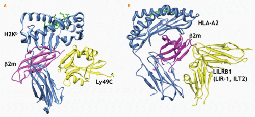
216,224 In the crystallographic structure of Ly49A complexed to H2Dd (2.3Å resolution), the lectin-like structure of Ly49A was confirmed217 (Fig. 17.4). Two interaction sites were seen between the lectin-like domain of Ly49A and H2Dd: site 1 involved the “left” side of the peptide-binding cleft of H2Dd and a wedge-like site 2 involved the undersurface of the peptide-binding cleft. The residues in Ly49A involved in binding either ligand site are overlapping. Mutational analysis revealed that Ly49A binds site 2 where it contacts α1, α2, and α3 of H2Dd and β2m.214,216,225 This site is near Asn80, an Asn-linked glycosylation site conserved in all MHC-I molecules, leaving open the issue of whether carbohydrates could affect the interaction, such as affinities or kinetic parameters, but this has not been studied in depth. These studies also provide a structural explanation for species-specific β2m requirements as revealed by functional studies.226 Thus, Ly49A recognizes site 2 in the MHC molecule in terms of trans recognition between the NK-cell receptor and target cell MHC-I molecule.


FIG. 17.4. Crystal Structures of Natural Killer (NK)-Cell Receptors in Complex with Their Ligands. A: Mouse Ly49C bound to H2Kb at site 2 (PDB ID = 3C8K).193,235 Ly49A interaction with site 2 of H2Dd is very similar.170 Site 1 of Ly49A-H2Dd interaction is approximately located where the H2Kb label is placed. B: Human LILRB1 (LIR1, ILT2) bound to human leukocyte antigen-A2 (PDB ID = 1P7Q).266 Figures were produced using the University of California, San Francisco Chimera package from the Resource for Biocomputing, Visualization, and Informatics at the University of California, San Francisco (www.cgl.ucsf.edu/chimera).1030 The structures are viewed from the side with the NK cell positioned at the top of the figure and the target cell surface at the bottom. The major histocompatibility complex molecules are oriented similarly.

Most but not all other Ly49 receptors are MHCI-specific inhibitory receptors. First noted by Southern blot analysis and cDNA cloning, genome sequence analysis revealed 15 complete Ly49 genes in C57BL/6 mice.199,200,201,202,227 There is evidence for alternative splicing and alternative transcriptional start sites for the Ly49 genes, though their importance has not been elucidated.202,228,229 Notably, Ly49C has broad specificity for H2 alleles, as revealed by tetramer staining, and is the only known inhibitory NKcell receptor specific for an H2b haplotype allele (H2Kb) in C57BL/6 mice, notwithstanding unconfirmed reports that Ly49I also binds H2Kb.168,213,230 Ly49C is recognized by two mAbs: 5E6, which also binds Ly49I, and 4LO3311, which has exquisite specificity for Ly49C.231,232,233 X-ray crystallographic studies have indicated that Ly49C binds H2Kb in a manner similar to Ly49A interaction with H2Dd (see Fig. 17.4).234,235 Only a site 2 interaction was seen with the contact residues, showing a similar but distinct topology to the Ly49A-H2Dd interaction. Interestingly, however, peptides bound to H2Kb clearly affect functional interactions with Ly49C236 and affinities as measured by SPR.234 However, Ly49C does not directly engage the peptide, indicating long-range effects. Finally, both Ly49A and Ly49C
appear to undergo conformational changes upon ligand binding, and a Ly49 dimer can engage two MHC-I molecules.235,237 Thus, Ly49 receptors bind their MHC ligands in a structurally related manner.

Recent studies suggest that Ly49 molecules also can bind MHC in a cis interaction between receptor and ligand on the NK cell itself.168,238 For example, an MHC ligand for Ly49A or Ly49C on the same cell prevents binding of MHC tetramer.168,238 If the cells are briefly exposed to mild acidic conditions, MHC-I expression is lost (due to disruption of the noncovalently linked MHC-I heterotrimer), and Ly49A binding to cognate MHC-tetramers is restored. This cis interaction is dependent on site 2 residues in the MHC molecule. These findings may help explain the observation that the presence of self-MHC ligands leads to downregulation of Ly49 expression, as previously noted on primary NK cells in MHC congenic mice.208,209,210 Cis interactions may also explain functional differences in NK cells in MHC-congenic mice or NK cells that do or do not express MHC ligands in Tg mice that are mosaic for MHC expression.239,240,241 Finally, there are biophysical data supporting a role for the relatively long, flexible stalk region of Ly49 receptors in allowing either trans or cis interactions.242 At the moment, the physiologic importance of cis interactions is incompletely understood but may be relevant to NK-cell tolerance and education, as discussed below.

Although they have not been studied as extensively as Ly49A and Ly49C, other inhibitory Ly49 receptors and their ligands have been identified202,230 and characterized with other MHC allele specificities, as detailed in a recent review.243 Moreover, they are structurally related.235,244 Individual NK cells may express multiple Ly49 receptors simultaneously,210,233,245 often (but not always) two or more, suggesting that individual NK cells may be inhibited by more than one MHC-I molecule.

Ontogenetic studies demonstrate that the total repertoire of Ly49 expression does not reach adult levels until sometime after 3 weeks of age, concomitant with attainment of full NK-cell cytolytic activity.246 Thereafter, the expression of Ly49 receptors is generally thought to be fixed and stable on an individual NK cell. Ly49E is expressed only on fetal NK cells, but NK cells in mice deficient in Ly49E are otherwise normal.247 In adult mice, developmental studies indicate that Ly49 receptors are first expressed on immature NK cells in the BM, before a phase of constitutive proliferation98 that appears to be modestly affected by MHC haplotype.168 There are only modest effects on the final “repertoire” of MHCspecific receptors expressed by splenic NK cells in different MHC environments.210,248

The expression of inhibitory Ly49 receptors appears to occur in a stochastic manner. There is evidence for monoallelic expression of Ly49 receptors (expression from one chromosome), initially described as “allelic exclusion,” a term that has fallen out of favor because it has a specific meaning and mechanism for TCRs and BCRs.249 At least some Ly49 genes possess bidirectional, overlapping promoters directed in opposite orientations.250 Transcription factors driving transcription in one direction prevent binding of other factors driving transcription in the opposite direction. Directionality and monoallelic expression may also be controlled by DNA methylation.251 A “probabilistic” model has been proposed to explain these findings that may also explain the stochastic expression of Ly49 genes and their stable expression. However, recent studies of Ly49 indicate highly variable transcriptional start sites, suggesting that the probabilistic model may not be correct.229 Other data indicate that TCF-1 but not LEF-1 in the T-cell factor/lymphoid enhancer family of DNA-binding proteins affects some but not all Ly49 receptor expression.252,253,254 Thus, the elements controlling Ly49 gene expression are incompletely understood.

Analysis of the Ly49 receptors thus far is largely based on examination of the C57BL/6 alleles, but the Ly49 receptors display extensive polymorphism. The Ly49 family is encoded in the NKC located on mouse chromosome 6 with the syntenic human region being chromosome 12p13.2195,203,255,256 (see Fig. 17.3). While the NKC also contains genes for other lectin-like receptors, the Ly49 genes are clustered with the exception of Ly49b. Corresponding to restriction fragment length polymorphic (RFLP) variants originally detected with the Ly49A cDNA,203 there is significant allelic polymorphism of the Ly49 cluster between inbred mouse strains with differences in gene number as well as alleles for the Ly49 genes.227,257,258,259 In contrast to C57BL/6J mice, genomic sequence analysis shows 8 putative Ly49 genes in BALB/c mice, 19 in 129 mice (of which at least 9 appear to be pseudogenes) and 22 in NOD mice. Array-based comparative genomic hybridization analysis of 21 mouse strains compared to the reference C57BL/6J strain indicated that these mice could be grouped into five clusters that correspond to or are predictive of restriction fragment length polymorphic patterns on Southern blot analysis.203,260 There are also multiple alleles for individual Ly49 family members.227,249,257,258,261 Thus, there is significant polymorphism of the Ly49 molecules at both the haplotype (gene numbers) and individual gene (alleles) levels, not unexpected because the Ly49 molecules bind highly polymorphic MHC-I molecules.

The MHC-I specificities have generally been well characterized for only a few Ly49 alleles. Interestingly, mAbs specific for one Ly49 allele may bind another molecule with a different function or specificity in another mouse strain,262,263,264 similar to what was recognized for mAb reactivity with different MHC alleles.265 Thus, the polymorphisms also raise practical issues when studying Ly49 molecules in different mouse strains.

Finally, it should be noted that Ly49 receptors may be expressed by other cells; some are selectively expressed on non-NK cells and some may have specificities for non-MHC ligands. NKT and other T-cell subsets may express Ly49 receptors but they have not been thoroughly studied.266,267 Ly49B and Ly49Q are not expressed on NK cells; rather, they are expressed on myeloid cells.268,269 Interestingly, Ly49Q recognizes H2Kb and positively regulates TLR signaling.270,271 On the other hand, Ly49E appears to recognize urokinase plasminogen activator, though physical binding has not been established.272 Interestingly, Ly49B, Ly49E, and Ly49Q are predicted to be distinct in fine structure from the
known MHC-I-specific Ly49 receptors on NK cells.235 Thus, while Ly49 receptors are predominantly NK-cell inhibitory receptors for MHC class I, they may have other roles that are less well understood; still other Ly49 receptors have activation function, as discussed subsequently.


Human Killer Immunoglobulin-like Receptors

In contrast to mouse NK cells, human NK cells can be cloned by limiting dilution in the presence of irradiated feeder cells, phytohemagglutinin, and IL-2, leading to establishment of short-term NK-cell clones that have differences in target killing and surface molecules. mAbs were isolated that reacted specifically with these clones; reactivity correlated with the capacity of the clones to kill certain tumors, and the mAbs affected cytotoxicity. This general approach led to the identification of the human NK-cell receptors.

A series of studies273,274,275 showed that the mAbs GL183 and EB6 identify serologically distinct 55 kDa or 58 kDa molecules, initially termed p58. These molecules had several features: 1) selective expression on overlapping NK cell subsets; 2) expression on NK cell clones correlated with expression of certain human leukocyte antigen (HLA) class I alleles on resistant targets; 3) a target susceptible to a given NK-cell clone bearing p58 molecules reactive with either mAb was made resistant by transfection of cDNAs encoding certain HLA-C molecules; 4) the otherwise resistant, HLA-C-transfected targets could be lysed in the presence of the appropriate anti-p58 mAbs. The mAb effect occurred with F(ab′)2 fragments, suggesting that the interaction between p58 and an HLA class I molecule on the target cell inhibits the NK cell. Thus, the p58 molecules displayed features consistent with a role as inhibitory human NK-cell receptors specific for MHC-I, analogous to the mouse Ly49A receptor that was being studied in parallel, as described previously.

Other studies noted that NK-cell specificity was skewed when the NK cells were grown in the presence of cells bearing allo-MHC determinants.276,277 This specificity correlated with reactivities that mapped to paired residues at position 77 and 80 in the .α1 domain of HLA-C. All known HLA-C molecules could be divided into two groups, one with Asn77-Lys80 (HLA-Cw2, -Cw4, -Cw5, -Cw6) and the other with Ser77-Asn80 (HLA-Cw1, -Cw3, -Cw7, -Cw8). Indeed, transfection analysis showed that p58 specificity for HLA-C molecules was related to expression of the EB6 epitope for the former (specificity 1, now termed HLA-C1), whereas the latter was related to the GL183 epitope (specificity 2, HLAC2) on the NK-cell clones.276,278,279 Thus, human NK-cell receptors showed promiscuous specificity that was dependent on residues 77 and 80 in HLA-C.

The NKB1 (p70) molecule was serologically similar to p58 molecules with regard to subset expression, and correlation of expression on NK-cell clones to specificity for HLA class I.280,281 In contrast to p58 molecules, however, NKB1 had a distinct Mr (70 kDa) and specificity for HLA-B. The NKB1+ clones were specifically inhibited by targets expressing transfected HLA-Bw4 molecules, and the anti-NKB1 mAb reversed the inhibition. Analysis of informative HLA-B alleles showed that this specificity was conferred by a region in the α1 domain overlapping the area on HLA-C recognized by p58 molecules.282 Finally, HLA-A3, -A11-specific receptors have similar properties to p58 and NKB1 except that they appear to be disulfide-linked dimers termed p140,283 whereas others have found that a monomeric HLA-A3-specific receptor resembles NKB1.284 Thus, representative alleles of all classical HLA class I loci are capable of inhibiting NK cells through p58/NKB1/p140 receptors although HLA-B and -C alleles dominate human NK-cell specificities, and it is not yet known if there are receptors reactive with each HLA allele.

When the cDNAs for the p58 and NKB1 molecules were cloned, they were surprisingly found to encode type I integral membrane proteins with Ig-like domains285,286,287 unlike the lectin-like Ly49 family of type II receptors. The Ig-like receptors are now collectively known as killer Iq-like receptors (KIRs) or CD158.288,289 The KIR nomenclature is based on whether the receptor has two or three Ig-like external domains (KIR2D or KIR3D, respectively), and possession of a long (L) or short (S) cytoplasmic domain. In general, the L forms are inhibitory because they contain ITIMs, whereas the S forms appear to be activation receptors (see following discussion). Each distinct receptor is also designated by a number. The KIR2DL1 (CD158a, p58.1) molecule bears the original EB6 epitope and is specific for HLA-C (Lys80, specificity 2), whereas KIR2DL2 (CD158b1, p58.2) and KIR2DL3 (CD158b2, p58) have the GL183 epitope and are specific for HLA-C (Asn80, specificity 1). (As detailed in the following, structural analysis supports grouping of HLA-C alleles into two mutually exclusive groups, HLA-C1 and HLA-C2, based on direct interaction of KIR2DL2/3 and KIR2DL1, respectively, with residue 80 of HLA-C, validating original functional groupings but simplifying HLA-C groupings to just residue 80.276,277) KIR2DL4 (CD158d, p49) reportedly binds HLA-G290 but displays both inhibitory and activation functions.291,292,293 KIR3DL1 (CD158e1, NKB1, NKAT3) is specific for HLA-A and HLA-B molecules with the Bw4 epitope.282 KIR3DL2 (CD158k, p140, NKAT4) has HLA-A3 and HLA-A11 specificity.283,284

There is unequivocal evidence that the KIR2DL and KIR3DL molecules are inhibitory HLA class I-specific receptors. In addition to the data with NK cell clones and mAbs mentioned previously, the following have been described: 1) KIR bind directly to HLA class I: soluble KIR2DL-Fc fusion proteins bind cells expressing the appropriate transfected HLA class I alleles.294,295 In addition, a soluble KIR2DL molecule containing only the extracellular domain binds specifically to its HLA-C ligand in solution.296 2) Gene transfer of KIR: KIR2DL specificity and inhibitory function were transferred when KIR2DL cDNAs were transiently expressed with vaccinia constructs in human NK-cell clones.294 Similarly, Tg expression of KIR2DL2 in mice conferred inhibition of rejection of BM expressing Tg HLA-Cw3.297 3) SPR measurements indicate that the KIRs bind their HLA ligands with Kd = ˜10 µM.298,299,300,301 Binding is affected by peptide bound by HLA molecule.300,301 Through histidine-rich domains, the KIRs bind Zn++, which affects KIR multimerization and binding kinetics to HLA ligands.302,303 4) Crystallographic studies demonstrate
KIR2DL1 (2.8 Å resolution) and KIR2DL2 (3.0 Å resolution) interactions with their cognate HLA ligands.299,304 Thus, KIR molecules are clearly MHC-I-specific inhibitory receptors on NK cells.

Interestingly, structural studies indicate that KIR molecules bind HLA class I molecules in a manner analogous to recognition of MHC by TCRs (Fig. 17.5). In particular, both KIR2DL1 and KIR2DL2 use surface loops near their interdomain hinge regions to bind their cognate HLA-C ligands (Cw4 and Cw3, respectively) with a footprint overlying the “right” side of the peptide-binding cleft (when viewed from the “top” in standard depictions of MHC-I molecules).
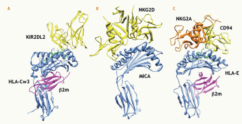
299,304 The receptors bind both α1 and α2 helices with interactions between KIR2DL1 and Lys80 of HLA-Cw4 and between KIR2DL2 and Asn80 of HLA-Cw3. These interactions with residue 80 of the HLA-C molecules were lost when the reciprocal residues were swapped, accounting for the previously described HLA-C groupings and KIR specificities in functional studies276,277 and mutational analysis indicating that residue 80 is more significant for KIR interaction than residue 77.305,306,307,308 Although neither KIR2DL molecule has extensive contacts with peptides bound to HLA-C, KIR2DL interactions with HLA-C imposes physical constraints on the p8 position of the peptide. This may account for observed peptide preferences in functional studies and antagonism of certain peptides on KIR inhibitory function.309,310 A recent structure (1.8 Å resolution) of KIR3DL1 complexed to HLA-B*5701 revealed a similar recognition strategy.301 Thus, KIRs and Ly49s bind their MHC ligands in markedly different ways, despite their analogous functions as MHC-specific inhibitory receptors.


FIG. 17.5. Additional Structures of Natural Killer-Cell Receptors in Complex with Their Ligands. A: Human KIR2DL2 with HLA-Cw3 (PDB ID = 1EFX).236 Two KIR molecules are apparent in the crystal structure with only one molecule (killer Ig-like receptor [KIR] A) contacting the human leukocyte antigen (HLA) molecule. In this view, KIR B molecule is not shown. B: Human NKG2D with MICA (PDB ID = 1HYR).412 C: Human CD94/NKG2A with HLA-E (PDB ID=3CDG).382 The figures were produced and oriented as described in Figure 17.4.

Also unlike the Ly49s, the KIRs are encoded in the LRC on human chromosome 19q13.4 that encodes many other Ig-like receptors (see Fig. 17.3)311; the KIR genes are clustered toward the telomeric end of the LRC.312 Interestingly, the mouse LRC on chromosome 7qA1 does not include genes for KIR-like molecules that instead are encoded on the X chromosome.313,314 On the other hand, like the Ly49s, the KIRs display remarkable polymorphism with at least 11 genes315,316,317 (see www.ebi.ac.uk/ipd/kir/index.html for updated database).

The human KIR locus also demonstrates considerable haplotype diversity with at least 27 different haplotypes,315,316,318 recently defined at the sequence level.317 While there has not been a consensus definition, two major types of haplotypes have been described.289 Group B contains one or more of the following: KIR2DL5, KIR2DS1, KIR2DS2, KIR2DS3, KIR2DS5, and KIR3DS1, whereas group A haplotypes have none of these genes. Reflecting extensive allelic polymorphism of individual genes, a large number of different KIR genotypes have been described, and they are distributed differently in the various ethnic populations.

As with the Ly49s, only a few KIR alleles have been well characterized with respect to HLA class I specificities. Nonetheless, these genetic variants have not only provided clues to new receptors and ligand specificities but also valuable links to the role of NK cells and their receptors in
disease pathogenesis (discussed in the subsequent clinical section), and more broadly, human evolution.


Convergent Evolution of Major Histocompatibility Complex-Specific Natural Killer-Cell Receptors

Despite the controversy surrounding the initial cloning of mouse Ly49s and human KIRs and leukocyte Ig-like receptors (LILRs), additional data have provided new interpretations of the distinctly different receptors used by mouse and human NK cells, respectively, to recognize MHC-I. Detailed genome sequence information is available on the NKC and LRC in mice, humans, and other species. In the mouse, MHC-specific NK-cell receptors with Ig-like domains have not yet been described, although there is conservation of several genes in the broader LRC on mouse chromosome 7312 (see Fig. 17.3). Activated mouse NK cells do express gp49b (Lilrb4), an Ig-like inhibitory receptor also expressed on other leukocytes, including mast cells.319,320,321,322 However, it is not expressed by resting NK cells and is not specific for MHC-I. Instead, it binds the integrin αvβ3 and appears more important for responses of other cells, such as neutrophil, eosinophil, and DCs.323,324,325,326,327,328 Mouse Kir3dl1 is on the X chromosome (see Fig. 17.3) and expressed in NK and T cells, but its function and ligand remain unknown.313,314 The Ly49 locus in humans consists only of LY49L (KLRA1) that is a pseudogene because of a point mutation that gives rise to a splicing abnormality.329 Thus, current data indicate that mouse NK cells do not express functional KIR orthologues while human NK cells do not express functional Ly49 orthologues.

One reason for this discrepancy may be that the corresponding orthologue is present in the genome but has not been identified. Indeed, genomic sequencing has revealed a multitude of candidate orphan receptors in the genome and specifically the LRC and NKC that have yet to be studied carefully.311,330 In that regard, identification and functional analyses of NK cell receptors led to the recognition that they are encoded in genomic regions containing gene clusters for related receptors that are expressed on other leukocytes, not just NK cells. Dissection of the expression and function of these receptors is a rich area of research that is beyond the scope of this chapter.

The alternative and currently favored view for the discrepancy is that mice and humans independently evolved analogous receptors to serve the same function. While both human and mouse NK cells express a conserved relatively nonpolymorphic lectin-like receptor, CD94/NKG2, it does not possess many of the features that are shared by mouse Ly49 and human KIRs:


  • both Ly49s and KIRs are constitutively and selectively expressed on naïve, unstimulated NK cells (with exceptions for rare populations of T cells);


  • both bind MHC-I molecules with intermediate affinity (KD = 2-10 µM);


  • binding to MHC is promiscuous;


  • MHC-bound peptides have only a modest effect, if at all, on recognition;


  • both use ITIMs to inhibit NK-cell activation;


  • they are expressed in a stochastic fashion on overlapping subsets of NK cells;


  • a single NK cell simultaneously expresses one or more of either type of inhibitory receptor (each may be functional);


  • once they are expressed, their expression appears to be stable;


  • both are germ-line encoded by small families of genes that are clustered in the genome;


  • both display impressive polymorphism, in terms of gene number and alleles for each gene;


  • both are related to molecules that lack ITIMs and instead are activation receptors; and


  • both are involved in NK-cell education by self-MHCI (see following discussion).

Thus, the mouse Ly49 receptors and human KIRs are analogous receptors in an apparently striking example of convergent evolution,331 whereby each species came up with a different genetic solution to provide extremely important functions for species reproduction and survival.

In other species, Ly49 and KIR genes have been analyzed primarily with respect to sequence and gene number.332 For example, the LY49L gene in baboons appears to be functional but the putative polypeptide lacks an ITIM.333 In rats, the Ly49 cluster appears to have markedly expanded with at least 25 genes,334,335 demonstrating one of the most rapid rates of gene expansion.336 Dog, cat, and pig appear to have only one Ly49, whereas horse represents the only known nonrodent mammal with several Ly49 genes.337 The chicken genome has several lectin-like receptor genes that are genetically linked to the MHC.338,339,340 On the other hand, multiple KIR genes have been described in primates and cattle.341,342,343,344 Rhesus macaques have a profound plasticity of KIRs with multiple genotypes and haplotypes345 encoding receptors that bind MHC-I.346,347 In rat, a KIRlike sequence has been reported,314 and dogs and cats lack functional KIRs.348 Interestingly, pigs and marine carnivores each possess a single Ly49 and KIR gene, but it is not clear if these are functional.348 Finally, several species have no readily identifiable Ly49 or KIR genes332 (eg, teleost fish instead possess a large number of novel immunetype receptors with sequence homology to mammalian LRC-encoded receptors349). Perhaps these species have had alternative convergent evolutionary stratetgies to preserve inhibitory MHC-I-specific receptors.

Additional studies of the Ly49-like and KIR-like molecules as well as potential new orthologues in other species will be of interest to evolutionary biologists for several reasons including prior description of NK-like cells in lower vertebrates and MHC genes that are coevolving.332,350 Moreover, in primitive chordates, NK-like, missing-self-like recognition affects histocompatiblity reactions351 but the molecular determinants of histocompatibility involves molecules unrelated to mammalian Ly49, KIR, or even MHC itself.352,353,354 Thus, evolutionary studies of NK-cell receptors and their ligands may provide unique insight into missingself and other histocompatiblity reactions.

While we have focused thus far on mouse Ly49 and human KIR as the major MHC-I-specific receptors, there are other well-described NK-cell receptors belonging to
either the lectin-like receptor or Ig-like receptor superfamilies. These receptors also have specificities for MHC-I molecules.


Human and Mouse CD94/NKG2

The analysis of CD94 (Klrd1) and NKG2 (Klrc, excluding NKG2D [Klrk1]) family of molecules was especially challenging and required insightful investigations. Identified by subtractive hybridization, the human NKG2 molecules are type II integral membrane proteins with external C-type lectin domains355 encoded in the NKC356 (see Fig. 17.3). Initial attempts to express NKG2 molecules on the cell surface were thwarted. Meanwhile, mAb reactivity suggested that CD94 was variably expressed on human NK cells as a disulfidelinked dimer (70 kDa NR, 43 kDa R),357 and both activation and inhibition functions for CD94 were described.358,359,360,361 Surprisingly, cDNA cloning revealed that CD94 has a short seven amino acid cytoplasmic domain, suggesting that it cannot signal on its own.360 Furthermore, anti-CD94 immunoprecipitates were not detectable from radiolabeled CD94 transfectants, despite easily detectable expression on FACS analysis with the same mAbs.362,363 These apparent discrepancies were resolved when it became clear that CD94 heterodimerizes with NKG2 molecules362; NKG2A is the 43 kDa molecule previously identified as Kp43 with anti-CD94 mAbs.364,365 While CD94 may be expressed as a homodimer, the NKG2 partner provides the signaling motif, whether activation or inhibition.364,366 (NKG2B is an alternatively spliced form of NKG2A. The rest of the NKG2 family is discussed in the following.)

The ligand specificity for CD94/NKG2 receptors was also initially thought to be promiscuous as interactions with many classical (class Ia) and nonclassical (class Ib) HLA molecules had been described.359,362,364,367,368,369,370 However, human CD94/NKG2 receptors directly recognize HLA-E, a MHC-Ib molecule homologous to mouse Qa-1.371,372,373 HLA-E (and Qa-1) is widely expressed with limited polymorphism.374,375,376 While HLA-E heavy chain is expressed with β2m and a peptide occupying its peptide-binding cleft, its peptide repertoire is largely derived from the leader sequences of MHC-Ia molecules, as previously noted for mouse Qa-1.377,378 HLA-E (or Qa-1) expression thus requires normal production of HLA-E (or Qa-1) and synthesis of certain MHC-Ia molecules. Mouse CD94/NKG2 recognizes Qa-1 that shares many features with HLA-E.379,380 These findings need to be considered in the context of the prevailing view at the time that mouse and human NK cells use structurally different receptors to recognize MHC-I molecules.381 Clearly, the CD94/NKG2 receptors and their ligands are conserved in humans and mice.

The crystal structure of human CD94/NKG2A bound to HLA-E was resolved to 2.5Å and 4.4Å resolution.382,383 Remarkably, CD94/NKG2A interfaces with HLA-E in a manner analogous to TCR recognition of peptide-loaded MHC-I,384 including TCR binding to HLA-E itself.385 Both innate and adaptive receptors for HLA-E lay across the peptide-binding cleft, though peptide itself plays a relatively minor role in binding CD94/NKG2A.382,383 Strikingly, this binding is distinct from that of Ly49 receptors to their classical MHC-I ligands (see previous discussion). On the other hand, this binding interface is similar to binding of another NKC-encoded, lectin-like receptor, the NKG2D activation receptor, to its MHC-like ligands (see following discussion). Thus, NKC-encoded, lectin-like receptors surprisingly use different strategies to contact their ligands, even though these ligands have structurally related MHC-I folds.

Despite its conservation between mice and humans, the role of CD94/NKG2 receptors in NK-cell function is still incompletely understood. For example, viruses encode peptides that bind and enhance expression of HLA-E, providing a CD94/NKG2A-dependent mechanism to avoid NK-cell attack.386,387 By contrast, human NK cells expressing CD94/NKG2C (an activation receptor) expand in response to cytomegalovirus (CMV)-infected targets.388 When a large number of human NK-cell clones were obtained from two normal individuals, CD94/NKG2 seemed to account for the majority of self-MHC-specific receptors on clones from one individual whereas KIRs dominated the self-specific receptors on clones from the other individual, suggesting that some individuals may depend on CD94/NKG2 for self-tolerance.389 Qa-1 and HLA-E can present peptides derived from other molecules, including the signal sequence of heat shock protein 60 (Hsp60) that is induced by a number of stimuli,390,391 a multidrug resistance transporter,392 or blastocyst MHC expressed in embryonic tissues.393 This may result in loss or gain of recognition by the inhibitory CD94/NKG2A receptor, suggesting intrinsic mechanisms to perturb inhibition by CD94/NKG2A in certain circumstances.

Yet, CD94 appears to be dispensable in certain strains of mice, such as DBA/2J, that do not appear to have any untoward NK-cell phenotype.394 This finding has been recapitulated in studies of a CD94 knockout mouse on the 129 strain background.395 Interestingly, CD94 knockout mice are more susceptible to ectromelia virus,396 as detailed below.

CD94/NKG2 molecules may be important in T-cell function. CD94/NKG2A is rapidly induced on antigenspecific CD8+ T cells during polyoma virus and other infections,397,398,399 and CD8+ T cells expressing CD94/NKG2A preferentially proliferate during persistent infection, suggesting that CD94/NKG2 receptors may play a role in memory T-cell responses,400 and that TCR specificity is correlated with CD94/NKG2A expression by human CTL.401 Indeed, CD94/NKG2A inhibits antigen-specific cytotoxicity in polyoma virus responses, although this effect is pathogen-dependent. CD94/NKG2A has been studied with other viral infections, including herpes simplex virus,402 murine CMV,403 gHV68, and influenza,404 for example. Although not all studies revealed functional consequences, the recurring theme is the appearance of CD94/NKG2A on previously activated CD8+ T cells. Thus, CD94/NKG2 receptors may regulate T-cell responses.


Other Human Immunoglobulin-like Receptors Specific for Major Histocompatibility Complex-I Molecules

The human LILR family is encoded in the LRC (see Fig. 17.3), just centromeric to the KIR genes. There are two general forms of these receptors, subfamily A that appears to be activation receptors, and subfamily B that has the ITIMs
characteristic of inhibitory receptors. The best characterized members, LILRB1 (also known as CD85j, Ig-like transcript 2 [ILT2], or leukocyte IG-like receptor 1 [LILR1]) is broadly expressed whereas LILRB2 (CD85d, ILT4, or LIR2) is not expressed on NK cells but is expressed by myelomonocytic cells, including DCs and monocytes. Both recognize HLA class I molecules405,406 with LILRB1 exclusively binding folded HLA class I molecules with β2m, whereas LILRB2 can bind both folded and free HLA class I heavy chains.407 Interestingly, a human CMV protein, UL18, binds LILRB1 with 1000-fold higher affinity than HLA molecules, implicating a role for LILRB1 in host defense.408 The ligands and functions of other LILRBs (LILRB3 [CD85a, ILT5, LIR3], LILRB4 [CD85k, ILT3, LIR5], LILRB5 [CD85c, LIR8], and LILRB6 [CD85b]) are as yet unknown but they may not be able to bind HLA due to structural constraints.409

LILRB1 has four Ig-like domains and binds a conserved region in the α3 domain of most, if not all, classical and nonclassical HLA class I molecules (HLA-A, -B, -C, -E, -F, and -G).408 Interestingly, the crystal structure of LILRB1 bound to HLA-A2 (3.4Å resolution) reveals that it binds MHC molecules under the peptide-binding domain where it contacts α3 and β2m, more akin to Ly49 engagement of MHC than KIR409 (see Figs. 17.4 and 17.5). Even though LILRB1 and LILRB2 have differing capacities to bind free HLA molecules, LILRB2 has an HLA class I binding site that overlaps with but is distinct from that for LILRB1.410 LILRB1 binds UL18 in a manner structurally similar to HLA class I binding.411

Interestingly, LILR genes demonstrate allelic polymorphisms,412 though less so than the adjacent KIR cluster.413 Nonetheless, polymorphisms in LILRB1 may affect receptor expression.414 Moreover, polymorphisms in LILR genes are associated with certain autoimmune diseases, such as rheumatoid arthritis.415


Major Histocompatibility Complex-Independent Natural Killer-Cell Inhibitory Receptors

As already mentioned, NK cells also express inhibitory receptors for non-MHC ligands, such as mouse gp49b, which binds the αvβ3 integrin,323 and there is a growing list of other such molecules.416 Some of these receptors will be discussed in a more appropriate context in the following sections. Most of these receptors contain cytoplasmic ITIMs so their inhibitory function can be predicted even if not directly tested, though some caution is required because the motifs may be involved in other signaling processes.

Human and mouse NK cells express the ITIM-bearing leukocyte-associated Ig-like receptor 1 (LAIR-1, CD305), which is an Ig-like molecule broadly expressed by most leukocytes and encoded in the LRC.417,418 Initial reports indicating that LAIR-1 binds epithelial cellular adhesion molecule were irreproducible.419,420 Instead, LAIR-1 binds multiple forms of collagen,421 which has been validated in crystallographic and biochemical studies.422 Interestingly, LAIR-1 mediates inhibition that is independent of Src homology 2 (SH2)-domain-containing phosphatases, and instead recruits C-terminal Src kinase (Csk),423 suggesting that Csk may be involved in inhibitory signaling. Although the in vivo context for functional interaction awaits further characterization, it is reminiscent of the broader reactivity of the Siglecs.

The CD33-related sialic acid binding Ig-like lectins (CD33rSiglecs) are type I receptors with varying numbers of Ig-like domains expressed on a broad array of cells and encoded in the “extended” LRC312,424,425 (see Fig. 17.3). Despite having sialic acid recognition in common, the Siglecs appear to show differences in carbohydrate recognition, depending on the specific glycan context.424 Human NK cells express Siglec-7 (p75, adhesion inhibitory receptor 1 [AIRM1]) and Siglec-10, whereas some mouse NK cells express a related Siglec-E.425 Siglec-7 has been most extensively studied. As expected, its cytoplasmic ITIM can recruit SHP-1 and inhibit NK-cell functions.426,427 Moreover, expression of its ligand on targets inhibits NK cells in a Siglec-dependent manner.428 However, the effects appear to be modulated by cis interactions between the Siglec receptor and its carbohydrate ligands on the NK cell itself,424,428,429 reminiscent of cis interactions between Ly49 and MHC ligands, as previously discussed.

The Ig-like receptors, termed paired Ig-like receptor (PILR)α and β, are not encoded in the LRC, rather on human chromosome 7 (mouse chromosome 5).430,431 Mouse PILRα is an ITIM-containing inhibitory receptor, whereas PILRβ is an activation receptor that couples to DAP12.430,432 Both receptors are expressed on NK (and other immune) cells and recognize CD99. Interestingly, sialylated O-linked glycans on CD99 are involved in recognition by PILRs.433

Thus, NK cells (and other leukocytes) express multiple inhibitory receptors that are capable of MHC-independent recognition. How they participate in NK-cell responses and contribute to MHC-dependent effects are beginning to be elucidated, and some appear to play a specific role in the context of activation receptors, as discussed in the following.


Natural Killer-Cell Activation Receptors

NK cells clearly kill MHC-I-deficient targets more efficiently than MHC-I-sufficient targets. However, this enhanced killing does not occur simply because a nonspecific default pathway is unleashed when MHC-I is absent. Instead, it is clear that susceptible targets express ligands for NK-cell activation receptors. In general, these receptors and their ligands were defined following description of the inhibitory receptors.


Approaches to Identification of Activation Receptors

Initial progress in elucidating NK-cell activation receptors was difficult. The approaches that yielded the molecular definition of the TCR, such as subtractive hybridization, mutagenesis of T-cell tumors, and anticlonotypic mAbs,434,435,436 were of limited success.437,438 Unlike the working paradigm of MHC restriction that guided the molecular identification of the TCR, the principles guiding NK-cell activation by targets were unclear. Breakthroughs in identifying NK-cell activation receptors thus required other approaches.

Some activation receptors were recognized because they were first identified on other cells, such as FcγRIII. Other
activation receptors were identified by genetic means, such as cDNA clones for molecules resembling the inhibitory receptors but lacking cytoplasmic ITIMs. Others were identified by a genetic positional cloning approach. Specific stimulation of NK cells through mAbs proved useful for the initial identification of candidate activation receptors and to validate the activation function of receptors identified by other means.

NK cells can be stimulated to mediate antibody-dependent cellular cytotoxicity (ADCC) through the FcγRIII (CD16) receptor that binds the Fc portion of the IgG coating a target. In a related way, anti-FcγRIII can also trigger through CD16 in a process termed “redirected lysis” or “reverse ADCC” because the antibody binds in the opposite orientation to ADCC. A few mAbs against other NK-cell surface molecules also can activate in the redirected lysis assay, highlighting a relatively unique functional property of the recognized molecules (and mAbs), because activation does not occur when most NK-cell surface molecules are cross-linked. First, popularized for analysis of anti-TCR antibodies,439 redirected lysis occurs when IgG reacts specifically with the NK-cell receptor, and its Fc portion binds a target cell Fc receptor (FcγR) that apparently provides bridging and cross-linking effects.439 Target lysis does not occur if FcγR binding on the target is prevented with FcγR-deficient targets, F(ab′)2 fragments of the anti-NK-cell receptor antibody, or anti-target cell FcγR Ab blockade. In the latter case, Fc regions must be removed to prevent inadvertent triggering of conventional ADCC via CD16 on the NK cell. Thus, the redirected lysis assay is a helpful experimental tool.

Gene transfer studies have been helpful adjuncts to study NK-cell receptors. NK cells are difficult to transfect, and there are few useful tumors with the notable exception of RNK-16, a rat NK tumor line.440 Viral vectors, such as vaccinia virus, have been useful for gene transfer with the caveat that functional experiments have to be performed within a small time frame before untoward effects occur.441 Recent use of lentiviral vectors also show promise for gene transfer into primary NK cells.442

Recent studies have exploited reporter cell assay systems similar to those used to identify TCR ligands.443 ITAMmediated signaling leads to inducible, nuclear factor of activated T cells (NFAT)-dependent expression of a reporter molecule, such as β-galactosidase or GFP. Even an inhibitory receptor can be used to activate the reporter cell by fusion of its extracellular domain to a suitable transmembrane and cytoplasmic domain containing ITAMs. Such reporter cells can then be used to detect ligands.444,445,446

In the following sections, we will describe NK-cell activation receptors with an emphasis on those with known ligands.


Activation Receptors Related to Major Histocompatibility Complex-Specific Inhibitory Receptors

Despite initial characterization as inhibitory receptors for MHC-I, the Ly49 family contains other members (ie, Ly49D and Ly49H in C57BL/6 mice) without cytoplasmic ITIMs. Instead, they are activation receptors containing charged transmembrane residues for association with DAP12 that facilitates expression and provides signal transduction capacity.447,448,449,450 Although Ly49H will be considered below in the context of viral infection, Ly49D has no known role in viral defense. A positional cloning approach indicated that Ly49D is the product of the Chok locus, which controls NKcell specificity for killing of a xenogeneic target, Chinese hamster ovary cells,159,451 due to recognition of a Chinese hamster MHC-I molecule.452 Interestingly, when Ly49D was transfected into RNK-16 cells, Ly49D can recognize H2Dd,440 but H2Dd tetramers do not bind Ly49D for unclear reasons, although potentially reflecting lower avidity.213,262 Several other Ly49 receptors have been identified in non-C57BL/6 mouse strains that have properties of activation receptors (charged transmembrane residues, no ITIMs) but they have been less well characterized,257,258 except for Ly49P in MA/My mice, which is involved in controlling viral infection that is related to MHC-I recognition453 (see subsequent details). Thus, some members of the Ly49 family are activation receptors with apparent specificity for MHC-I, potentially with less avidity.

Molecular cloning of the KIR family also led to the identification of two domain receptors (also known as p50) or three domain Ig-like receptors with short cytoplasmic domains lacking the ITIM.454,455 These molecules are now known as the KIR2DS or KIR3DS, respectively, with numbers for specific molecules (ie, KIR2DS1 [CD158h, p50.1], KIR2DS2 [CD185j, NKAT5, p50.2, clone 49], KIR2DS3 [NKAT7], KIR2DS4 [CD158i, NKAT8, clone 39], KIR2DS5 [CD158g, NKAT9], and KIR3DS1 [CD158e2]). Expression of these molecules may be difficult to determine because mAbs for KIR2DL molecules cross-react with KIR2DS molecules.456 KIR2DS molecules can associate with DAP12 and activate NK cells in the redirected lysis assay.454,457 KIR2DS and KIR3DS molecules can recognize HLA class I molecules with specificities similar to corresponding inhibitory KIRs, apparently with lower avidity.458,459,460,461 However, KIR2DS4 and HLA-C alleles and KIR3DS1 and HLA-B alleles influence their respective interactions.462,463 Thus, further analysis is needed to support the hypothesis that activating forms of KIRs bind HLA alleles less well than the inhibitory receptors as a potential explanation for dominance of inhibition over activation (see Fig. 17.2D).

It remains possible that the activating forms of the Ly49s and KIRs have other ligands and perhaps their MHC specificities are instead somehow related to their physiologically relevant ligand. Indeed, this has been demonstrated for Ly49H, an activation receptor that recognizes a virusencoded ligand with an MHC-I-like fold.444,446,464 Moreover, KIR2DS4 may recognize a non-MHC ligand.465 Thus, further analysis is required for understanding the role of Ly49 and KIR activation receptors in MHC-I recognition and with respect to their inhibitory counterparts.

In addition to NKG2A, the NKG2 family also contains NKG2C, NKG2E, and NKG2F, which are products of different genes.355,466 (NKG2B is an alternatively spliced isoform of NKG2A, whereas NKG2H is an alternatively spliced isoform of NKG2E.) NKG2C and NKG2E lack cytoplasmic ITIMs and contain charged transmembrane residues for association with DAP12.467 While the role of NKG2F is unknown
because it lacks an external domain and remains inside the cell, associated with DAP12 but not with CD94,468 NKG2C and NKG2E form functional heterodimers with CD94.364 Like CD94/NKG2A receptors, these heterodimers recognize HLA-E or Qa-1, but unlike CD94/NKG2A receptors, they activate NK cells.469,470 Interestingly, the inhibitory form binds with higher affinity to HLA-E than the activating form.382,383,471,472 There also appears to be some peptide preference between the different functional forms382,383,473 that may be relevant in certain physiologic situations. Thus, the CD94/NKG2 receptors may discriminate between subtle differences in their MHC-Ib ligands.


FcγRIII (CD16)

Frequently overlooked but perhaps the first molecularly defined activation receptor on NK cells is FcγRIII (CD16), through which NK cells mediate ADCC against IgG-coated targets.474,475 Unlike other Fcγ receptor-bearing effector cells, NK cells are generally thought to express only one of the known Fcγ receptors that binds IgG with low affinity,476 although others suggest that human NK cells may express FcγRII isoforms.477 There are two human FcγRIII isoforms with identical extracellular domains.476 Human NK cells express only FcγRIIIA., which is a transmembrane molecule, whereas FcγRIIIB has a glycosylphosphatidyl-inositol (GPI) linkage and is expressed by neutrophils. In mice, only the transmembrane isoform (FcγRIII) is present478 and displays 95% sequence conservation with muFcγRII. The recently described FcγRIV is a newly recognized orthologue of human CD16A but is not expressed on mouse NK cells.479,480 There are species differences in CD16 binding to mouse IgG isotypes; mouse IgG3 mAbs bind human CD16 the most efficiently (3 > 2a > 2b >> 1), whereas they bind mouse CD16 with the lowest affinity (2b > 2a > 1 >> 3).476 In the laboratory, a rabbit antimouse Ig polyclonal Ab, whose Fc portion binds strongly to both human and mouse CD16, could be added to facilitate Fc receptor binding.

The transmembrane FcγRIII molecules are physically associated with FcεRIγ and less commonly with CD3ζ.481 FcγRIII can also associate with γζ heterodimers. The associated chains are required for optimal cell surface expression of FcγRIII and for signal transduction. After cross-linking, FcγRIII activates biochemical events that are reminiscent of T-cell activation, leading to granule exocytosis and cytokine production.161,482,483,484,485 In vivo, ADCC may be useful in host defense against pathogens or infected cells if Abs are bound to their surface, triggering not only killing but also cytokine production and other NK-cell responses. Although NK cells are generally thought to participate early in a primary immune response (see subsequent discussion), the delay required for isotype switching to IgG production suggests that CD16 cross-linking on NK cells plays a role in secondary immune responses in vivo.

Regardless, ADCC is remarkably similar to natural killing and was important for the initial establishment of the concept of NK-cell activation receptors.161 Yet, CD16 is not required for NK-cell target recognition because human CD16-CD3- lymphocytes can still mediate natural killing.486 Moreover, CD3ζ is phosphorylated upon CD16 ligation but not when NK cells are exposed to NK-sensitive targets. Deficiency of γ chain abrogated ADCC but not natural killing.474 Thus, CD16 is not involved in natural killing.

CD16-related artifacts must be considered when studying NK cells. Flow cytometry experiments may be flawed if CD16 binding is not taken into account. To eliminate this possibility, F(ab′)2 fragments should be used. Alternatively, blockade of FcγRIII binding may be sufficient with protein A or G (that bind Fc region on Ig) or anti-FcγRIII mAbs, such as unlabeled mAb 2.4G2 (ATCC HB 197) that reacts with both mouse FcγRII and FcγRIII.478 Similarly, as discussed previously, antibody blockade experiments should be done with caution if the antibody specifically reacts with the target because ADCC may be stimulated.


NKG2D

NKG2D (KLRK1) was first cloned from human NK cells as a cDNA related to NKG2A and C.355 However, NKG2D is distinct from other NKG2 molecules for several reasons. There is only limited sequence homology between NKG2D and other NKG2 molecules (28% amino acid identity for the lectin-like domain) whereas other NKG2 molecules are closely related to each other (70% identity). Rather than heterodimerizing with CD94, NKG2D is expressed as a disulfide-linked homodimer on all NK cells in humans and mice. In humans, NKG2D is also expressed on all γδTCR+ and CD8+ T cells, whereas in mice, NKG2D is expressed on most NKT and γδTCR+ T cells but not on resting CD8+ T cells.81,82,83 However, essentially all activated mouse CD8+ T cells express NKG2D. In both humans and mice, CD4+ T cells do not express NKG2D, but it is found on a subset of CD4+ T cells in patients with rheumatoid arthritis.84 Finally, NKG2D has functional properties and ligand specificities that distinguish it from the other NKG2 molecules, indicating that NKG2D should not be considered as a member of the NKG2 family.

NKG2D does not have any known cytoplasmic motif and was first shown in humans to preferentially associate with a signaling chain termed DAP10, encoded by a gene localized 130 bp away from the gene for DAP12.487 DAP10 does not have any ITAMs; instead, it contains a YxxM motif for recruitment of PI3K.487 This motif is similar to that found in CD28, and functional studies indicate that NKG2D can act as a costimulatory molecule on T cells.488,489,490 Moreover, DAP10 has a site for recruitment of Grb2.491 Thus, NKG2D may provide qualitatively different signals, resulting in different cytokine production, for example,490 than activation receptors associated with ITAM-signaling chains.

Other studies have suggested that NKG2D functions as a primary activation (triggers alone) rather than costimulatory receptor (does not stimulate unless it synergizes with another receptor) on NK cells.80,492 Such studies need to be reconsidered in light of several factors. 1) Many studies of NKG2D function use targets that are poorly killed by NK cells. When these targets are transfected with NKG2D ligands, killing is enhanced in an NKG2D-dependent manner. However, such studies do not distinguish whether NKG2D functions as a primary activation receptor or as a costimulatory receptor (analogous to CD28 requirement for
full activation of T cells) as the same experimental outcome is anticipated in either case. 2) When cross-linking is done with immobilized mAbs alone and CD16 coengagement on NK cells is avoided, NKG2D functions as a costimulatory receptor on mouse IL-2-activated NK cells.80 3) In mice but not humans, there are two alternatively spliced isoforms of NKG2D.34,493 A long form (NKG2D-L) contains a 13 amino acid extension at the amino terminus (cytoplasmic domain) as compared to the short form (NKG2D-S). Resting NK cells predominantly express NKG2D-L that preferentially associates with DAP10. However, activation of NK cells with cytokines causes a transient increase in NKG2D-S that associates with ITAM-containing DAP12 as well as DAP10. However, others have found that NKG2D-L can associate with DAP12, albeit to a lesser degree, and that both isoforms are present in resting NK cells.494 Regardless, in the absence of DAP10, mouse NKG2D can associate with DAP12,35 allowing it to signal akin to a primary activation receptor. Thus, mouse NKG2D is an unusual example of a receptor with the same extracellular domain but with potentially different functional outcomes (primary activation versus costimulation), depending on its associated partner chain.

Only gold members can continue reading. Log In or Register to continue

Stay updated, free articles. Join our Telegram channel

Aug 29, 2016 | Posted by in IMMUNOLOGY | Comments Off on Natural Killer Cells

Full access? Get Clinical Tree

Get Clinical Tree app for offline access