Cr
BUN
eGFR
1.5
30
50
1.5
35
40
1.4
30
51
1.8
36
30
1.4
20
41
1.3
25
37
How the Diagnosis Was Made
The patient had osteoporosis and a high fracture risk, and despite stable BMDs on Actonel, sustained a hip fracture. She had several risk factors for a fracture, which included being female, her Caucasian descent, age, menopausal status, history of withdrawal of hormone replacement therapy, and the use of SSRIs and TCA. Per the Kidney Disease Outcomes Quality Initiative (KDOQI) of the National Kidney Foundation, the patient also had stage 3 (moderate) chronic kidney disease (eGFR = 30–59), likely due to her hypertension.
Lessons Learned
1.
This is a classic case in which a patient suffering from osteoporosis and being effectively treated with an antiresorptive medication, such as a bisphosphonate, sustains a hip fracture. Of note is that bisphosphonates and other skeletal osteoprotective therapies reduce, but do not eliminate fracture risk. Certain therapies have been shown to reduce hip fracture by ~40 %. The fracture in the patient should therefore not be seen as failure of therapy, as patients on statins are not exempt from heart attacks.
2.
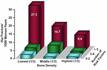
Figure 30.1 shows that no matter what a patient’s BMD is, three or four risk factors increase the risk of fracture [1]. This was noted even in patients within the highest tertile of BMD values, which included those with normal BMDs. This was evident on the patient FRAXTM calculations (20 % and 9 % for all osteoporosis-related and hip fractures, respectively). However, FRAXTM may have in fact underestimated the patient’s fracture risk, particularly as she had three further risk factors for a fracture, namely acute withdrawal of hormone replacement therapy, chronic kidney disease, PPI use, and TCA/SSRI use. Therefore, aggressive therapy was mandated.


Fig. 30.1
Effect of the number of risk factors and bone mineral density on hip fracture risk. Redrawn from [1]
3.
No matter what one’s BMD is, the older one gets, the higher the risk of fracture is at all sites. This effect of age is in part due to an increased propensity to fall in older individuals. However, there occur changes in structural and material properties of bone as one gets older (Table 30.2). These changes alter bone strength and increase its propensity to fracture. The implication of this is that if there is a T-score of −2.6 in a patient aged 80 years, his/her fracture risk is considerably higher than a patient who is 55 years of age with the same T-score. In other words, while a 14.1 % absolute fracture risk over 10 years, per FRAXTM, requires a T-score of −3.0 at age 50 years, about the same fracture risk (14.6 %) at age 70 years just requires a T-score of −1.5. Therefore, the requirement of a low T-score to produce a given risk of fracture diminishes as one gets older, so that older patients, both men and women, fracture at considerably conserved T-scores.
Table 30.2
Changes in structural and material properties of bone with age leading to reduced bone strength
Structural properties |
Thinning of cortical bone |
Thinning and loss of trabecular structures |
Increased cortical porosity |
Material properties |
Reduced bone mineral density |
Altered collagen crosslinks and phosphorylation pattern
Stay updated, free articles. Join our Telegram channel
Full access? Get Clinical Tree
 Get Clinical Tree app for offline access

|

