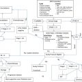
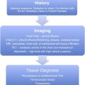
Stage III non–small cell lung cancer represents a heterogeneous group of patients who are best managed with a multidisciplinary approach, including evaluation for surgical, radiation, and chemotherapeutic options.
Key points
- •
Non–small cell lung cancer is a serious health condition requiring multidisciplinary input from surgical oncology, radiation oncology, and medical oncology.
- •
Appropriate staging studies are required to develop the optimal strategy for an individual patient. This strategy includes positron emission tomography/computed tomography scan and endobronchial ultrasonography or mediastinoscopy for locally advanced disease to determine resectability.
- •
Chemoradiation is the standard approach in stage III lung cancer, with surgery as an option in some cases. Concurrent chemoradiation has been proved to be superior to sequential therapy.
- •
Cisplatin doublet therapy is considered standard for chemotherapy selection.
- •
Pathologic review of biopsy samples now includes testing for EGFR gene mutations and EML4-ALK gene rearrangements.
Introduction
According to the American Cancer Society, there were an estimated 222,520 new cases of lung cancer in 2010. It is the second most prevalent cancer in men and women, behind prostate cancer and breast cancer, respectively. It is responsible for the most cancer-related deaths in both men and women. Lung cancer was responsible for 157,300 deaths in 2010. The mortality data are impressive when compared with other cancers ( Table 1 ).
| Cancer | Incidence | Mortality |
|---|---|---|
| Lung | 222,520 | 157,300 |
| Colorectal | 142,570 | 51,370 |
| Breast | 209,060 | 40,230 |
| Prostate | 217,730 | 32,050 |
| Pancreatic | 43,140 | 36,800 |
| Colorectal, breast, prostate, pancreatic combined | 612,500 | 160,450 |
There are nearly as many deaths from lung cancer yearly as there are from the combined total of colorectal, breast, prostate, and pancreatic cancers. As a contributing factor to the high mortality, many patients present with advanced disease. According to the National Cancer Database report on lung cancer, 30% of patients with non–small cell lung cancer (NSCLC) present with locally advanced, stage IIIA/B disease, and 40% of patients present with metastatic disease at diagnosis.
This article reviews staging and clinical evaluation of patients with lung cancer, with a focus on those with locally advanced disease. Patients should have routine staging studies, including pathology review, history, and physical examination, computed tomography (CT) of the chest and upper abdomen (including adrenals), positron emission tomography (PET) or integrated PET-CT, bronchoscopy, evaluation of the mediastinum by mediastinoscopy or endobronchial ultrasonography (EBUS)/endoscopic ultrasonography as recommended by National Comprehensive Cancer Network (NCCN) guidelines plus brain magnetic resonance imaging (MRI). In addition, for some locally advanced, but potentially resectable cancer, chest MRI may delineate the depth of invasion better (Pancoast tumor, pericardial invasion, involvement of vertebral bodies).
Most patients with stage III disease are recognized based on clinical staging, especially because the addition of integrated PET-CT to the staging armentarium has increased the accuracy of preoperative evaluation. A few patients are found on pathologic review to have unsuspected N2 or N3 disease; this usually constitutes microscopic metastasis that is too small to be fluorodeoxyglucose (FDG)-avid and, therefore, PET-negative. Once the pretreatment clinical staging has been completed, patients should be discussed at a multidisciplinary tumor board, which includes but is not limited to medical oncology, radiation oncology, and a dedicated thoracic surgeon. In addition, it improves patient care and communication if representatives from pulmonary medicine, pathology, social services, and radiology are present.
Surgical evaluation
Advanced local and regional cancer includes the following: any T stage with ipsilateral (N2; stage IIIA) or contralateral (N3; stage IIIB) or any scalene or supraclavicular node involvement (N3; stage IIIB). In addition, tumors that are more than 5 cm (T2b, T3>7.0 cm) or any tumor that directly invades resectable structures (T3 invasion) should be considered locally advanced and require a multimodality approach to increase the cure rate. Some tumors that invade the mediastinum, heart, great vessels, trachea, recurrent laryngeal nerve, esophagus, vertebral body, or carina (all T4) may be resectable. Some lobe nodules (formerly considered T4; TNM Classification of Malignant Tumours, Sixth Edition ) are now surgical disease (T3; stage IIB if node-negative).
Special consideration should be given to separate tumor nodule(s) in a different ipsilateral lobe to that of the primary. In the TNM Classification of Malignant Tumours, Sixth Edition , this condition was considered incurable stage IV disease (M1); in the current edition, after reviewing more than 100,000 patients with lung cancer and stratifying them according to survival, this condition is now considered T4 disease and potentially curable by bilobectomy or pneumonectomy. Most locally advanced lung cancer constitutes stage IIIA and B disease.
Stage IIIA (positive ipsilateral N2 nodes, T3 invasion [N1 or N2], T4 extension, and N1 disease [+N2, considered stage IIIB]) is considered for multimodality treatment (ie, neoadjuvant chemoradiation followed by restaging and possible resection). Consideration of possible resection requires special attention on how to stage the mediastinum. Based on the findings that the positive predictive value of integrated PET-CT is only in the range of 60%, patients need cytologic or pathologic confirmation of N2 disease. Mediastinoscopy has been considered the gold standard for this purpose. It provides pathologic evaluation of FDG-avid lymph nodes in the paratracheal compartment (including subcarinal, ie, nodal station 7); at least 3 nodal stations should be biopsied during mediastinoscopy. Surgical mediastinoscopy is not repeatable; after neoadjuvant chemoradiation and restaging (integrated PET-CT), FDG uptake in the mediastinum may be caused by tumor necrosis and not recalcitrant disease. EBUS has enormous value. It only provides cytologic evaluation, but is repeatable before and after induction chemoradiation. Because surgical mediastinoscopy is still considered the gold standard of N2/N3 evaluation, mediastinoscopy can be reserved for the final evaluation after neoadjuvant therapy before surgical resection.
Stage IIIA disease is overall a heterogeneous group; on the one end of the spectrum, it encompasses patients with single nodal station microscopic disease, and on the other hand, patients with bulky multistation involvement (ie, subcarinal plus levels 2 and 4 on the right and levels 5, 6, and 7 and L2, L4 on the left). Surgical therapy alone has a poor outcome. The exception to this claim is nonclinical N2 disease, which means micrometastasis in a single nodal station on final pathologic review after lobectomy and thoracic lymphadenectomy, with negative CT and integrated PET-CT beforehand. The Intergroup trial INT0139 confirmed that surgery (lobectomy, but not pneumonectomy) has a role after induction concurrent chemoradiotherapy. Based on several studies, the 5-year survival for stage IIIA NSCLC can vary between 3% (clinical N2 disease; bulky, multiple stations) and 34% (single-station N2 involvement, microscopic disease). Anatomic resection for advanced lung cancer can be performed either with a minimally invasive approach (video-assisted thoracic surgery [VATS]) or through a traditional posterolateral thoracotomy. Because more experience has been gained with VATS, this approach can be offered even after induction chemotherapy and radiation therapy. Many trials require an open approach (ie, thoracotomy) and coverage of the bronchial stump with an intercostal muscle flap to prevent the complication of a bronchopleural fistula. It has been shown in the adjuvant setting that if the VATS approach is used, chemotherapy can be given earlier because of the expedited recovery from a minimally invasive approach. This strategy may translate into an overall survival benefit, because more patients are likely to finish adjuvant therapy.
With the contemporary staging (integrated PET-CT, EBUS), 85% of advanced lung cancers are assigned the correct stage before treatment. Only a few undergo upfront surgical resection and on pathologic examination are upstaged to a higher stage. Surgery does play a role in the treatment of advanced lung cancer as part of a multimodality approach. Stage IIIA disease, the most common stage of advanced NSCLC for which surgery could be considered, is heterogeneous and, therefore, survival rates vary widely (depending on N2 disease burden).
For patients with clinical stage IIIA disease (T1–3, N2), the standard of care is definitive concurrent chemoradiation (category I recommendation in the NCCN guidelines). For stage IIIB, the standard of care is definitive chemoradiation followed by chemotherapy.
Once the standard for chemoradiation therapy was established, several studies were performed to evaluate concurrent versus sequential treatment. One of the key studies was LAMP (Locally Advanced Multimodality Protocol), a randomized phase II study. It was closed prematurely because of limited accrual but did provide some important data and median follow-up of approximately 40 months. The study enrolled 257 patients with unresectable stage III NSCLC and randomized the patients to 1 of 3 arms: sequential chemotherapy followed by radiation therapy, induction chemotherapy followed by concurrent chemoradiation, or concurrent chemoradiation followed by consolidative chemotherapy. The median survival for the 3 groups was 13, 12.7, and 16.4 months, respectively. The differences were not statistically significant different between the arms, although the data suggest an improved outcome in the third group.
Radiation therapy is a treatment modality that directs ionizing radiation toward tumors, mostly malignant, with the objective of tumor cell kill. The goal of radiation therapy is to deliver a maximally safe dose to the target volume and minimize dose to surrounding normal structures; the target volume is delineated by the radiation oncologist using diagnostic imaging. The total dose is divided into multiple treatments, or fractions, so that the same dose is delivered each day.
In NSCLC, radiation therapy is used either in the postoperative setting, as definitive treatment with or without chemotherapy, or in a palliative manner. In patients with early-stage and potentially resectable tumors who are medically inoperable, stereotactic ablative radiation therapy may be a reasonable treatment option. This technique delivers a high dose per fraction over a shorter period and fewer treatments.
In locally advanced NSCLC, the standard of care is concurrent chemoradiation therapy. These patients typically receive daily treatment, 5 days per week, with the same dose per fraction for approximately 6 weeks. Patients requiring radiation therapy for palliation of symptoms, such as obstruction of the mainstem bronchus, are commonly treated over 1 to 3 weeks.
Radiation therapy is delivered by either teletherapy or brachytherapy. Teletherapy typically uses a linear accelerator, which is situated approximately 100 cm from the patient; the large machine emits radiation and is directed toward the target. Teletherapy is the primary method of treatment delivery in patients with locally advanced lung cancer. Brachytherapy uses a radioactive source, which is juxtaposed against, or within, the tumor. An example of brachytherapy in lung cancer is endobronchial radiation, delivered with the assistance of a bronchoscope, in patients with an obstructing intraluminal mass.
Treatment planning for external beam radiation therapy with teletherapy is CT-based. A CT simulation is performed with the patient in the treatment position, thus simulating how the actual treatment is delivered. Patients are often supine with arms raised above their head, resting on the treatment table, so that the arms do not obstruct any portion of the torso. A wing board is a common device used at the time of simulation; it is placed under the patient’s upper body and has a spot for a head rest as well as adjustable bars to grasp with the hands. A customized mold can also be created for the torso to help ensure that the patient is lying in the same position daily. Once the scan has been completed, the patient is marked with small but permanent tattoos as a positioning verification tool that is checked each day when setting the patient up for treatment. The images are linked to the treatment-planning software, and the physician delineates the target volumes, and surrounding normal tissue structures, on each axial CT slice. Next, a treatment plan is generated to deliver a maximally safe dose to the tumor and minimize dose to the surrounding critical structures; Figs. 1 and 2 show an example of 2 treatment fields in a patient with locally advanced NSCLC.


