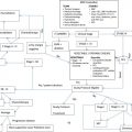
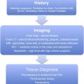
The standard treatment of limited-stage small cell lung cancer (SCLC) is concurrent cisplatin and etoposide with thoracic radiation therapy, whereas treatment of extensive-stage disease is typically chemotherapy alone with a platinum compound plus etoposide. Surgical resection of early disease is generally reserved for patients with small, node-negative disease. Prophylactic cranial irradiation reduces the development of brain metastases and prolongs survival in patients with both limited-stage and extensive-stage disease who have responded to chemotherapy. Further understanding of the molecular underpinnings of SCLC is necessary to develop better treatment options and improve outcomes for patients.
Key points
- •
Small cell lung cancer is an aggressive malignancy with a propensity for rapid growth and early metastases.
- •
Concurrent chemotherapy and thoracic radiation therapy are the standard treatment option for patients with limited-stage small cell lung cancer.
- •
Surgical resection is an option for very early disease (typically small lesions with uninvolved lymph nodes), but there are conflicting results in the literature on its usefulness.
- •
Extensive-stage small cell lung cancer is treated with combination chemotherapy, which improves survival and quality of life; the usual first-line treatment regimen is etoposide plus a platinum compound.
- •
Prophylactic cranial irradiation prolongs survival in patients with limited-stage and extensive-stage disease and should be considered in all patients who respond to initial chemotherapy.
Introduction
More than 226,000 cases of lung cancer will be diagnosed in 2012, with more than 160,000 deaths attributed to this diagnosis, making it the second most common cancer and the most common cause of cancer death in the United States. The epidemiology of lung cancer has been shifting, given the decline in cigarette smoking in this country; although small cell lung cancer (SCLC) was previously a common histologic subtype, it now represents approximately 13% of all cases. Also because of changes in smoking trends, SCLC used to be a disease found predominantly in men, but currently it is seen equally in men and women.
SCLC has a distinct natural history and treatment response pattern compared with non–small cell lung cancer (NSCLC). It tends to have a rapid doubling time and high propensity for early metastases. In contrast to NSCLC and many other solid tumors, it is commonly very chemosensitive initially, although it almost always recurs after a period of response. Given the various specialists involved in the care of patients with SCLC, it is clear that the evaluation and treatment of this disease require a multidisciplinary approach.
SCLC background
Pathologic Diagnosis
SCLC exists along a continuum of other neuroendocrine neoplasms of the lung, with carcinoid at one end of the spectrum, given its indolent nature, and large cell neuroendocrine carcinoma and SCLC at the other end, given their rapid growth and aggressive behavior. Histologically, SCLC typically appears as small round or oval blue monotonous cells with hyperchromatic nuclei, a salt-and-pepper chromatic pattern, and foci of necrosis. The mitotic count is frequently high, indicating the rapid growth kinetics that is seen clinically. Almost all SCLCs stain positively for keratin, thyroid transcription factor 1, and epithelial membrane antigen. Markers of neuroendocrine differentiation are also commonly seen, including chromogranin A, synaptophysin, CD56, and neuron-specific enolase. A subset of NSCLC also stains positive for neuroendocrine markers, so histologic as well as immunohistochemical evaluation is important in diagnosis: tumors that appear to be NSCLC by light microscopy but stain with neuroendocrine markers may be classified as having neuroendocrine differentiation, whereas tumors with neuroendocrine histologic features as well as staining pattern are considered SCLC or large cell neuroendocrine carcinoma.
Staging
SCLC can be staged based on the TNM staging system that is used for NSCLC, but more commonly it is divided into just 2 stages: limited-stage SCLC (LS-SCLC) and extensive-stage SCLC (ES-SCLC) based on the Veterans Administration Lung Group’s classification introduced in 1957. In this staging system, LS-SCLC is defined as disease that is confined to the ipsilateral hemithorax and is safely encompassable within a radiation field (including potentially contralateral mediastinal and ipsilateral supraclavicular nodes), whereas ES-SCLC is considered disease that is not encompassable within a radiation field, typically beyond the ipsilateral hemithorax, including malignant pleural or pericardial effusion or distant metastases. Recently, there has been an impetus toward using TNM staging for SCLC, given the prognostic information it provides as well as the usefulness when surgical resection is being considered ; however, most practitioners continue to use the limited or extensive staging classification, because treatment options tend to be the same within just those 2 groupings.
To fully stage patients, those with LS-SCLC should have a positron emission tomography (PET) scan to rule out occult nodal or metastatic disease, which occurs in more than 30% of patients, as well as brain imaging, given the propensity of SCLC to travel to the central nervous system. More than half of patients with SCLC are diagnosed with extensive-stage disease, a point at which their disease is incurable. This pattern has not changed significantly over the past few decades, but may change in the future with the new lung cancer screening guidelines. Patients with ES-SCLC may benefit from brain imaging because the incidence of asymptomatic brain metastases is common at this stage; PET scans may not be necessary once patients are already determined to have distant disease.
Signs and Symptoms
Most SCLCs present with bulky hilar and mediastinal lymphadenopathy; extrinsic bronchial compression often occurs, but the presence of an endobronchial component is rare. The symptoms at presentation can often be attributed to this pattern of disease and commonly include dyspnea, cough, chest pain, and pulmonary infections. Because patients commonly present with metastatic disease, presenting symptoms are often attributable to involved sites: bone pain, neurologic symptoms, and systemic symptoms such as anorexia, weight loss, and fatigue.
Paraneoplastic Syndromes
Paraneoplastic syndromes are associated with SCLC but their presence is relatively uncommon. Patients with syndrome of inappropriate antidiuretic hormone present with low serum sodium levels, which may or may not be symptomatic. Cushing syndrome can be caused by adrenocorticotropic hormone production. Several neurologic syndromes also associated with SCLC, including encephalomyelitis and sensory neuropathy from anti-Hu antibodies, and Lambert-Eaton syndrome (usually resulting in proximal muscle weakness) from antibodies against voltage-gated calcium channels and SOX proteins. Anti-Ri, Ma and Ta antibodies, among others, have also been associated with paraneoplastic syndromes. Although treatment of the underlying malignant disease frequently improves the paraneoplastic endocrine syndromes, often the neurologic syndromes remain active even with successful treatment of the malignancy and may require immunosuppression.
Limited-stage disease
Overview
Long-term cure is possible in patients with LS-SCLC, but only in a small subset of patients: in 1998, the population 5-year survival rate was 10%, up from 4.9% in 1973. In a landmark randomized trial published in 1999, a 5-year overall survival (OS) rate of 25% was achieved. Significant research has been undertaken to prolong survival and increase the cure rate, with modest success in recent years.
The Role of Surgery
Although surgical resection of early-stage disease is the standard treatment of NSCLC, surgery has a more controversial role in the management of SCLC ( Table 1 ). Two randomized, prospective trials have examined the use of surgical resection of SCLC, one performed by the British Medical Research Council (BMRC) and the other by the Lung Cancer Study Group (LCSG).
| Study | N | Trial Design | Treatment | Survival Outcome | Note |
|---|---|---|---|---|---|
| BMRC | 166 | Prospective, randomized | Surgical resection vs TRT | Mean survival of 199 d vs 300 d ( P = .04); 5-y survival of 1% vs 4% (NS) | Only 20% of patients in the surgery arm and 12% in the TRT arm received CT |
| LCSG | 328 | Prospective, randomized | CT → surgical resection → TRT vs CT → TRT | Median survival 15.4 mo vs 18.6 mo (NS) | Excluded patients with peripheral nodules as the only site of disease |
| Rea et al, 1998 | 104 | Prospective, single-arm | Surgical resection | 5-y survival (%): stage I 52.2; stage II 30; stage III 15.3 | All patients received CT (induction for stage III or adjuvant for stage I or II) followed by TRT |
| IASLC | 339 | Retrospective, multicenter database analysis | Complete R0 surgical resection | 5-y survival (%): IA 56; IB 57; IIA 38; IIB 40; IIIA 12; IIIB 0 | Most patients presumably received platinum-based CT |
| Brock et al, 2005 | 82 | Retrospective, single-institution analysis | Surgical resection | 5-y survival of 42% (62% among those who also received platinum-based CT) | 77% of patients received induction or adjuvant CT |
| Rostad et al, 2004 | 29 | Retrospective, Norway Cancer Registry analysis | Surgical resection | 5-y survival of 44.9% | Only stage IA and IB tumors were included (<2% of patients with SCLC); 62% received adjuvant CT |
The BMRC trial randomized 166 operable patients to resection versus thoracic radiation therapy (TRT). In the surgery arm, 48% of patients had a pneumonectomy with complete resection achieved, 34% underwent a thoracotomy without resection, and 18% did not undergo surgery (because of clinical decline before surgery or patient refusal). In the radiation group, 85% of patients underwent the prescribed course, whereas 11% were given a palliative course and 4% did not receive radiation because of disease deterioration or refusal. Only 20% and 12% of patients in the surgery and radiation arm, respectively, received cytotoxic chemotherapy. The results of this trial favored the radiation arm, with mean survival of 300 days compared with the surgery group, which had a mean survival of 199 days ( P = .04). Although not statistically significant, the 5-year and 10-year survival also favored radiation (4% survival at 5 and 10 years in the radiation group vs 1% at 5 years and 0% at 10 years in the surgery group). Even among the 34 patients who underwent a complete resection, long-term outcomes were poor: only 2 (6%) were alive at 2 years and none was alive at 5 years.
The LCSG trial looked at a different question: whether surgical resection was beneficial after induction chemotherapy compared with chemotherapy alone in LS-SCLC. Three hundred twenty-eight patients with limited-stage disease (excluding supraclavicular lymph nodes) were enrolled; those with peripheral nodules were excluded. All patients received cyclophosphamide, doxorubicin, and vincristine for 5 cycles, and those who achieved an objective response and were considered operable (a total of 146 patients) were randomized to surgery followed by radiation to the chest and brain versus radiation alone. Median survival was 15.4 months in the surgery group and 18.6 months in the nonsurgery group ( P = .78). Two-year survival for the entire population was 20%. Because of small sample sizes in each of the TNM stages, it was not possible to obtain comparative survival analysis of the surgical versus the nonsurgical group by stage, but among those who underwent surgical resection, there appeared to be a longer median survival in those with T1-2, N0 disease compared with those with T3 disease or nodal involvement.
Although the BMRC and LCSG trials are the only source of prospective randomized data for surgery versus radiation in SCLC, there are several points that shed doubt on applying the results to all patients. The BMRC trial was performed in the 1960s, without advanced imaging such as computed tomography and PET, likely putting many patients with occult metastatic disease into the operable category. In addition, few patients received chemotherapy, whereas currently adjuvant chemotherapy is routinely administered in addition to resection. Thus, patients overall did poorly, with a mean survival of less than 1 year in both groups and few long-term survivors. However, given the statistically significant results of radiation, resulting in a higher mean survival, this trial set the practice standard of avoiding surgery in SCLC for many years. The LCSG study did include treatment with chemotherapy and radiation, but excluded patients with peripheral nodules, which is the patient population that might be expected to benefit from surgical resection.
Since the results of these trials were published, several other retrospective and single-arm prospective studies have emerged that suggest a benefit to surgical resection of early-stage SCLC (many of which have also included induction or adjuvant chemotherapy), with 5-year survival ranging from 27% to 68%. The benefit to surgery in most of these trials appeared to be in patients without nodal disease (ie, stage I patients). This finding has swayed the practice standard toward resection for select patients (particularly those with small peripheral lesions and no nodal involvement who likely have a different disease biology than those with the more typical SCLC presentation of central, bulky disease), although definitive prospective data confirming the benefit to resection have not been published.
In practice, the question of whether to resect SCLC rarely presents itself. Patients more commonly present with unresectable or advanced disease, or occasionally present with a small nodule that is resected and incidentally is found to be of small cell histology. In the latter situation, adjuvant chemotherapy should be administered when feasible: in 1 retrospective study, adjuvant chemotherapy (plus prophylactic radiation to the brain and mediastinum in a subset of patients) after surgery resulted in a median survival of 20 months, longer than in historic controls of surgery alone. In the rare circumstance that a patient presents with a biopsy-proven SCLC that is amenable to resection, a multidisciplinary approach is crucial, given the various treatment options and the paucity of definitive data.
The Role of Radiation and Chemotherapy
Generally, LS-SCLC is not considered a resectable disease. Therefore, standard of care for these patients is concurrent chemotherapy and TRT, which was shown to be beneficial compared with chemotherapy alone in 2 meta-analyses in the early 1990s (see Box 1 for details of chemotherapy regimens). The meta-analysis by Pignon and colleagues used data from 13 trials to compare patients with LS-SCLC who were randomized to receive chemotherapy alone or chemotherapy plus TRT. The investigators found a relative risk of death of 0.86 ( P = .001) and a 5.4% increase in OS at 3 years with the inclusion of radiotherapy (14.3% vs 8.9%). The greatest benefit was found in patients younger than 55 years, with an 8.2% increase in 3-year survival rate with the addition of radiation. The other meta-analysis by Warde and Payne included 11 trials that compared chemotherapy with and without radiotherapy in limited-stage disease. This study also showed a benefit with the addition of TRT, with an odds ratio of 1.53 and an overall improvement in 2-year survival of 5.4% ( P <.05).
LS-SCLC
- •
Cisplatin 80 mg/m 2 intravenously (IV) day 1 and etoposide (EP) 100 mg/m 2 IV days 1, 2, 3, every 28 days (with concurrent TRT)
- •
Cisplatin 60 mg/m 2 IV day 1 and EP 120 mg/m 2 IV days 1, 2, 3, every 28 days (with concurrent TRT)
ES-SCLC (Initial Therapy)
- •
Cisplatin 75 to 80 mg/m 2 IV day 1 and EP 100 mg/m 2 IV days 1, 2, 3, every 21 days
- •
Carboplatin area under the curve (AUC) 5 to 6 IV day 1 and EP 100 to 140 mg/m 2 IV days 1, 2, 3, every 21 days
- •
Cisplatin 60 mg/m 2 IV day 1 and irinotecan 60 mg/m 2 IV days 1, 8, 15, every 28 days
- •
Cisplatin 30 mg/m 2 IV days 1, 8 and irinotecan 65 mg/m 2 IV days 1, 8 every 21 days
- •
Carboplatin AUC 5 IV day 1 and irinotecan 50 mg/m 2 IV days 1, 8, 15, every 28 days
ES-SCLC (Second-Line Therapy)
- •
Sensitive disease (relapse at >6 months)
- ○
Retreatment with initial regimen
- ○
- •
Sensitive disease (relapse at 3–6 months)
- ○
Topotecan 2.3 mg/m 2 /d by mouth days 1 to 5, every 21 days
- ○
Topotecan 1.5 mg/m 2 /d IV days 1 to 5, every 21 days
- ○
Irinotecan 100 mg/m 2 IV, weekly
- ○
Cyclophosphamide 1000 mg/m 2 IV/doxorubicin 45 mg/m 2 IV/vincristine 2 mg IV, every 21 days
- ○
Paclitaxel 80 mg/m 2 IV, weekly for 6 weeks, every 8 weeks
- ○
Docetaxel 100 mg/m 2 IV, every 21 days
- ○
Gemcitabine 1000 mg/m 2 IV, days 1, 8, 15, every 28 days
- ○
Vinorelbine 30 mg/m 2 IV, weekly
- ○
- •
Refractory disease (relapse at <3 months or no response to initial regimen)
- ○
Clinical trial
- ○
Consider other second-line regimen listed above
- ○
Note: other dosing and schedules exist for several regimens.
Stay updated, free articles. Join our Telegram channel

Full access? Get Clinical Tree


