Fig. 17.1
An unsuspected paraspinal mass in a patient with a history of toxic multinodular goiter and progressive low back pain. A paraspinal mass is shown at L3, with complete obliteration of the L3 vertebral body by tumor

Fig. 17.2
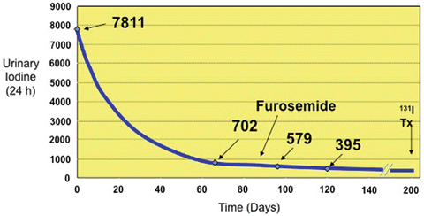
Prolonged clearance of iodine in a patient awaiting therapy with radioiodine following multiple exposures to iodinated contrast
Review of How the Diagnosis Was Made
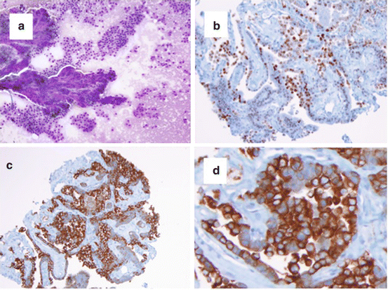
The diagnosis of papillary thyroid cancer is generally straightforward. Thyroid FNA is both sensitive and specific for the detection of papillary thyroid cancer within a thyroid nodule. However, occasionally the first presentation for thyroid cancer is with either locoregional or distant metastases. This patient’s paraspinal mass led to the discovery of an otherwise unsuspected thyroid cancer. CT-guided biopsy revealed a follicular lesion and was subjected to special immunohistochemical staining (Fig. 17.3) to pinpoint the primary source. Specifically, TTF-1 staining was positive, indicating either pulmonary or thyroid etiologies, and thyroglobulin staining was strongly positive, confirming metastatic thyroid cancer. Ultimately, thyroidectomy confirmed the presence of a small innocuous appearing primary papillary thyroid cancer.


Fig. 17.3
Histology and special staining of tumor obtained following CT-guided biopsy of a 4 cm paraspinal mass. (a) Typical appearance of papillary thyroid cancer; (b) TTF1 immunostaining; (c) thyroglobulin immunostaining, low magnification; (d) thyroglobulin immunostaining, high magnification
Lessons Learned
1.
Papillary thyroid cancer may have unusual presentations. When metastatic to bone, thyroid cancer may present with a large paraspinal mass, as occurred in the present case. Failure to perform special staining might have resulted in a misdiagnosis of adenocarcinoma of unknown primary and the patient would not have received therapy with radioiodine. Likewise, had the patient presented with colon or lung cancer metastatic to spine, he may not have been considered for the extensive tumor extirpation and spine stabilization performed in this case, which were only undertaken after consideration of the comparatively favorable prognosis and efficacy of treatment for thyroid cancer.
2.
Exposure to iodinated contrast results in a delay of the delivery of effective radioiodine therapy. This patient’s protracted and complicated hospital course necessitated exposure to iodinated contrast during the performance of diagnostic studies deemed critical to the patient’s acute management. The resultant iodine overload required a prolonged period of iodine restriction and introduction of furosemide therapy, which has been shown to augment urinary excretion of iodine in animal studies, to facilitate attainment of a low iodine state in preparation for radioiodine therapy.
3.


Spinal metastases from thyroid cancer present unique therapeutic challenges. Bone tissue provides a rich environment for metastatic disease, with high blood flow and elaboration of local growth factors in response to tumor damage to bone. Approximately 2 % of papillary thyroid cancer patients experience bone metastases compared to 7–20 % of patients with follicular thyroid cancer. The spine and pelvis are the most frequently involved sites, followed by the rib cage. Approximately half of these patients present with bone pain, 23 % are asymptomatic, and the remainder present with local edema, fracture, or cord compression. Patients with spinal metastases from thyroid cancer tend to present with radicular pain and less commonly, paresis or paraplegia. Paraspinal masses due to thyroid cancer metastases may be quite large and hypervascular. A 2012 series of 22 patients with spinal cord compression due to metastatic thyroid cancer showed clinical improvement in all patients following surgical intervention [1]. Patients with bone metastases from differentiated thyroid cancer tend to have higher serum thyroglobulin levels and 40–80 % have visible uptake on radioiodine imaging. Treatment modalities considered for bone metastases from thyroid cancer include surgery to remove the focus (extirpation), tumor embolization, radioiodine, external beam radiotherapy, and bisphosphonate therapy. Complete tumor extirpation has been shown to positively affect overall survival. In the present case, extensive spine stabilization was required to prevent acute cord compression in the course of surgical extirpation and radioiodine therapy. In general, patients with radioiodine uptake within bone metastases tend to be younger and have improved survival, although radioiodine therapy per se has not always been shown to improve survival in these patients. It has been argued that radioiodine may not be able to deliver a tumoricidal dose to boney metastases. In one example in the literature, dosimetric evaluation showed that a dose of 400 mCi 131I would deliver a radiation dose of 3,500 cGy to a thoracic spine metastasis, yet a 10,000 cGy would be needed for a tumoricidal effect. Tumor embolization provides prompt relief of pain and improvement in neurological symptoms in many patients without affecting long-term survival. Likewise, external beam radiation therapy can deliver high doses of radiation to a metastatic deposit in bone, resulting in pain relief but no demonstrable effect on survival. Bisphosphonate therapy for the treatment of bone metastases is discussed in another case in this section.
Stay updated, free articles. Join our Telegram channel

Full access? Get Clinical Tree


