The classification applies only to malignant mesothelioma of the pleura. There should be histological confirmation of the disease. The regional lymph nodes are the intrathoracic, internal mammary, scalene and supraclavicular nodes. The pT and pN categories correspond to the T and N categories. Note pM0 and pMX are not valid categories.
PLEURAL MESOTHELIOMA (ICD‐O‐3 C38.4)
Rules for Classification
Regional Lymph Nodes
TNM Clinical Classification
T – Primary Tumour
TX
Primary tumour cannot be assessed
T0
No evidence of primary tumour
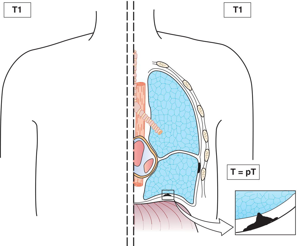
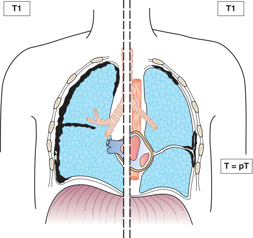
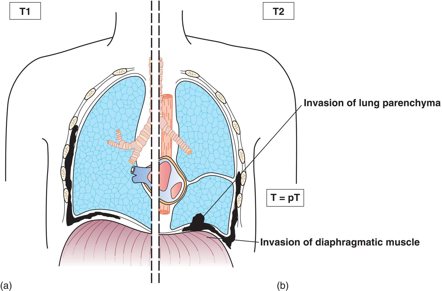
T1
Tumour involves ipsilateral parietal pleura only, with or without involvement of visceral, mediastinal or diaphragmatic pleura (Figs. 288, 289, 290a)
T2
Tumour involves the ipsilateral pleura (parietal or visceral pleura), with at least one of the following:
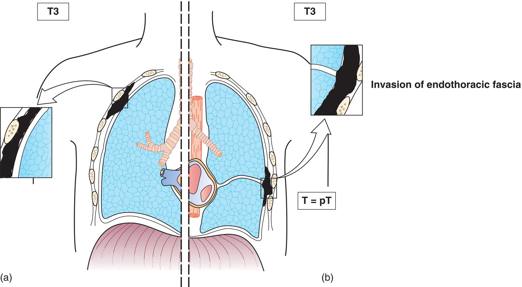
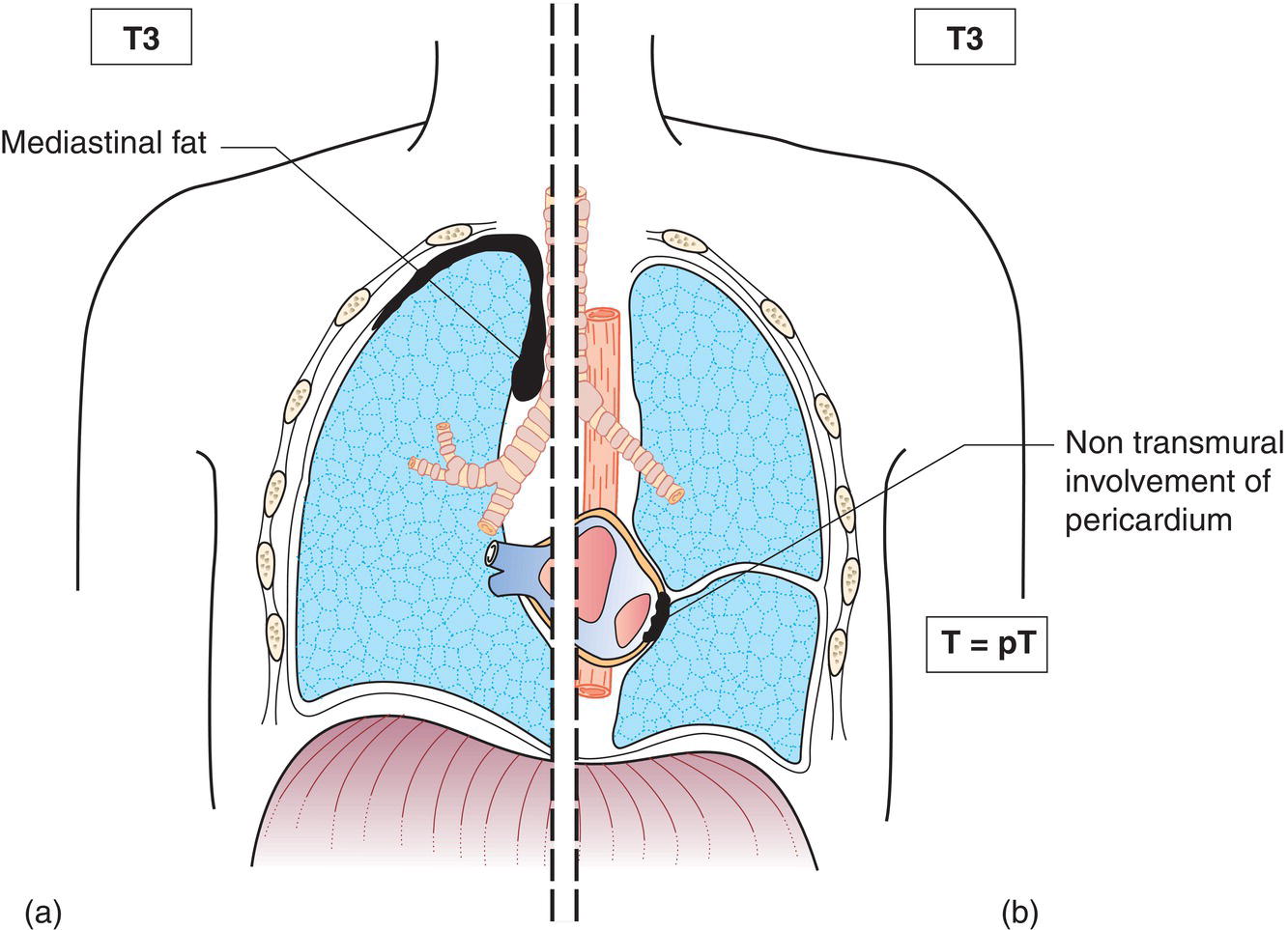
T3
Tumour involves ipsilateral pleura (parietal or visceral pleura), with at least one of the following:
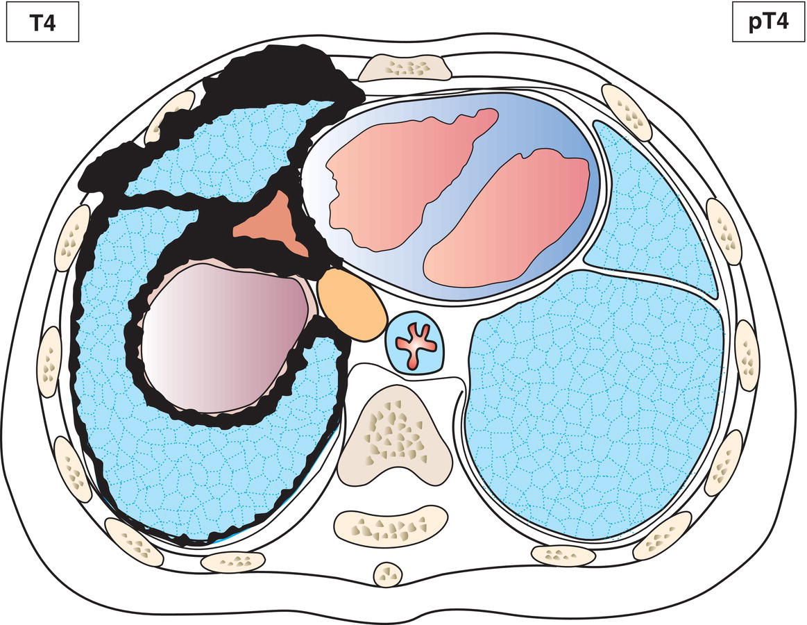
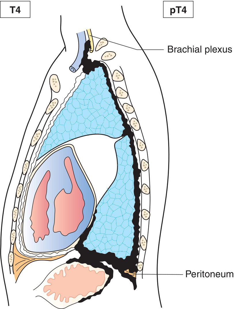
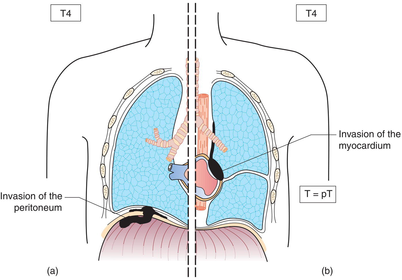
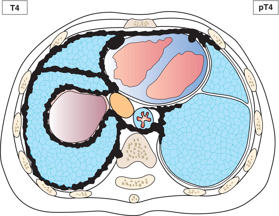
T4
Tumour involves ipsilateral pleura (parietal or visceral pleura), with at least one of the following:
N – Regional Lymph Nodes
NX
Regional lymph nodes cannot be assessed
N0
No regional lymph node metastasis
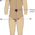
N1
Metastases to ipsilateral intrathoracic lymph nodes (includes ipsilateral bronchopulmonary, hilar, subcarinal, paratracheal, aortopulmonary, paraesophageal, peridiaphragmatic, pericardial fat pad, intercostal and internal mammary nodes)
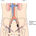
N2
Metastases to contralateral intrathoracic lymph nodes. Metastases to ipsilateral or contralateral supraclavicular lymph nodes
M – Distant Metastasis
M0
No distant metastasis
M1
Distant metastasis
pTNM Pathological Classification
pM1
Distant metastasis microscopically confirmed
Summary
Related posts:


Stay updated, free articles. Join our Telegram channel

Full access? Get Clinical Tree


