, Hans Guski2 and Glen Kristiansen3
(1)
Carl-Thiem-Klinikum, Institut für Pathologie, Cottbus, Germany
(2)
Vivantes Klinikum Neukölln, Institut für Pathologie, Berlin, Germany
(3)
Universität Bonn, UKB, Institut für Pathologie, Bonn, Germany
Ki-67:
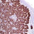
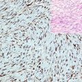
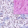
Ki-67 is a nonhistone nuclear protein expressed in active cell cycles. The expression of Ki-67 begins in the G1 phase and persists during the active phases of cell cycle throughout the S, G2, and M phases. Ki-67 is undetectable in the G0 phase or in the initial stage of the G1 phase and during DNA repair. The expression of Ki-67 strongly correlates with the intensity of cell proliferation and tumor grade. In routine histopathology, Ki-67 is an important marker for the assessment of cell proliferation. The Ki-67 index is an important criterion for tumor diagnosis (benign, borderline, malignant, low- or high-grade tumor). Furthermore, it is a helpful marker to differentiate between atrophy, thermal alterations, and dysplasia. The irregular accumulation of Ki-67-positive cells in different tissue types would suggest a tendency of the cells to escape the regulation mechanisms. In stratified squamous epithelium, the expression of Ki-67 in more than 30% of the full thickness of the epithelium above the suprabasal layers signifies an abnormal or dysplastic behavior of the epithelium. The Ki-67 index is also an important parameter to distinguish between high-grade and low-grade lymphoma.
p53:
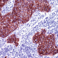
p53 is a nuclear phosphoprotein encoded by the TP53 gene located on chromosome 17p13, which in turn encodes several isoforms of the p53 protein. p53 is a tumor-suppressor protein that binds to DNA inducing the synthesis of the p21 protein, which regulates the genomic stability and binds to the cell division-stimulating protein cdk2. The p21-cdk3 complex hinders the cells to pass through to the next phase of cell division, which can activate the transcription of different preapoptotic genes and initiate the apoptosis.
Mutations within the TP53 cause the overexpression and accumulation of mutated p53 protein not able to bind DNA to stimulate the p21 synthesis acting as a stop signal in the cell cycle, consequently causing an uncontrolled proliferation of involved cells.
The overexpression of p53 is associated with different neoplastic and preneoplastic lesions. The detection of p53 by immunohistochemistry can be useful to differentiate between dysplastic or neoplastic changes usually positive for p53 and reactive changes negative for p53.
Stay updated, free articles. Join our Telegram channel

Full access? Get Clinical Tree


